Anatomy Of The Eyelid
Understanding these relationships is essential in eyelid surgery. It is 3 mm higher in asians.
The human eyelid features a row of eyelashes along the eyelid margin which serve to heighten the protection of the eye from dust and foreign debris as well as from perspiration.

Anatomy of the eyelid. Anatomy of the eyelids. Ullieach eyelid is divided by a. The thickness of skin at 7 mm above the eyelashes was 860305 µm.
Its key function is to regularly spread the tears. Skin and subcutaneous tissue. This can be either voluntarily or involuntarily.
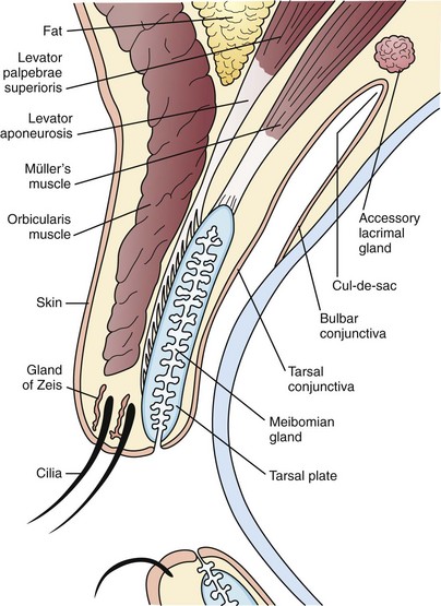
The description above only offers a superficial overview of the anatomy. The eyelid consists of five main layers superficial to deep. Overview of external anatomy.
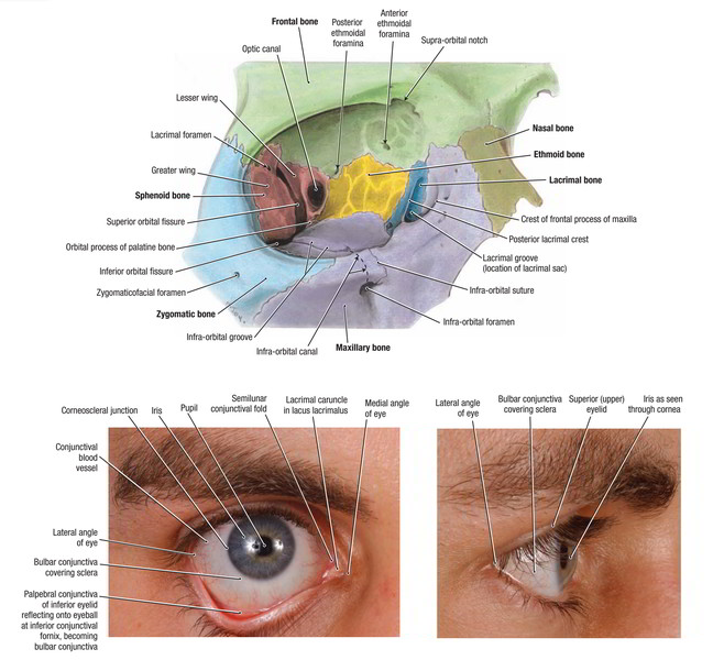
The eyelids comprise of an upper and lower eyelid joined at the medial and lateral canthi. Palpebral means relating to the eyelids. The adjacent forehead and midface influence correct eyelid positioning.
In youths the upper lid margin rests at the upper limbus while in adults it rests 15 mm below the limbus. Eyelid and orbit anatomy anatomy of the eyelid. The horizontal length of the fissure is 30 to 31 mm.
Ulliits key function is to regularly spread the tears and other secretions on. The average aperture of the eyelids measures about 30 mm in horizontal width and approximately 10 mm in vertical height. The lids move through the action of a circular lid closing muscle the orbicularis oculi and of the lid raising muscle the levator of the upper lid.
Anatomy of eyelid 1. Impulses for closing come by way of the facial seventh cranial nerve and for opening by way of the oculomotor third cranial nerve. Eyelid anatomy including thickness measurements was examined in numerous age groups.
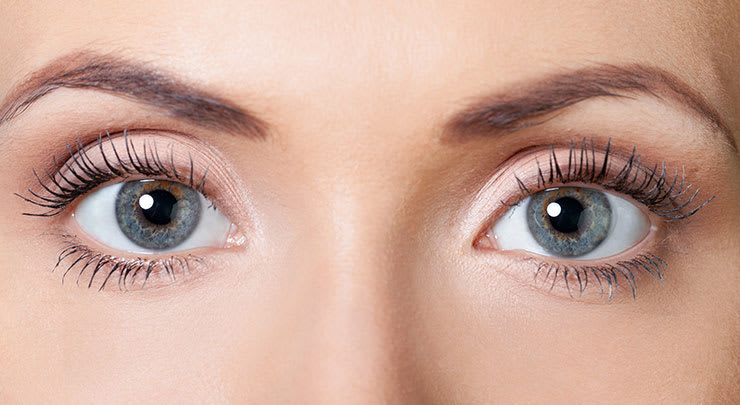
The eyelid is primarily made of skin. The upper and lower eyelids meet at an angle of approximately 60 degrees medially and laterally. An eyelid is a thin fold of skin that covers and protects an eye.
Skin and subcutaneous tissue. The eyelids provide globe protection contribute to tear production and distribute tears. The thickest part of the upper eyelid is just below the eyebrow 1127238 µm and the thinnest near the ciliary margin 32049 µm.
Ullian eyelid is a thin fold of skin that covers and protects an eye. In primary position of gaze the upper eyelid margin lies at the superior corneal limbus in children and 15 to 20 mm below it in the adult. The upper eyelid starts at the eye and extends up words joined the skin.
The levator palpebrae superioris muscle retracts the eyelid exposing the cornea to the outside giving vision. Free powerpoint templates anatomy of eyelid. The lateral canthal angle is 2 mm higher than the medial canthal angle in europeans.
The lower eyelid margin rests at the level of the lower limbus. The upper and lower eyelids along with the upper and lower puncta oppose the globe.
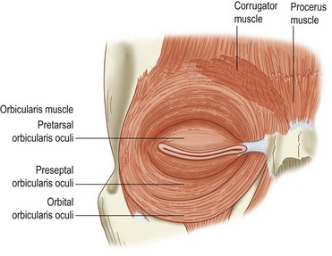 Eyelid Anatomy And Function Clinical Gate
 Figure 1 From Functional Considerations In Aesthetic Eyelid
Ptosis Pediatrics Clerkship The University Of Chicago
Eyelid Malpositions An Overview
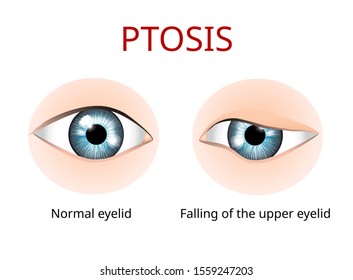 Eyelid Anatomy Images Stock Photos Vectors Shutterstock
 Eyelid Anatomy Ophthalmology Review
 Eyelid And Its Clinical Anatomy By Adrija Basu On Prezi
 Anatomy Of Lower Eyelid Of Humans Human Eye Anatomy Parts Of
 Amazon Com Anatomy Eye Surgery Eyelid Print Sra3 12x18
 Anatomy Of The Upper And Lower Eyelids Plastic Surgery Key
 Upper Eyelid Anatomy 2019 Update
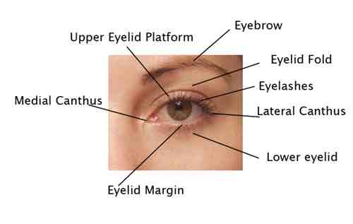 Facial Anatomy Plastic Surgery Beverly Hills Lidlift
 Eyelid And Orbital Anatomy Lecture At Keck School Of
 Eyelid Anatomy And Disorders 978 613 9 46141 7
 Lower Eyelid Surgery Vero Beach Lower Eyelid Surgery Melbourne
 An Easy Guide To Your Eye S Anatomy Lenstore Co Uk
Anatomy Of Eyelid Authorstream
 Lower Eyelid An Overview Sciencedirect Topics
 Easy Notes On Eyelids Learn In Just 4 Minutes Earth S Lab
 Anatomy Of The Eye Columbia Eye Clinic
 Upper Eyelid An Overview Sciencedirect Topics
 Anatomy Of The Eyelid Medical Illustration Medivisuals
 Upper Eyelid Anatomy 2019 Update





Belum ada Komentar untuk "Anatomy Of The Eyelid"
Posting Komentar