Maxillary First Molar Anatomy
It is the posterior tooth with the highest endodontic failure rate and unquestionably one of the most important teeth. The maxillary first molar is the tooth located laterally from both the maxillary second premolars of the mouth but mesially from both maxillary second molars.
The Permanent Maxillary Molars Dental Anatomy Physiology
Maxillary first molar anatomy the root morphology of the maxillary first molar includes a concavity leading into the furcation area between the two buccal roots figure 1.

Maxillary first molar anatomy. There are usually four cusps on maxillary molars two on the buccal and two palatal. 1 the palatal root is usually convex however it could contain shallow concavities. It should be noted that the left maxillary first molar had the same root canal anatomy figure 2.
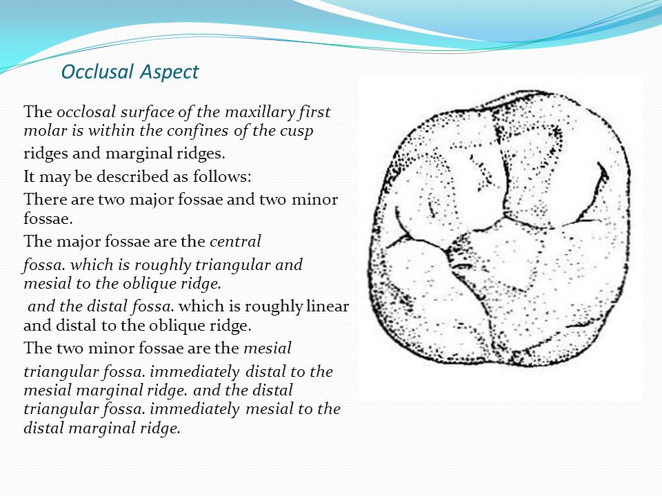
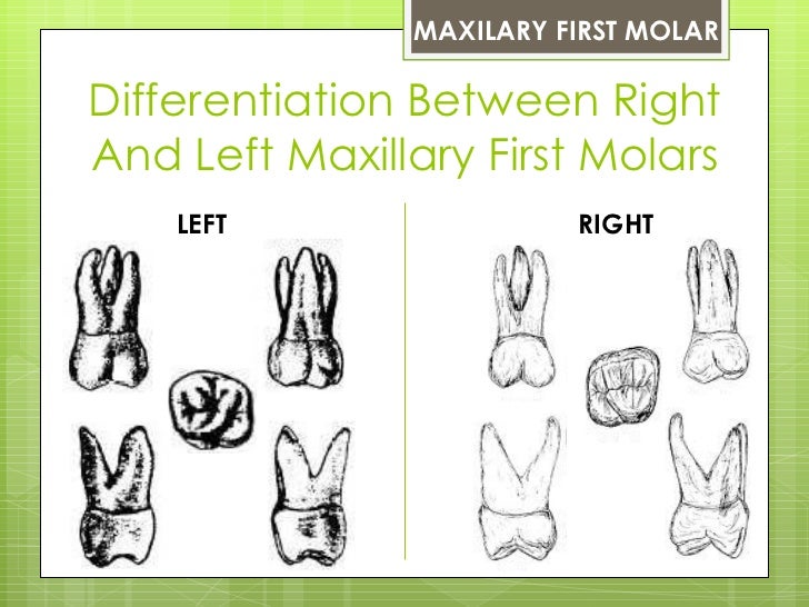
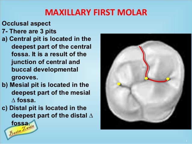
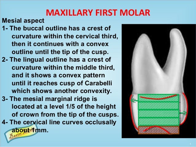
Maxillary first molar the tooth largest in volume and most complex in root and root canal anatomy the 6 year molar is possibly the most treated least understood posterior tooth. This tutorial describe the coronal anatomical features of the maxillary first molar from all aspects. The initial radiographic evaluation showed the presence of occlusal amalgam fillings and recurrent caries on the mesial surface as well as a two rooted maxillary first molar figure 1.
The buccal to lingual dimension. Which is larger the buccal surface of the maxillary first molar or the adjacent premolar. For more dental anatomy videos please see my channel.
The maxillary first molar is the human tooth located laterally away from the midline of the face from both the maxillary second premolars of the mouth but mesial toward the midline of the face from both maxillary second molars. The function of this molar is similar to that of all molars in regard to grinding being the principal action during mastication commonly known as chewing. Introductionthe permanent maxillary first molar the maxillary first molar is the tooth located laterally away from the midline of the face from both the maxillary second premolars of the mouth but mesial toward the midline of the face from both maxillary second molars.
Which is wider on the maxillary first molar the buccal to lingual dimension or the mesial to distal dimension.
 Maxillary First Molar Wikipedia
 Maxillary Molar Root Canal Morphology And Anatomy Key
 Pdf Evaluation Of Complex Mesiobuccal Root Anatomy In
 Step By Step Protocol Restoring The First Upper Molar
 Maxillary First Molar Doh114 Oral Anatomy Histology
 Maxillary Permanent First Molar Morphology
Occlusal Aspect The Mandibular First Molar Is Somewhat
 Permanent Molar Anatomy Images At Carrington College Studyblue
 Primary Dentition An Overview Of Dental Anatomy
The Permanent Maxillary Molars Dental Anatomy Physiology
 Hand Instrumentation Of First Molar Teeth Dimensions Of
 Human Dentition Dental Anatomy Physiology And Occlusion
The Permanent Maxillary Molars Dental Anatomy Physiology
Evaluation Of Anatomy And Root Canal Morphology Of The
 The Permanent Maxillary First Molar
 Review Of The Reported Cases For Maxillary First Molars With
 Maxillary Molars Flashcards Quizlet
 Maxillary First Molar Second Mesiobuccal Mb2 Root Canal
 Maxillary First Molar Second Mesiobuccal Mb2 Root Canal
Management Of A Maxillary First Molar Having Atypical
 Dental Anatomy Permanent Molars
 Analysis Of The Internal Anatomy Of Maxillary First Molars
The Permanent Maxillary Molars Dental Anatomy Physiology
Molars Dental Technology How To Tips


Belum ada Komentar untuk "Maxillary First Molar Anatomy"
Posting Komentar