Radiography Anatomy
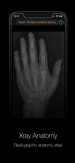
E anatomy is an award winning interactive atlas of human anatomy. It is meant for health care personnel who in their daily work are responsible for producing and interpreting radiographs be it radiologists or other medical specialists general practioners or radiological technologists working in rural areas.
 Review Of Normal Anatomical Landmarks And Variations
It is the most complete reference of human anatomy available on web ipad iphone and android devices.

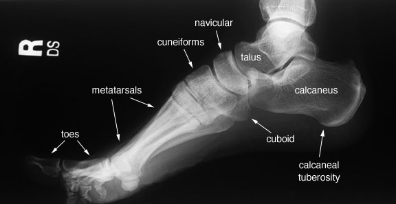
Radiography anatomy. Sarah nemanic dvm phd ms dacvr. From there the text discusses the basic principles of the paralleling technique use of xcp film holding devices processing panoramic radiography troubleshooting both technique and processing errors production of x rays image characteristics normal abnormal and common radiographic anatomy and radiology biology and prevention. Radiographic anatomy of the skeleton.
In the living subject it is almost impossible to study anatomy without also studying some physiology. Radiographic anatomy fractures and luxations involving the temporomandibular joint. Radiographic anatomy is a branch within the discipline of anatomy which involves the study of anatomy through the use of radiographic films also known as x rays.
Anatomy is the study classification and description of the structure and organs of the human body whereas physiology deals with the processes and functions of the body or how the body parts work. Explore over 5400 anatomic structures and more than 375 000 translated medical labels. Medical students typically spend some time studying radiographic anatomy during their general educations and certain medical specialists may go on to study it extensively such as radiographers orthopedic surgeons and dentists.
Radiography in a later stage also ultrasonography. The radiographic anatomy of owls is similar to other birds. Ct mri radiographs anatomic diagrams and nuclear images.
The x ray film represents two dimensional image of a three dimensional object due to the summary projection of different anatomical structures onto a planar surface. The radiographic anatomy and patient positioning. It requires certain skills.
We would like to show you a description here but the site wont allow us. Radioanatomy x ray anatomy is anatomy discipline which involves the study of anatomy through the use of radiographic films.
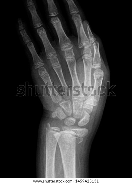 Wrist X Ray Anatomy Radiology Radiographic Stock Photo Edit
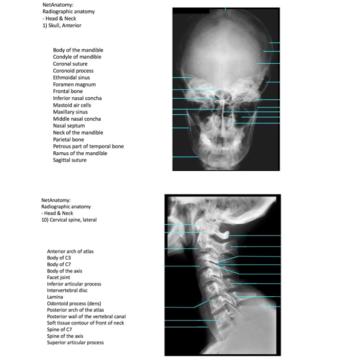
![]() Anatomy Study Guides For Radiology Technologists
Anatomy Study Guides For Radiology Technologists
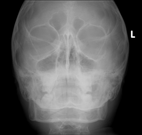
 Radiographic Anatomy Mandible Pa Radiology Medical
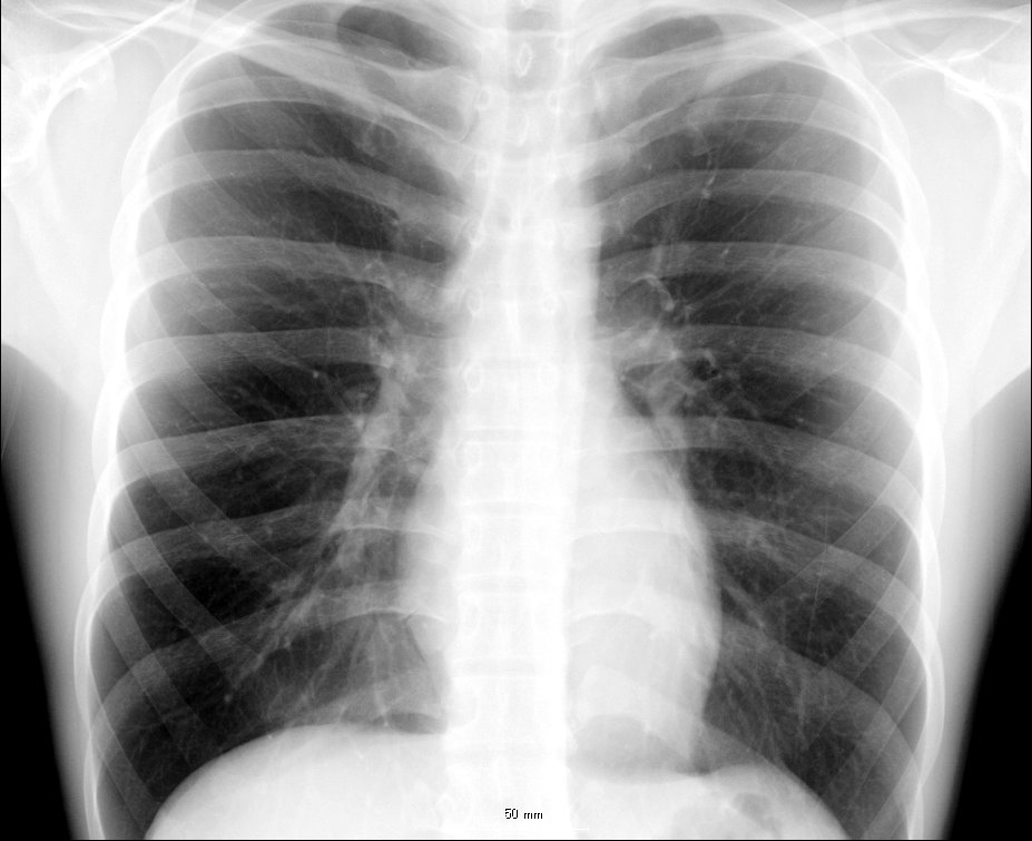
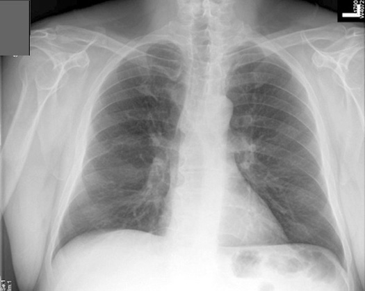
 The Radiology Assistant Chest X Ray Basic Interpretation
 Manus X Ray Anatomy Radiology Radiographic Stock Photo Edit
 Medical Imaging Technology Radiographic Anatomy Of Shoulder
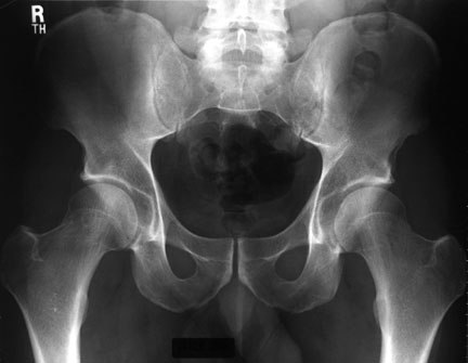
 Male Versus Female Pelvis Labeled Radiographic Anatomy
 Radiographic Anatomy Knee Ap Medical Anatomy
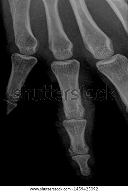
 Radiographic Anatomy Hand Lateral Radiology Medical
 Radiographic Anatomy Pelvis Ap Female My Next Practical I
![]() Medical Imaging And Radiological Anatomy X Ray Ct Mri
Medical Imaging And Radiological Anatomy X Ray Ct Mri
 Radiologic Anatomy Wayne State University School Of
 Radiographic Anatomy Of Adult Pelvis Orthopaedicsone
 Radiographic Anatomy Wikipedia
 Examples Of Clipped Anatomy For Both Examples The
 Normal Radiographic Anatomy Of The Wrist Radiology Case
 Radiology Anatomy
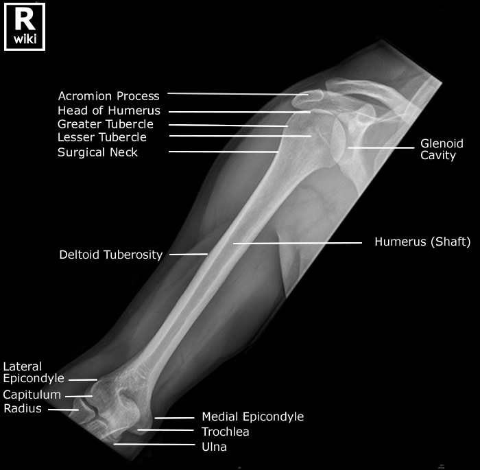 Radiographic Anatomy Of The Humerus Radiologypics Com
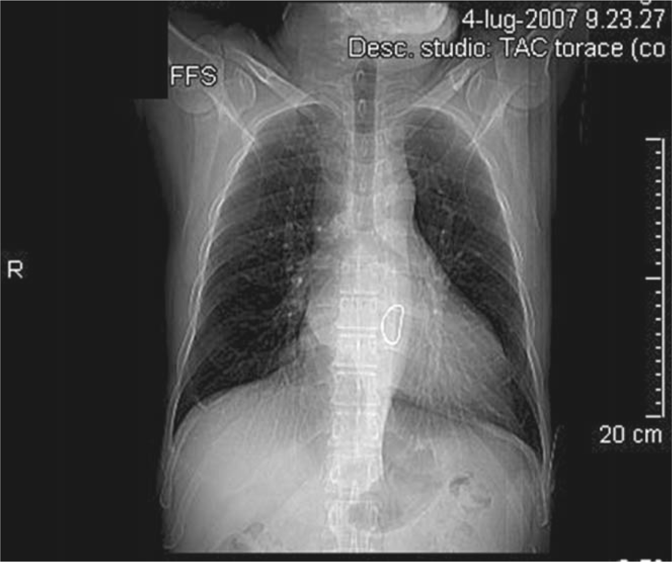 X Ray Anatomy Of The Heart Springerlink
 Radiographic Anatomy An Overview Sciencedirect Topics
 An Atlas Of Interpretative Radiographic Anatomy Of The Dog
Belum ada Komentar untuk "Radiography Anatomy"
Posting Komentar