Right Foot Anatomy
Click on a link to get sagittal view t1 axial view t2fatsat coronal view t2fatsat sagittal view t2fatsat. The talus which is the.
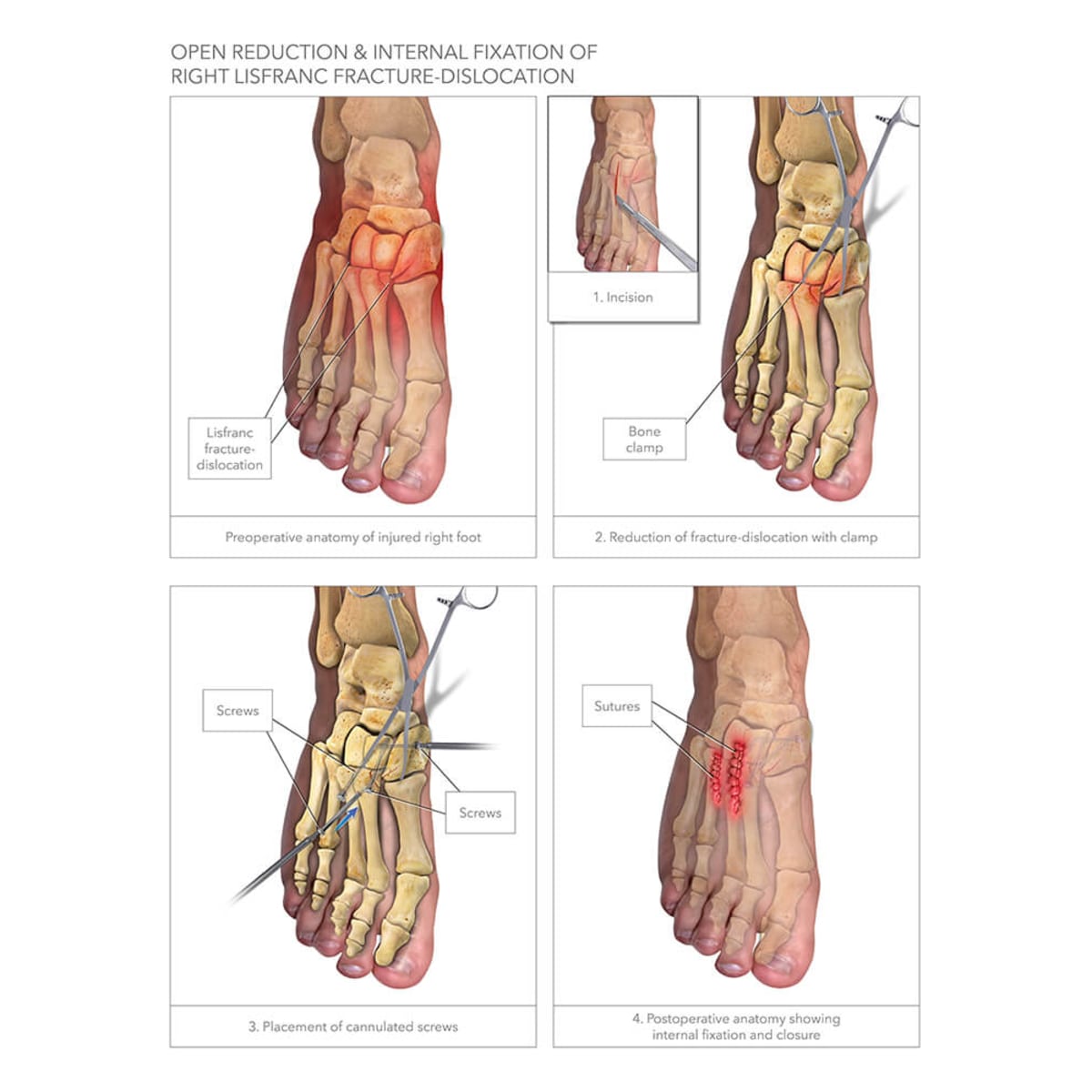 Right First Second Tarsal Metatarsal Tmt Joint Fusions
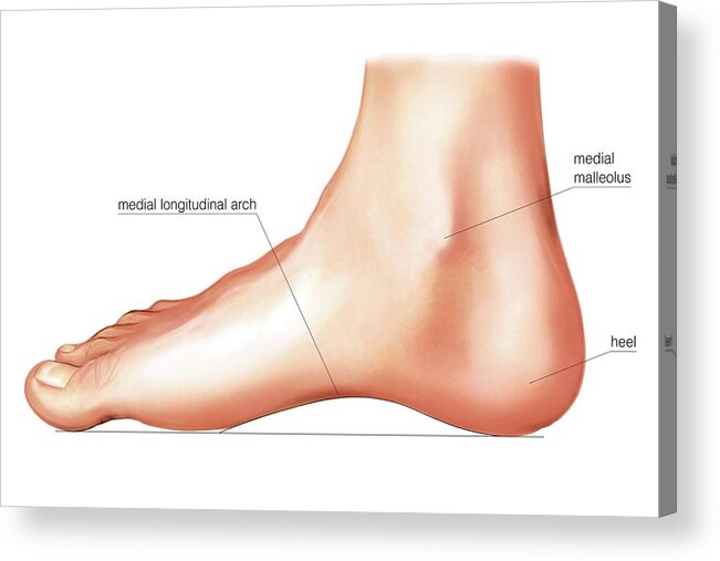
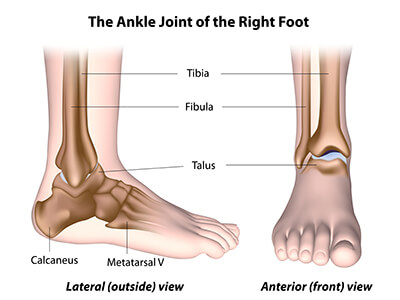
The hindfoot forms the heel and ankle.

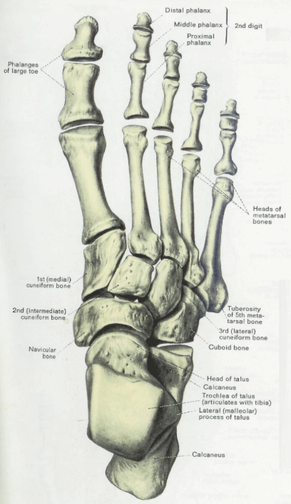
Right foot anatomy. The foot consists of thirty three bones twenty six joints and over a hundred muscles ligaments and tendons. Foot and ankle anatomy is quite complex. The cuneiform bones the navicularis and the cuboid all of which function to give your foot.
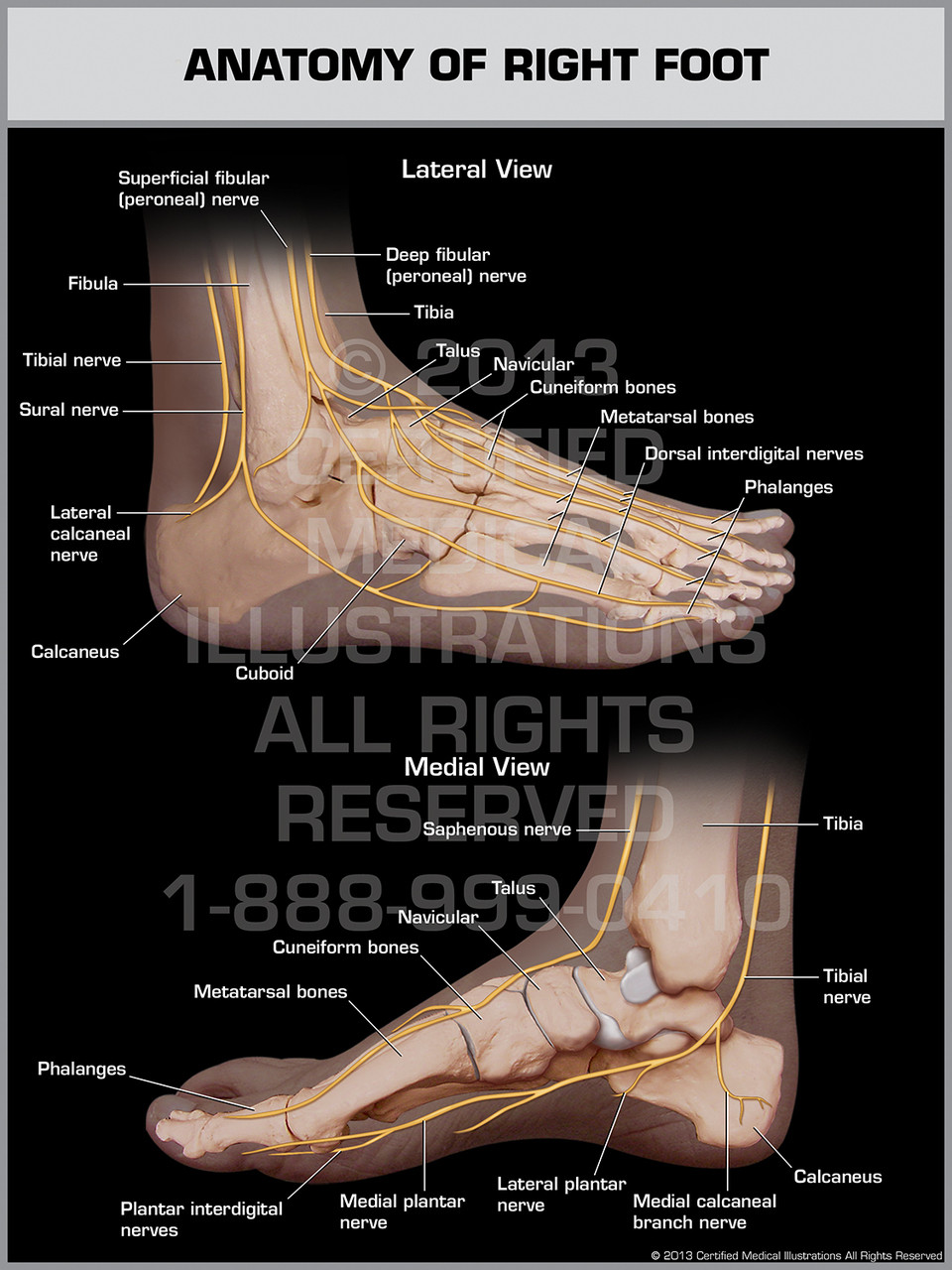
The foot is an extremely complex anatomic structure made up of 26 bones and 33 joints that must work together with 19 muscles and 107 ligaments to execute highly precise movements. The midfoot is a pyramid like collection of bones that form the arches of the feet. The contributors to this site are all board certified orthopaedic surgeons who specialize in treating patients with foot and ankle problems.
Learn about the anatomy of the foot. At the same time the foot must be strong to support more than 100000 pounds of pressure for every mile walked. Often a foot x ray is also requested for the investigation of osteomyelitis arthritides or.
The talus bone supports the leg bones. The other bones of the foot that create the ankle and connecting bones include. This webpage presents the anatomical structures found on ankle mri.
The forefoot contains the five toes phalanges and the five longer bones metatarsals. The calcaneus which is the bone in your heel. Mri of the ankle.
The metatarsals which run through the flat part of your foot. These all work together to bear weight allow movement and provide a stable base for us to stand and move on. Remember to check the whole film though.
The feet are divided into three sections. Foot radiograph an approach foot radiographs are commonly performed in emergency departments usually after sport related trauma and often with a clinical request that states lateral border pain. The phalanges which are the bones in your toes.
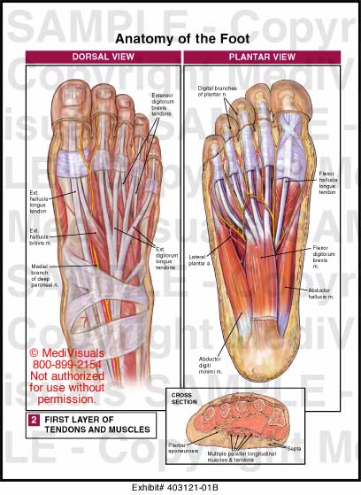
The foot is comprised of many bones joints tendons and ligaments including the plantar fascia and the achilles tendon.
 Obama S Checkup Healthy But Ouch That Right Foot The
 Foot And Ankle Musculoskeletal Key
 Lower Leg Ankle And Foot Dutton S Orthopaedic
![]() Foot Reflexology Essential Oils Jade Balden
Foot Reflexology Essential Oils Jade Balden
 Dorsal View Of The Bones Of The Right Foot Purposegames
 Anatomy Regions Of The Right Foot Acrylic Print
 Dorsal And Plantar View Of Right Foot Google Search Foot
 Understanding And Caring For Your Feet Breaking Muscle
 08126 01x Normal Blood Supply To The Right Foot Anatomy
 Anatomy Of The Foot What Is Special About It
 Talus Bone Wikipedia
 Dorsal View Of The Right Foot Showing The Major Muscles Tendons And Nerves
 Line Drawing Of The Left And Right Foot Soles Stock Vector
 Muscles Of The Lower Leg And Foot Human Anatomy And
 Foot Ankle Preservation Baltimore Md Towson Orthopaedics
 Drawing To Show The Bones Of The Right Foot Dorsal Or Top View
 Anatomy Of Right Foot
 Side View Of Human Right Foot Muscles Anatomy Model Isolated
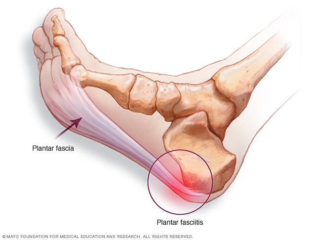 Plantar Fasciitis Symptoms And Causes Mayo Clinic
 Muscles Of The Lower Leg And Foot Human Anatomy And
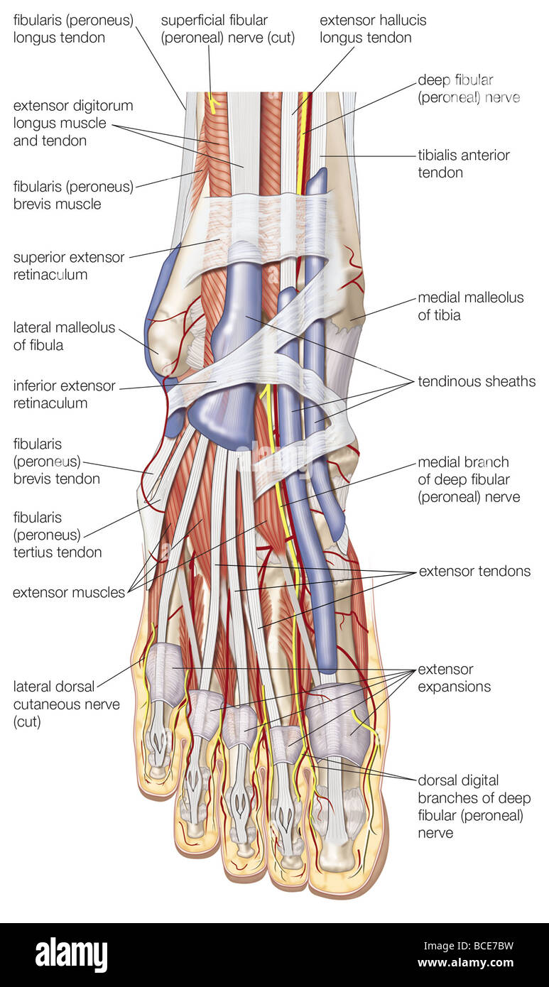 Dorsal View Of The Right Foot Showing The Major Muscles
Bones Of The Right Foot Superior View Diagram Quizlet
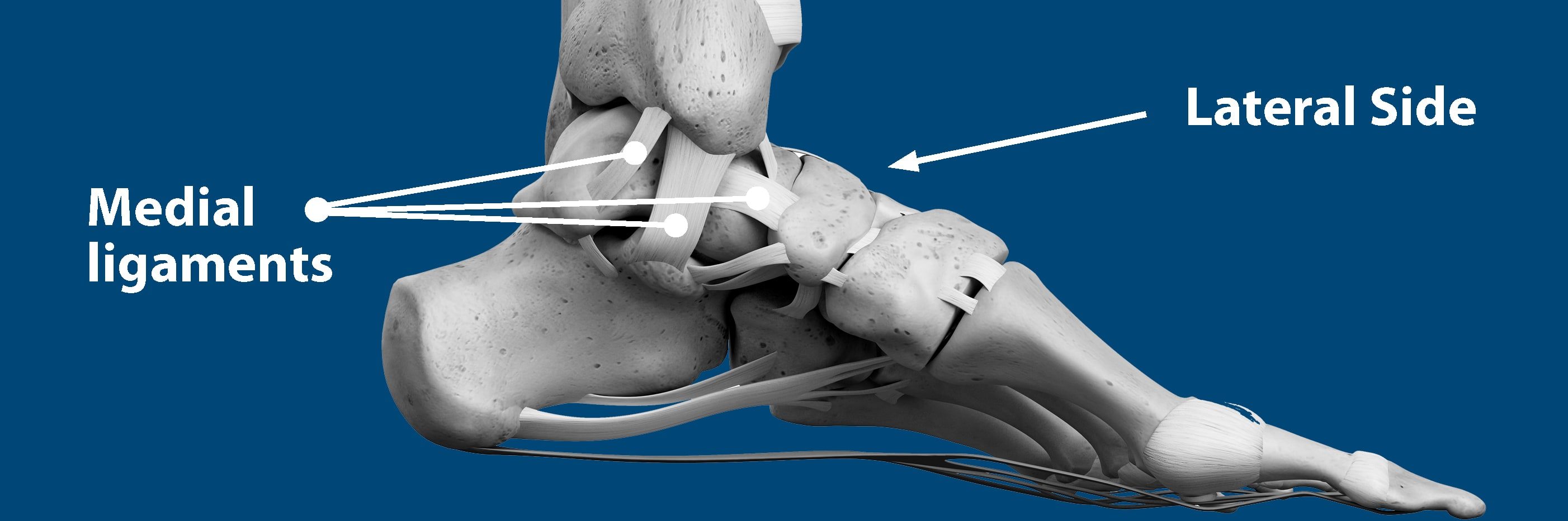 Sprained Ankle Florida Orthopaedic Institute
Belum ada Komentar untuk "Right Foot Anatomy"
Posting Komentar