Chest Xray Anatomy
If the ctr is 50 on either a posterior anterior pa or an anterior posterior ap view then the heart size is within normal limits. Adapted here for independent study.

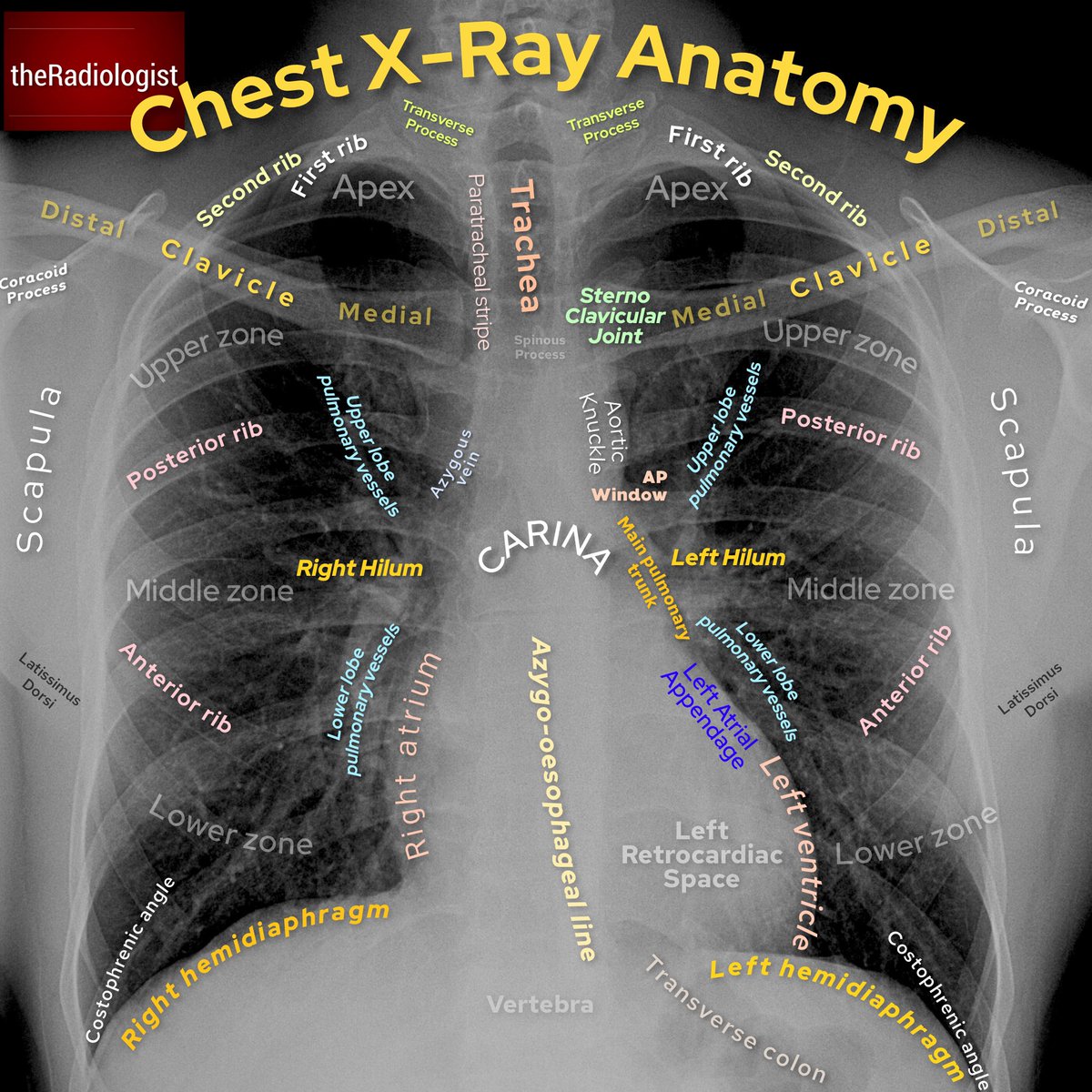
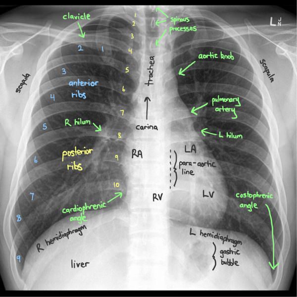
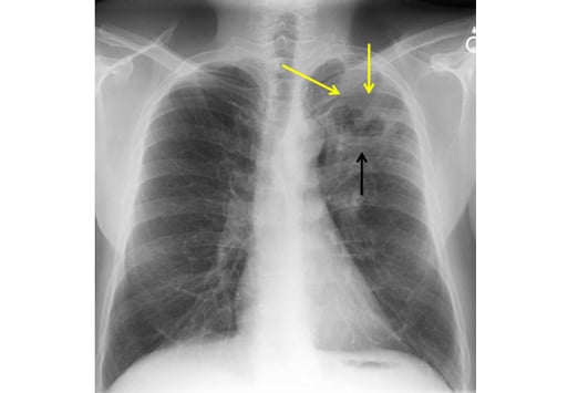
The large airways are visible on most good quality chest x rays.

Chest xray anatomy. It is the most complete reference of human anatomy available on web ipad iphone and android devices. The heart size should be assessed on every chest x ray. X ray of the chest also known as a chest radiograph is a commonly used imaging study and is the most frequently performed imaging study in the united states.
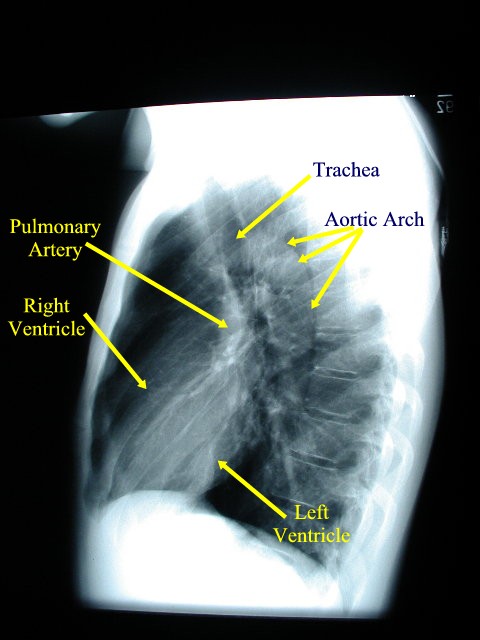
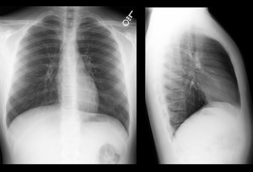
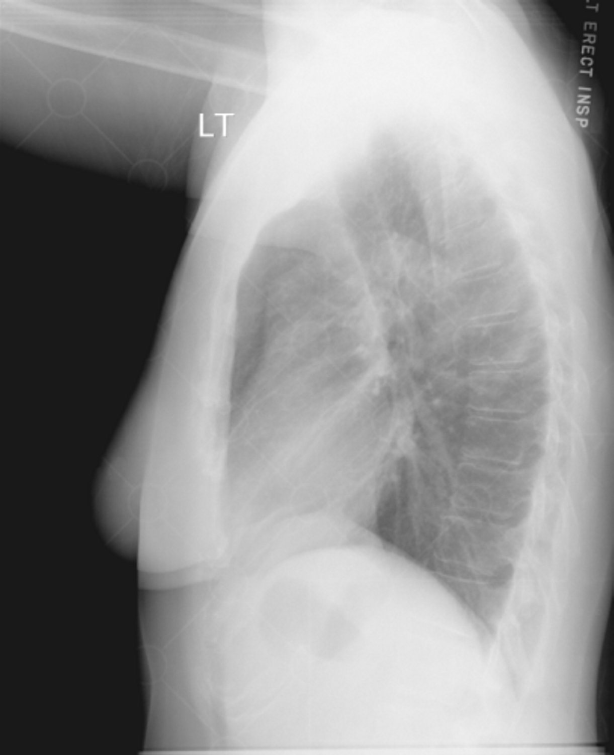
Explore over 6700 anatomic structures and more than 670 000 translated medical labels. This is because an ap view will exaggerate the heart size due to magnification. Lateral radiographs can be particularly useful in assessing the retrosternal and retrocardiac airspaces.
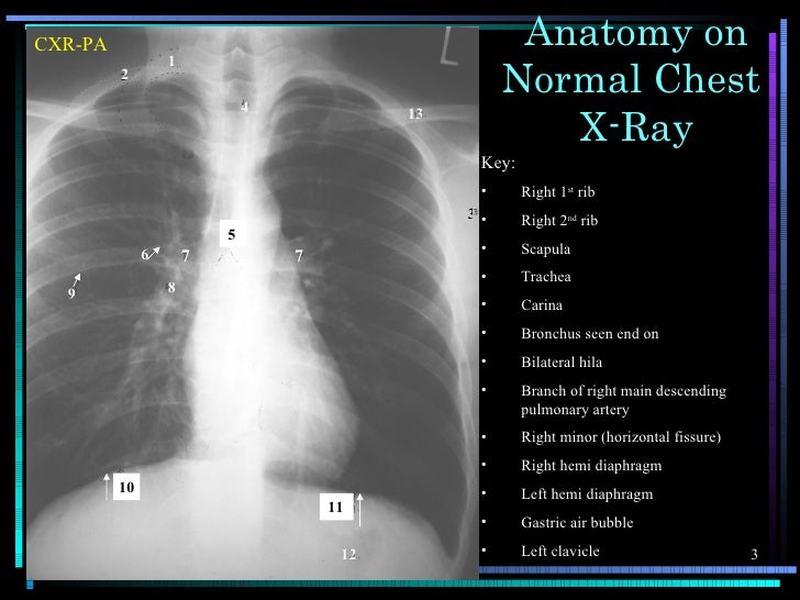
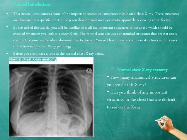
Chest x rays can diagnose pneumonia lung masses and broken ribs. The lateral chest view examines the lungs bony thoracic cavity mediastinum and great vessels. Chest x ray anatomy many structures of the chest are readily visible on a chest x ray but others are difficult to see.
Gillian lieberman forthe harvard medical school human body lecture series. For pediatric chest radiograph see chest radiograph pediatric. They contain air and so are of lower density blacker than the surrounding soft tissues.
Chest x ray anatomy 1. It is almost always the first imaging study ordered to evaluate for pathologies of the thorax although further diagnostic imaging laboratory tests and additional physical examinations may be necessary to help confirm the diagnosis. Available in 11.
Other anatomical structures such as the pleura only become clearly visible when abnormal. The lateral chest view may be performed as an adjunct to a frontal chest radiograph in cases where there is diagnostic uncertainty. E anatomy is an award winning interactive atlas of human anatomy.
The chest radiograph also known as the chest x ray or cxr is thought to be the most frequently performed radiological investigation globally ref. Living anatomy of the chest for 1st year medical students original version compiled by dr. Principles of reading 1.
Ct mri radiographs anatomic diagrams and nuclear images. The trachea branches at the carina into the left and right main bronchi and these can often be followed as they branch beyond the hila and into the lungs. Miscellaneous such as pacemakers catheters etc.
In fact some important structures such as the phrenic nerve are not visible at all. A chest x ray test is a very common non invasive radiology test that produces an image of the chest and the internal organs. However a pa view is required to confidently diagnose cardiac enlargement.
 Chest Radiograph Wikipedia
 Theradiologist On Twitter Basics Chest X Ray Anatomy And

 Basic Chest X Ray Anatomy
 Reading Chest X Rays Anatomy Labelled Reading A Chest X
 Doctor Examining A Lung Radiography Doctor Looking Chest X
 Chest X Ray Anatomy

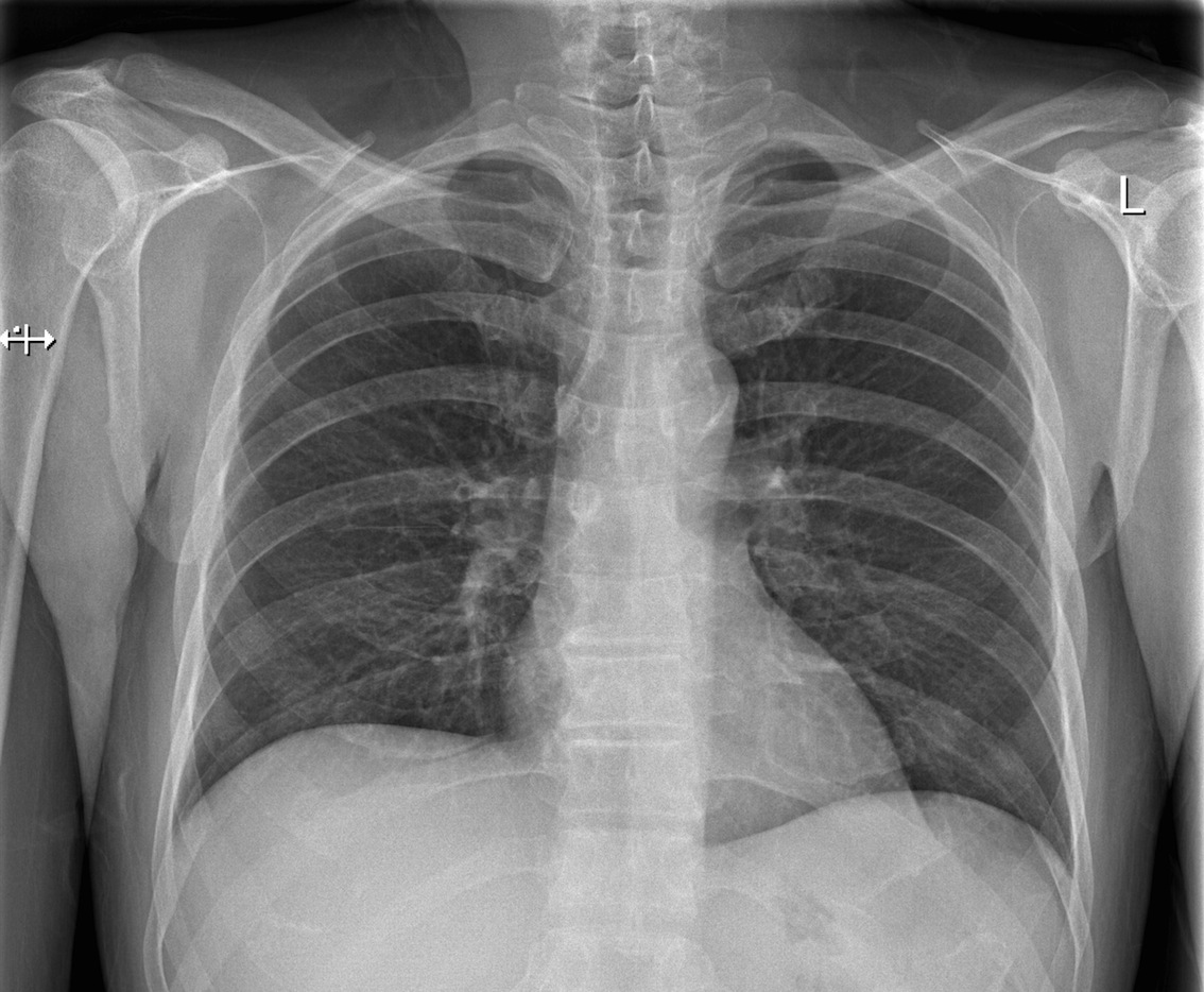
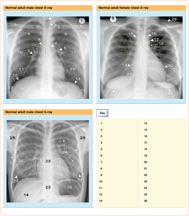
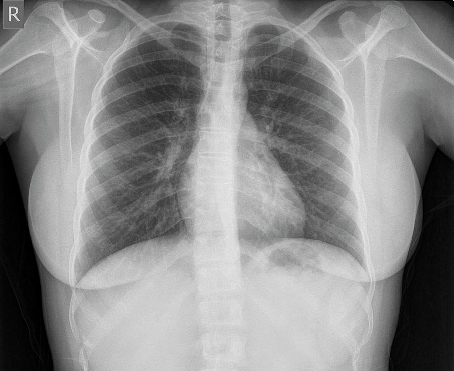 Normal Healthy Chest X Ray
Film Critique Part 1 Chest
 Practice Chest X Ray Interpretation
 X Ray Image Of The Chest Showing The Internal Anatomy Of The
 Interpreting Chest X Rays Ct Scans And Mris Respiratory


 Chest X Ray Anatomy
 How To Interpret A Chest X Ray Lesson 2 A Systematic Method And Anatomy
 Film Critique Part 1 Chest
 Anatomical Features In Two Chest X Ray Images And Their
 Interpreting Chest X Rays Ct Scans And Mris Respiratory

 Anatomy Of A Chest Xray
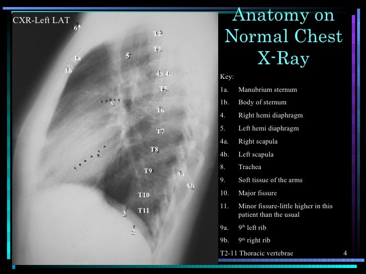
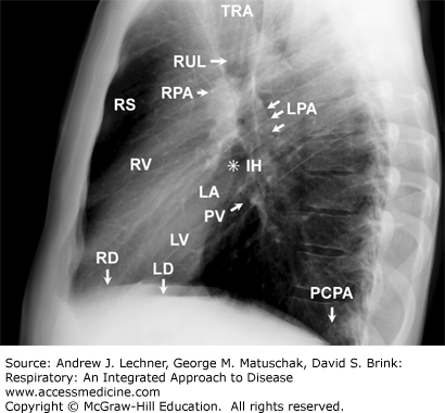
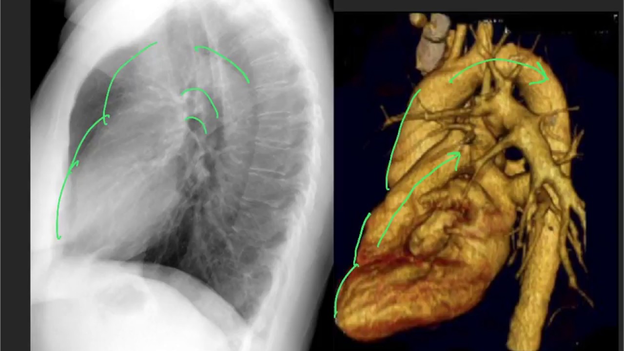 Anatomy On The Lateral Cxr
 X Thorax Startradiology
 Chest X Ray Anatomy How To Interpret Chest X Ray 2
 Chest X Ray Anatomy Flashcards Quizlet
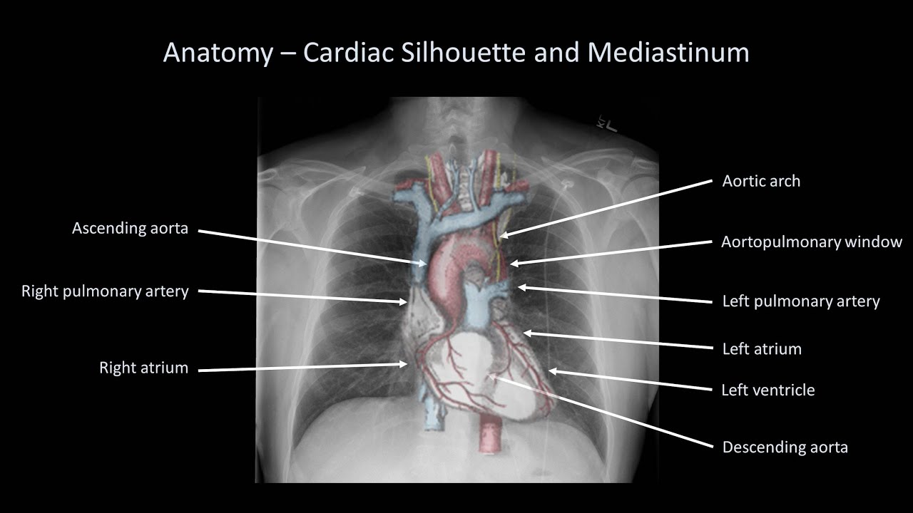
 Chest Xray Annotation Cardiac Anatomy Radiology Student
 Digital Radiography Image Artifacts Radiology Suny
Belum ada Komentar untuk "Chest Xray Anatomy"
Posting Komentar