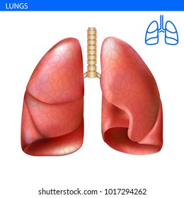
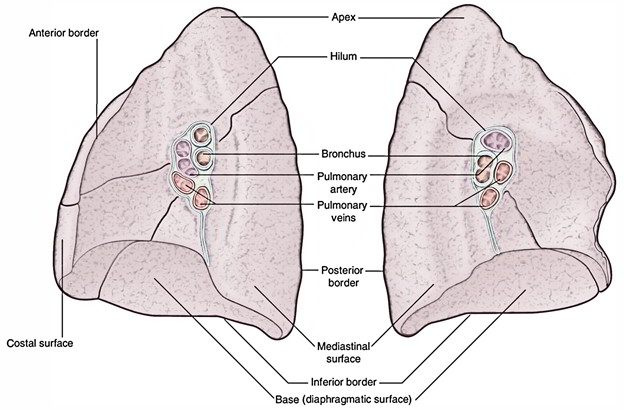
Hilum Lung Anatomy
Abnormalities in the hilum are usually noted on imaging. This image added by admin.
:max_bytes(150000):strip_icc()/72420503-56a5cf7d5f9b58b7d0de8b0d.jpg) Hilum Of The Lung Definition Anatomy And Masses
Hilum Of The Lung Definition Anatomy And Masses
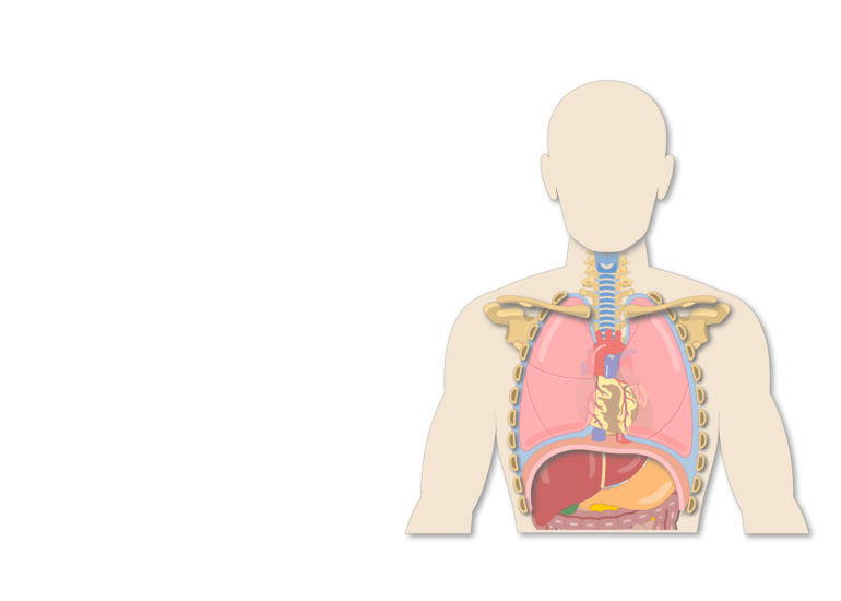
In human respiratory system.
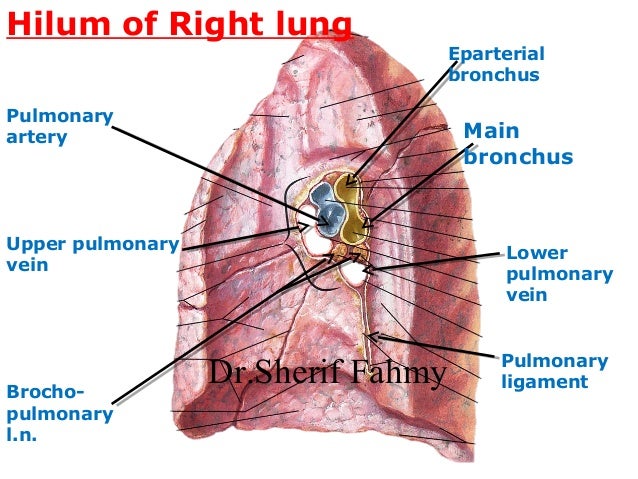
Hilum lung anatomy. Hilum of the lung. The structures of the lung root are embedded in the connective tissue and surrounded by extension. Hilum of the right lung.
Anatomy and abnormalities anatomy of the hilum. Plural hili is a depression or fissure where structures such as blood vessels and nerves enter an organ. The hilar region of.
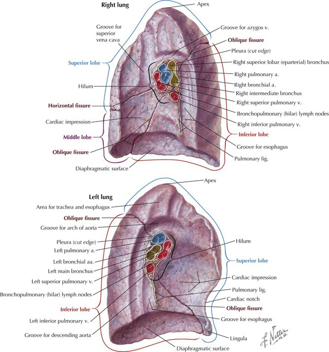
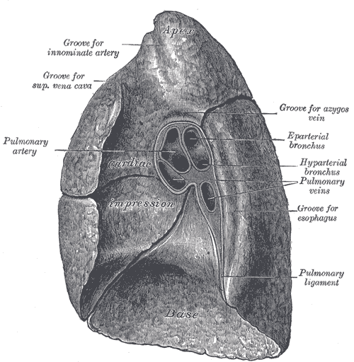
Gross anatomy with the mediastinum at the hilum a circumscribed area where airways blood and. The lung hila or roots are found on the medial aspect of each lung. This concavity is deeper in the right lung due to the higher position of the right dome overlying the liver.
Lung roots lie opposite to t5 t7 vertebrae. Both the right and the left lung have a hilum which lies roughly midway down. Describe the root and hilum of lungs.
Hilum anatomy in human anatomy the hilum ˈhaɪləm. It rests on the dome of the diaphragm and has a concave shape. The hilum of the lung is a wedge shaped section in the central area of the lung that permits arteries veins nerves bronchi and other structures to enter and exit.
You can click the image to magnify if you cannot see clearly. Lung root consists of the structures passing to and from the hilum of the lung to the mediastinum. The lung hilum where structures enter and leave the lung is located on this surface.
Structure of lung in lung to its apex is the hilum the point at which the bronchi pulmonary arteries and veins lymphatic vessels and nerves enter the lung. The right hilum is caudally related to the terminal azygos vein and posteriorly related to the right atrium and superior vena cava. Tests to evaluate the hilum.
Both human lungs have a hilar region meaning both lungs have an area called the hilum. The structures within the right hilum are arranged such that the principal bronchus is posteriorly related to the pulmonary artery. Plural hila sometimes formerly called a hilus ˈhaɪləs.
The left and right lung roots are similar but not identical. Hilus of dentate gyrus part of hippocampus that contains the mossy cells. The base of the lung is formed by the diaphragmatic surface.
We think this is the most useful anatomy picture that you need. Gross anatomy left hilum in the left hilum the left pulmonary artery occupies the upper part.
 Respiratory System Lungs Anatomy Flashcards Quizlet
 Hilum Anatomy Wikipedia
![]() Lungs Anatomy Structure Supply Kenhub
Lungs Anatomy Structure Supply Kenhub
 Non Small Cell Lung Cancer Staging Stages Of Lung Cancer
 Left Lung Images Stock Photos Vectors Shutterstock
 Thorax Basicmedical Key
 Pulmonary Cavities Anatomy An Essential Textbook 1st Ed
 Lungs Pleura Anatomy With Blanck At University Of South
Summary Netter S Anatomy Lecture Lungs Dbs 8110 Studocu
Hilum Of Lung
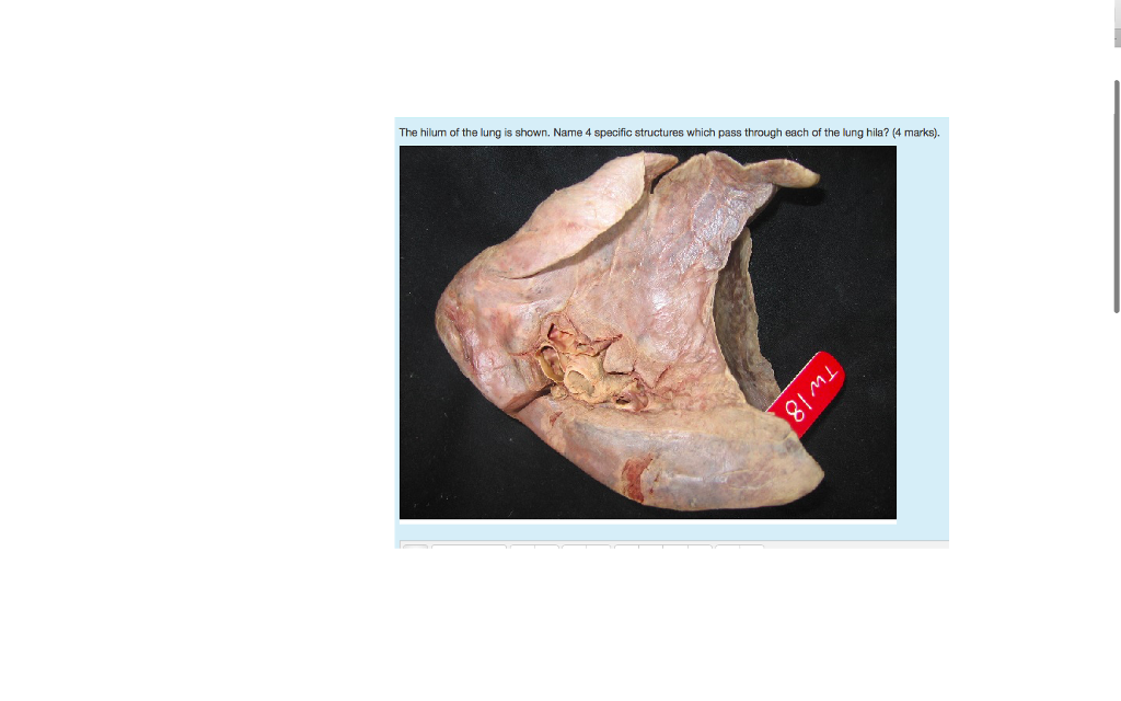 Solved The Hilum Of The Lung Is Shown Name 4 Specific St
 Lung Cancer Dr Yousef Noaimat Md Fccp Consultant
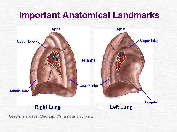
 Lungs Anatomy Shapes And Surfaces Of The Lungs
 Anatomy Of The Lung Flashcards Quizlet
 Easy Notes On Lungs Learn In Just 4 Minutes Earth S Lab
 Lung Relationships Respiratory Medbullets Step 1
 What Is The Function Of The Hilus Is The Word Hilum The
 Anatomy Descriptive And Applied Anatomy 1190 The Organs
 The Lungs Anatomy Of The Thorax
Untitled
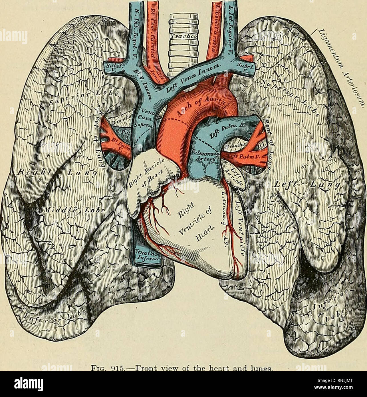
 Pulmonary Vascular System And Pulmonary Hilum Sciencedirect
/iStock_000006469946_Large-56a5c5575f9b58b7d0de6a59.jpg) Hilum Of The Lung Definition Anatomy And Masses
Hilum Of The Lung Definition Anatomy And Masses

![]() Hilum Of The Lung Anatomy And Clinical Aspects Kenhub
Hilum Of The Lung Anatomy And Clinical Aspects Kenhub
 Root Of The Lung Wikipedia
 Medial View Of Left And Right Lung Anatomy
Belum ada Komentar untuk "Hilum Lung Anatomy"
Posting Komentar