Fundus Anatomy
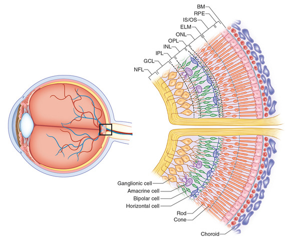
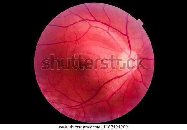
The tapetal area is in the superior half of the fundus and the nontapetal area is in the inferior half of the fundus as well as the periphery of the superior fundus. The red curving structures are blood vessels which enter the retina through the nerve.
 The Stomach Structure Neurovasculature Teachmeanatomy
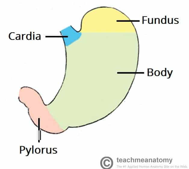
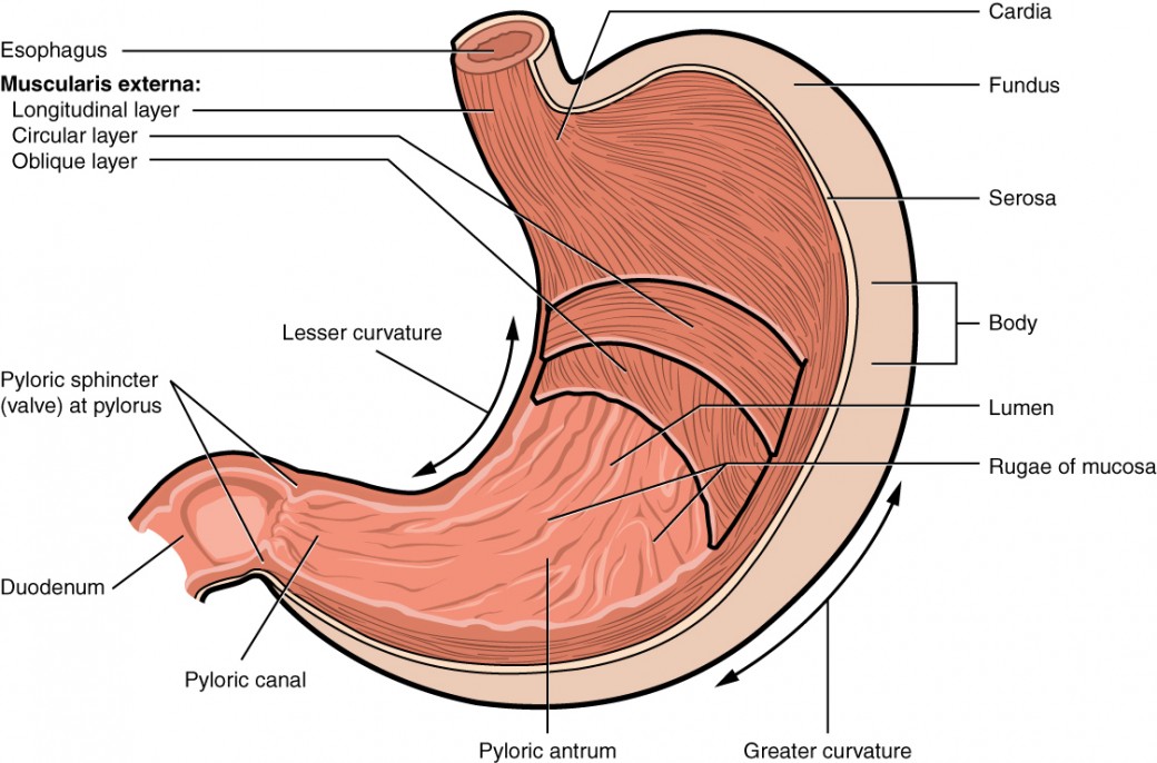
The body is the main central region of the stomach.

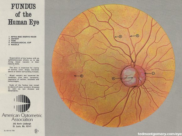
Fundus anatomy. The pylorus from greek meaning gatekeeper is the lower section of the stomach. The optic disc is typically centrally located but will vary in its location. Fundus anatomy the base of a hollow organ or that part of the organ farthest from its opening.
Fundus in the anatomy of stomach it is the uppermost portion forming the upper curvature of the organ. The portion of an organ most remote from its opening. The fundus of the stomach.
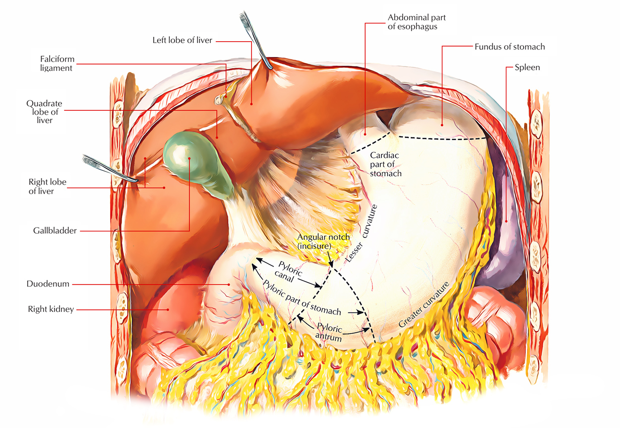
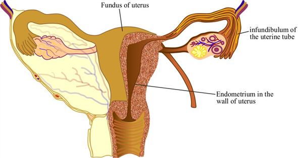
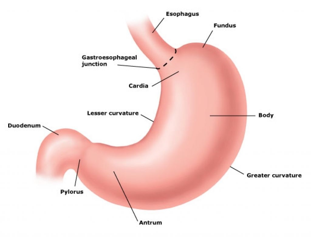
Located inferior to the diaphragm above and to the left of the cardia is the dome shaped fundus. Each fundus has no sign of disease or pathology. The fundus is the topmost portion of the uterus and is known as the roof of the uterine cavity.
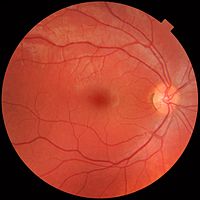
This fundus photograph shows the normal appearance of the retina. This page describes normal retinal anatomy. During pregnancy this is usually when the fertilized egg implants.
The fundus and uterus can stretch significantly throughout pregnancy without any trauma or damage. When chemical digestion takes place in the stomach stomach gases are produced. The cardia fundus body and pylorus.
There are four main regions in the stomach. Anatomical structure bodily structure body structure complex body part structure a particular complex anatomical part of a living thing. The fundus from latin meaning bottom is formed in the upper curved part.
He has good bone structure. Normal ocular anatomy the fundus is typically divided into the tapetal and nontapetal fundus area. The cardia or cardiac region is the point where the esophagus connects to the stomach and through which food passes into the stomach.
Readers must therefore always check the product information and clinical procedures with the most up to date published product information and data sheets provided by the manufacturers and the most recent codes of conduct and safety regulations. Sections the cardia is where the contents of the esophagus empty into the stomach. The whitish circle is the nerve that connects the retina to the brain.
The larger part base or body of a hollow organ. Refer to this page for comparison with the retinal disease pages. Fundus photographs of the right eye left image and left eye right image as seen from the front so that left in each image is to the persons right and the persons nose would be between the two images.
Oxford university press makes no representation express or implied that the drug dosages in this book are correct.
 Anatomy Of The Eye Medical Illustration Medivisuals
 Ch27 Uterine Anatomy
 Phrygian Cap Anatomy Wikipedia
 Center For Uterine Fibroids
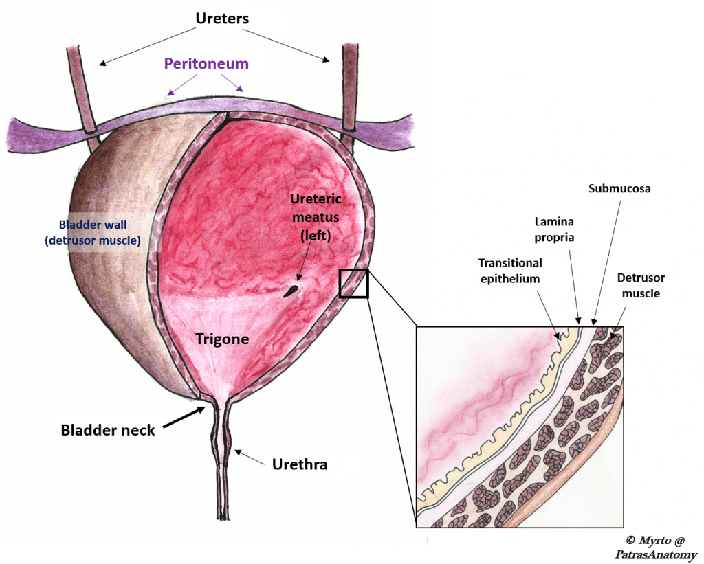 The Urinary Bladder Structure Function Nerves
 Anatomy Of The Eye External Eye Eyelids Lacrimal Gland
 Easy Notes On Stomach Learn In Just 4 Minutes Earth S Lab
 Stomach Fundus More Anatomy
 Fundus Oculi
 Introduction Fundus Photography
 Solved Describe The Location Of Each Of The Following
 Eye Fundal Anatomy 1 Vet Notes
 Gastric Fundus An Overview Sciencedirect Topics
 The Stomach Anatomy And Physiology Ii
 Retina Optic Nerve Through Ophthalmoscope Anatomy The
 Vector Illustration Of Anatomy Of The Fundus
 Normal O
 The Best Free Fundus Drawing Images Download From 9 Free
 Normal Retinal Anatomy The Retina Reference
 Orbit Eye Atlas Of Anatomy
 Monochromatic Fundus Photography Ophthalmic Photographers
 Human Eye Anatomy Taking Images Mydriatic People
 Normal Fundus Eye Images For Both Left And Right Eye
 Shows Abnormal Findings Caused By Diabetic Retinopathy And
 Figure 20 Ocular Fundus Photo Of A Child With A Pale Optic
 Stomach Wikipedia
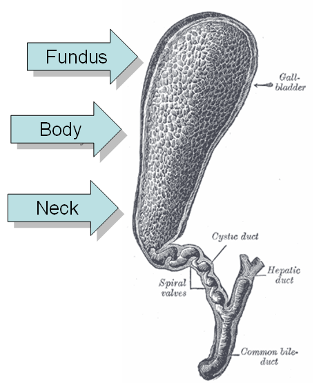
 Gallbladder Radiology Reference Article Radiopaedia Org
 Uterus Wikipedia
 The Fundus Oculi Of Birds Especially As Viewed By The
 Normal Fundus
 Fundus Eye Wikipedia


Belum ada Komentar untuk "Fundus Anatomy"
Posting Komentar