Sinus Of Valsalva Anatomy
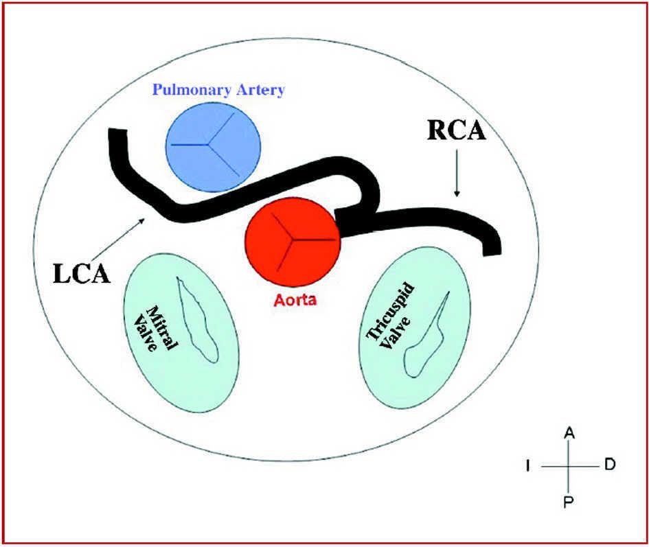
Right coronary or anterior. The anterior aortic sinus gives rise to the right coronary artery.
 The Geometrical Modeling Of Aortic Root Complex Ugurlucan M
A congenital lack of continuity in the media between the aorta and annulus fibrosus of the aortic valve may initiate aneurysm formation or less frequently infection or degeneration processes may affect an aortic sinus.
Sinus of valsalva anatomy. In typical anatomy there are three sinuses. Medical definition of sinus of valsalva. The sinus of valsalva more readily known as the aortic sinus refers to the three pouches behind the valves of the heart known as the semilunar valves which come from the aorta.
Left coronary or left posterior. Each aortic sinus can also be referred to as the sinus of valsalva the sinus of morgagni the sinus of mehta the sinus of otto or petits sinus. Usually no vessels arise from the right posterior aortic sinus which is therefore known as the non coronary sinus.
Any one of the pouches of the aorta and pulmonary artery which are located behind the flaps of the semilunar valves and into which the blood in its regurgitation toward the heart enters and thereby closes the valves called also aortic sinus. Gives rise to right coronary artery. The anatomy of the aortic root has been studied in different species and common structural features of importance have been detailed.
These sinuses form part of the functional aspect of the corresponding aortic valve and pulmonary valve. The sinuses of valsalva are dilations related to both the aortic root of the ascending aorta and the root of the pulmonary trunk. This cardiac lesion can be congenital or acquired.
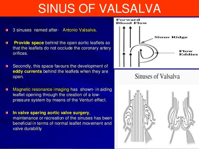
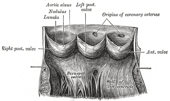
The findings confirm the universal presence of a well marked ridge limiting the distal extent of the aortic sinuses. A rupture of a sinus of valsalva is a rare cardiac anomaly mostly occuring in aneurysmal dilated sinuses. The sinuses of valsalva also known as aortic sinuses are the anatomic spaces at the aortic root bounded internally by the aortic valve leaflets and externally by outward bulges of the aortic wall.
A ct scan may be helpful in diagnosing an aneurysm of the sinus of valsalva.
 Annular Rupture During Tavi Radcliffe Cardiology
 Ruptured Sinus Valsalva Heart Valve Heart
Aorta And Aortic Valve Basic Principle Of 2d Echo Assessment
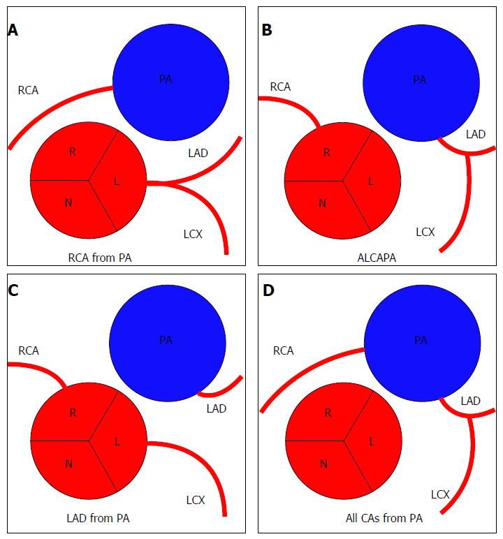
 Congenital Coronary Artery Anomalies With Origin In The
 Sinus Of Valsalva Aneurysm
 Sinus Of Valsalva Aneurysm Repair Dr Ian Nicholson
 The Aorta Diseases Of The Aorta Springerlink
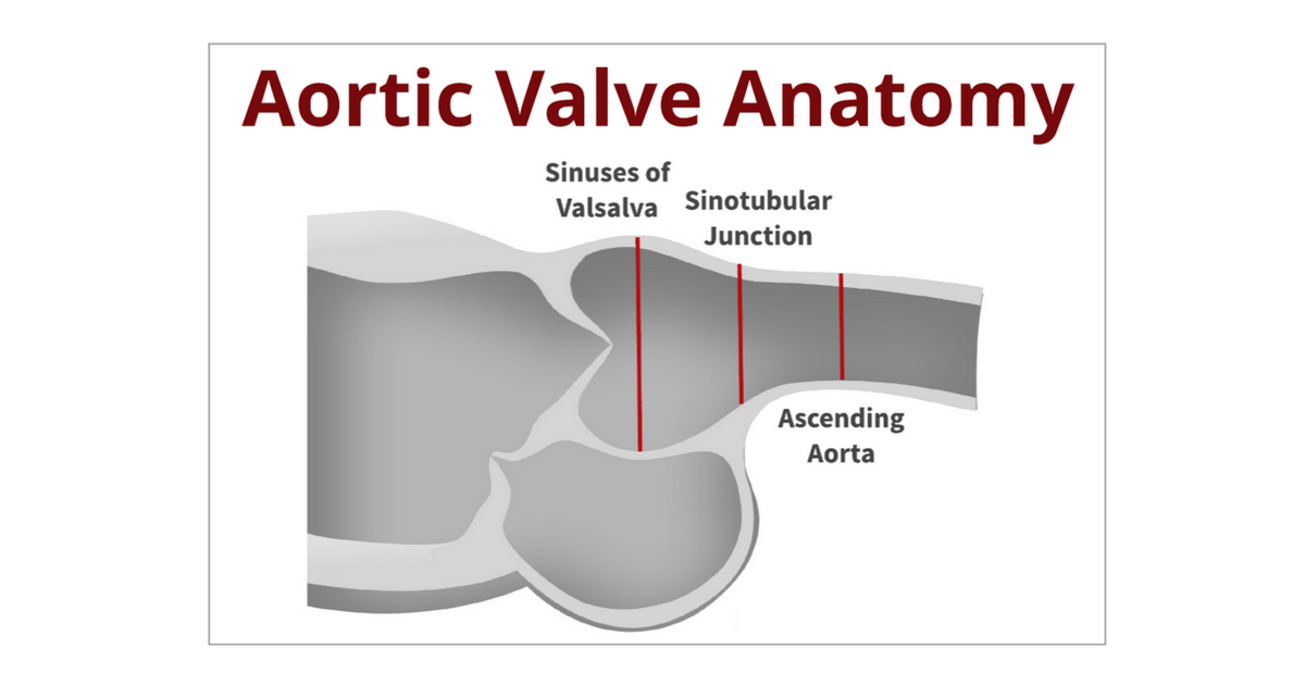
The Anatomy Of The Sinus Of Valsalva
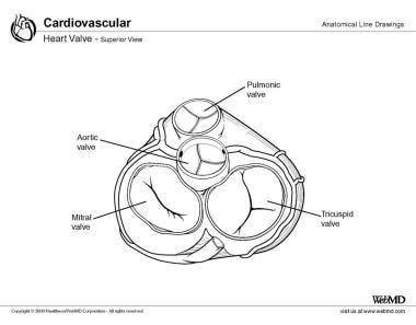 Aortic Valve Anatomy Overview Gross Anatomy Microscopic
 Correlating Possible Predisposing Demographics And Systemic
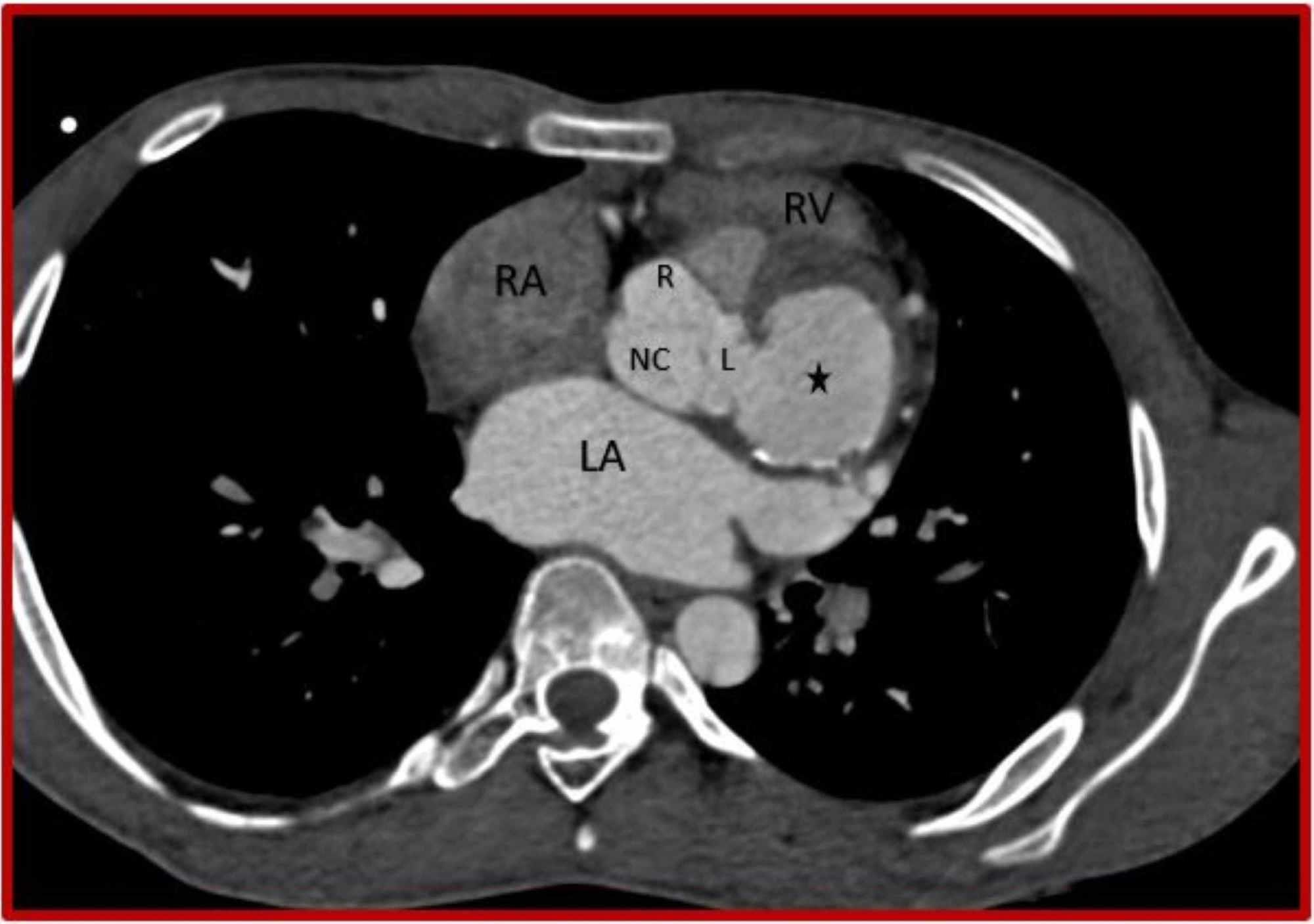
Multi Detector Ct Angiography Of The Aortic Valve Part 1
 Aneurysm Of Sinus Of Valsalva Wikipedia
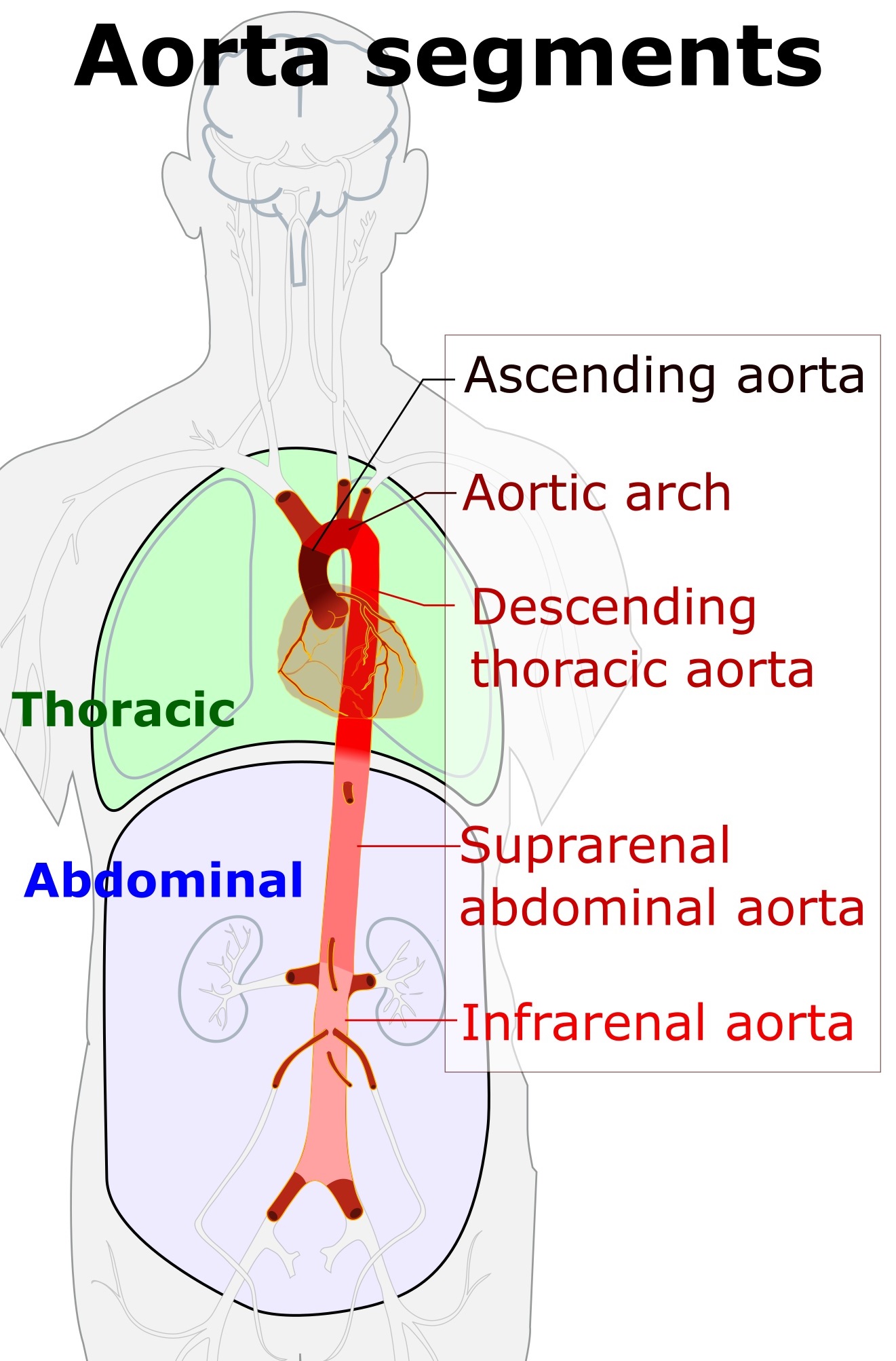 Aorta Wikipedia
 Aortic Sinus Wikipedia
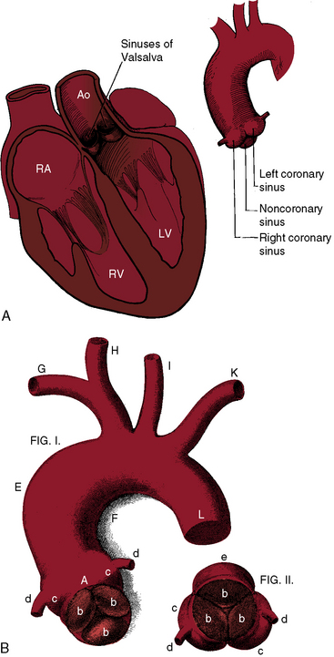
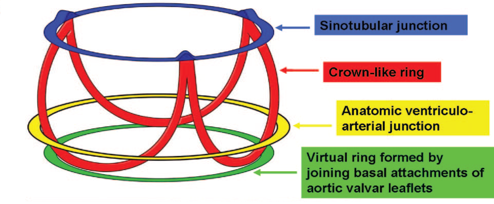 Anatomy And Function Of Normal Aortic Valvular Complex
 The Geometrical Modeling Of Aortic Root Complex Ugurlucan M
 Coronary Artery Anomalies Overview The Normal And The Abnormal
 Back To The Basics Aortic Valve Anatomy
 Right Coronary Artery An Overview Sciencedirect Topics
 Sinus Of Valsalva Aneurysms Review Of The Literature And An
 Cureus Rupture Of Sinus Of Valsalva Aneurysm Into
 Congenital Aneurysms Of The Sinuses Of Valsalva Thoracic Key
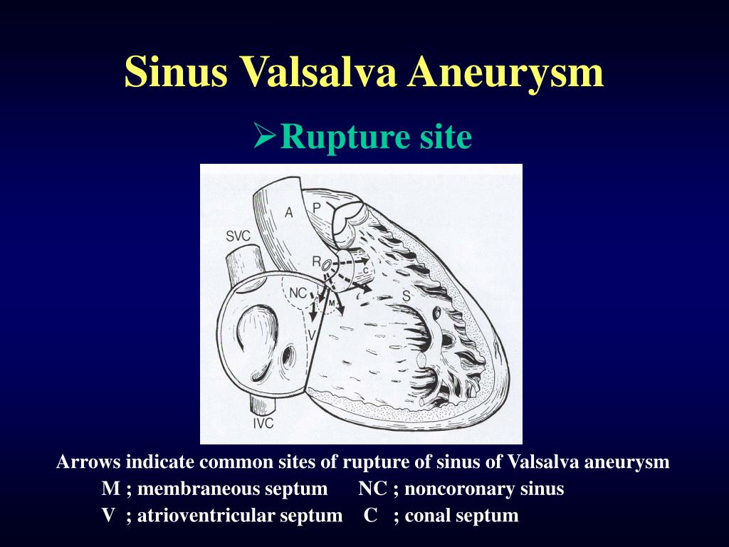 Ppt Sinus Valsalva Aneurysm Powerpoint Presentation Free
Aortic Root Radiology Reference Article Radiopaedia Org
 Aortic Anatomy
 Angiographic Features Of Ruptured Sinus Of Valsalva Aneurysm


Belum ada Komentar untuk "Sinus Of Valsalva Anatomy"
Posting Komentar