Anatomy Of Chest X Ray
Living anatomyradiology is ideally suited to image anatomy in the living patientlabeled plain. Chest x ray can be.
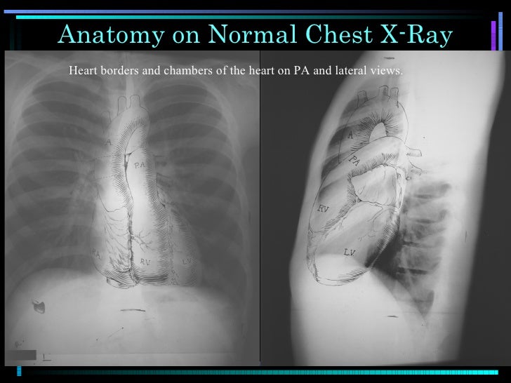
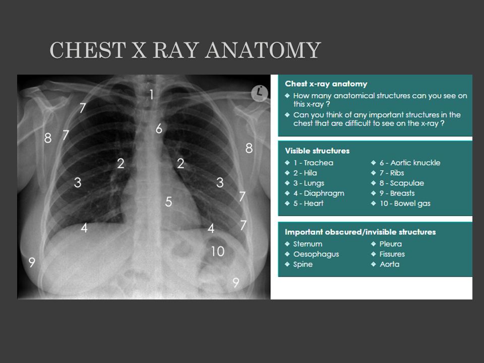
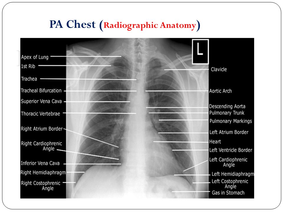 Chest Radiography Lecture 2 Ppt Video Online Download
Chest radiographs are the most common film taken in medicine.
Anatomy of chest x ray. Each of these anatomical structures should be viewed using a systematic approach. The large airways are visible on most good quality chest x rays. There are also important structures that are obscured or become visible only when abnormal.
They contain air and so are of lower density blacker than the surrounding soft tissues. Depending on its density each organ within the chest cavity absorbs varying degrees of radiation producing different shadows on the film. A description of a systematic method for examining a chest x ray and a review of the relevant thoracic anatomy.
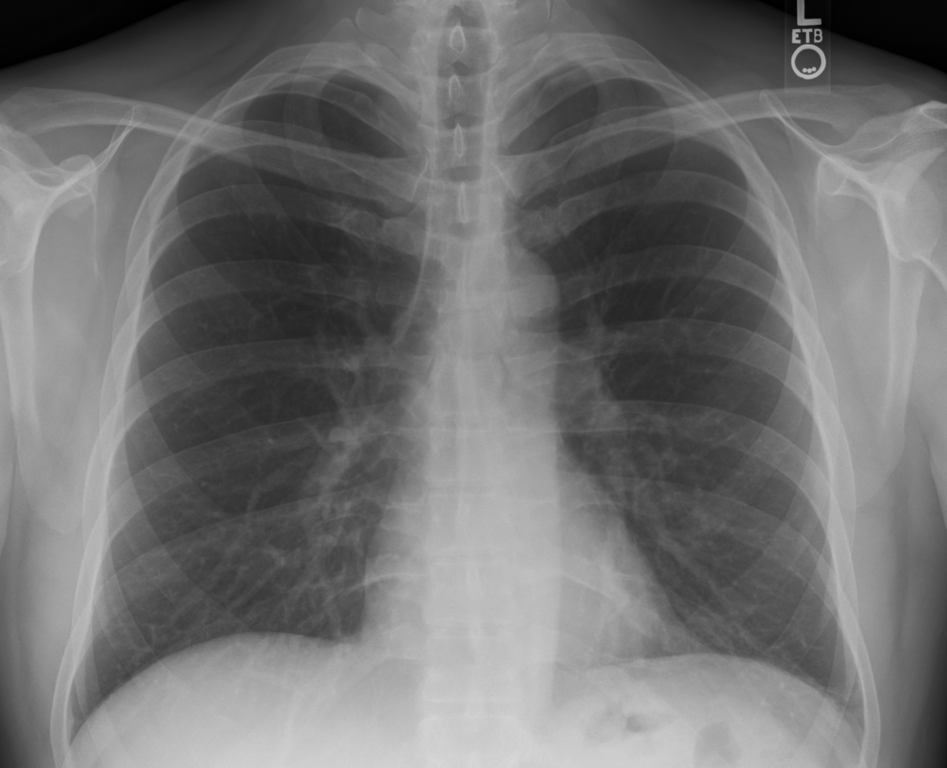
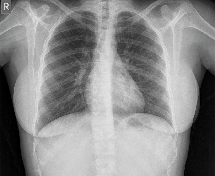
Key points some anatomical structures in the chest should be assessed on every chest x ray. A chest radiograph called a chest x ray or chest film is a projection radiograph of the chest used to diagnose conditions affecting the chest its contents and nearby structures. Right 1st rib 3 right.
Chest x ray images are black and white with only the brightness or darkness defining the various structures. The mean radiation dose to an adult from a chest radiograph is around 002 msv for a front view and 008 msv for a side vi. Cxr pa anatomy on 1 2 2 4 normal chest x ray 13 key.
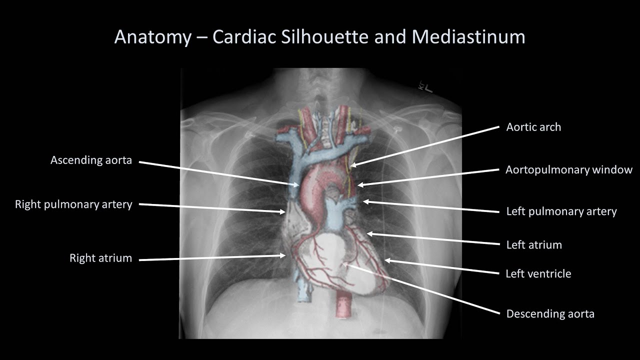
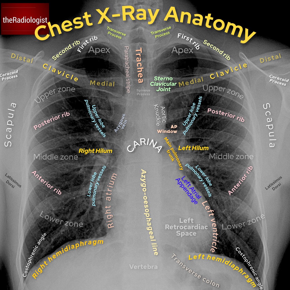
Anatomy of a chest x ray. The trachea branches at the carina into the left and right main bronchi and these can often be followed as they branch beyond the hila and into the lungs. Cardiovascular anatomy of the mediastinum on a lateral chest radiograph.
Case discussion an understanding of the cardiovascular structures that contribute to the normal cardiomediastinal silhouette is essential in chest radiograph interpretation. How to interpret a chest x ray lesson 2 a systematic method and anatomy. Chest x ray anatomy 1.
The radiologist needs to know both the structures within the mediastinum forming the mediastinal margins and the lobes of the lungs forming the margins of the lungs along the mediastinum and chest wall. Chest x ray is also referred to as a chest radiograph chest roentgenogram or cxr. Living anatomy of the chest for 1st year medical students original version compiled by dr.
Chest radiology anatomy mediastinum lungs. Like all methods of radiography chest radiography employs ionizing radiation in the form of x rays to generate images of the chest. Posted by radiologypics.
 How To Interpret A Chest X Ray Lesson 2 A Systematic Method And Anatomy
 The Radiology Assistant Chest X Ray Basic Interpretation
 View Large Accessmedicine Mcgraw Hill Medical
 Medzcool On Twitter Can You Identify And Name All These
 Practice Chest X Ray Interpretation
 Film Critique Part 1 Chest
 Thorax Radiography An Overview Sciencedirect Topics
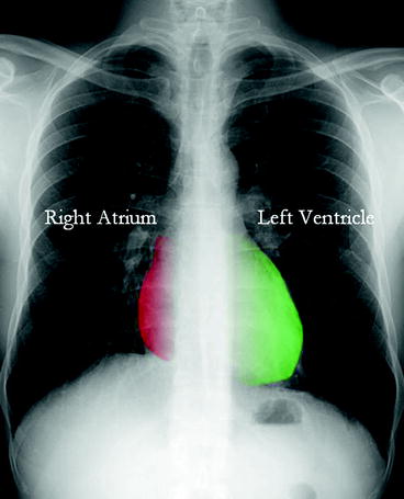
 Cardiomediastinal Outline On Chest X Ray Human Anatomy
 Thorax Wikipedia
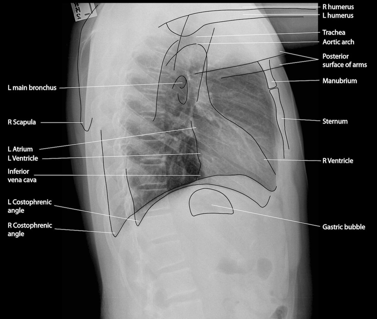
![]() Normal Chest X Ray Anatomy Tutorial Kenhub
Normal Chest X Ray Anatomy Tutorial Kenhub
 X Thorax Startradiology
 The Radiology Assistant Chest X Ray Basic Interpretation
 Cardiomediastinal Anatomy On Chest Radiography Annotated
 Interpreting Chest X Rays Ct Scans And Mris Respiratory
 Chest X Ray Anatomy
 Coccidioidomycosis Chest X Ray A D A M Interactive
 Anatomy And Radiology
 File Chest Xray Pa 3 8 2010 Png Wikimedia Commons
 Chest X Ray Anatomy Flashcards Quizlet
World S Best Chest X Ray Anatomy Stock Pictures Photos And
 Anatomy Of A Chest X Ray How To Read A Chest X Ray Part 1
 X Thorax Startradiology
 Normal Healthy Chest X Ray
 Normal Lung Anatomy
 Interpreting A Chest X Ray Stepwards
 Interpreting Chest X Rays Ct Scans And Mris Respiratory
 Uams Anatomy X Ray Quiz
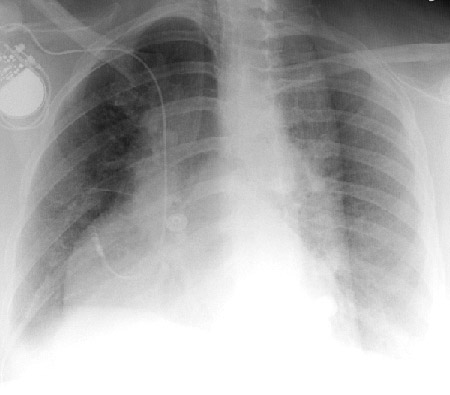
A Chest Xray Film Of A Patient With Severe Pneumonia Stock
 Diagnostic Imaging Normal Chest Anatomy On Xr Ppt Video
 Theradiologist On Twitter Basics Chest X Ray Anatomy And
 Normal Chest X Ray Terminology And Radiographic Anatomy
A Chest Xray Of A Blunt Chest Wall Injuried Stock Photo Download Image Now
 Normal Chest X Ray Litfl Medical Blog Labelled Radiology

Belum ada Komentar untuk "Anatomy Of Chest X Ray"
Posting Komentar