Chest Venous Anatomy
Learn online with high yield video lectures by world class professors earn perfect scores. Anatomical illustrations this e anatomy module presents an illustrated anatomy of the lungs trachea bronchi pleural cavity and pulmonary vessels.
 Anatomy Thorax Review Of Critical Care Medicine
Study for your classes usmle mcat or mbbs.
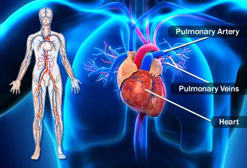
Chest venous anatomy. Although plain film radiography was once considered the gold standard for chest evaluation both multidetector computed tomography and magnetic resonance imaging are becoming indispensable for delineation of the different drainage patterns of the thoracic venous system. The chest is the major hub of the circulatory system it houses the heart lungs and other major organs that need large amounts of blood flow. Watch the video lecture arteries and veins anatomy of the heart boost your knowledge.
Venous anatomy or variations thereof can also be crucial to surgical colleagues for operative planning and follow up. Although plain film radiography was once considered the gold standard for chest evaluation both multidetector computed tomography and magnetic resonance imaging are becoming indispensable for delineation of the different drainage patterns of the thoracic venous system. Anatomy of the chest and the lungs.
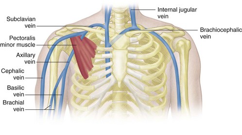
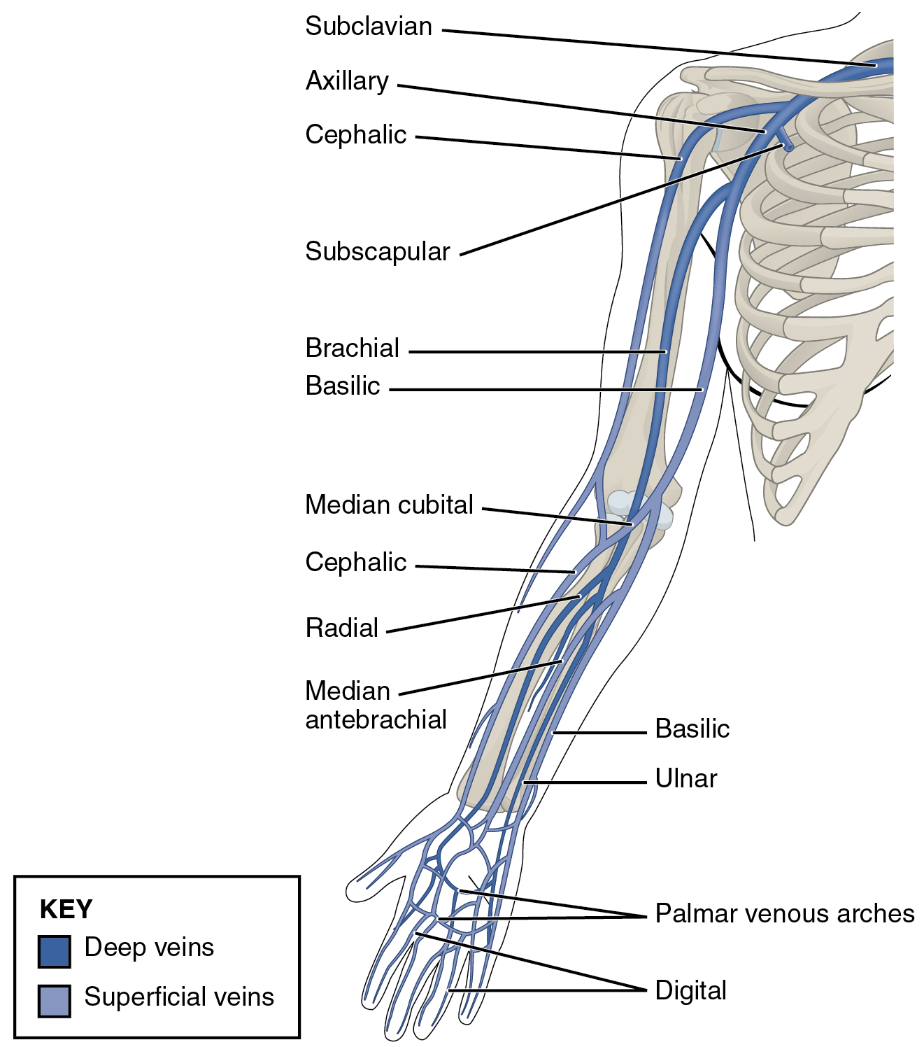
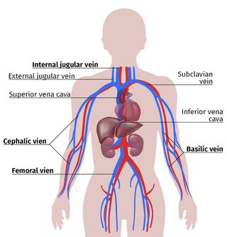
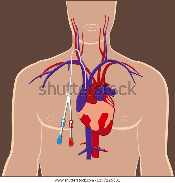
This thoracic and pulmonary anatomy tool is especially designed for students of anatomy medical and paramedical studies. Save time study efficiently. In general the veins preferred for placement of central and peripheral venous access catheters are the internal jugular veins in the neck the axillary and subclavian veins in the chest the cephalic and basilic veins in the upper extremities and the superficial femoral and common femoral veins in the lower extremities.
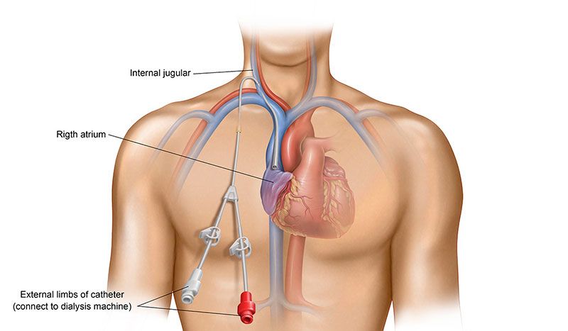
Venous catheters placed caudad to this landmark and cephalad to the right superior cardiac silhouette or no more than 29 cm caudad to the tracheobronchial angle result in catheter tips within the svc. Clinical implications this article provides a practical approach to the clinical implications and importance of understanding the collateral venous anatomy of the thorax. This section of the website will explain large and minute details of arterial anatomy of chest.
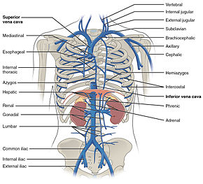
The normal anatomy of the azygos and hemiazygos systems is described in heitzmans excellent text on the mediastinum basically both systems are thoracic continuations of the ascending lumbar veins and provide venous drainage for intercostal and paravertebral veins within the posterior aspect of the thorax. As the heart pumps inside the center of the chest. Venous anatomy or variations thereof can also be crucial to surgical colleagues for operative planning and follow up.
Try now for free.
![]() Cephalic Vein Anatomy And Clinical Points Kenhub
Cephalic Vein Anatomy And Clinical Points Kenhub
 Us Guidance Venous Access Puncture In Chest Port Imlantation
 Visual Guide To Vein And Artery Problems
 Azygos Vein Radiology Reference Article Radiopaedia Org
 Central Venous Access Device Insertion Deranged Physiology
 1000 Venous Anatomy Stock Images Photos Vectors
 Venous Sonography Of The Upper Extremities And Thoracic
 Coronary Venous Anatomy And Anomalies Sciencedirect
 3 Venous Drainage Of The Rectum And Anal Canal Note The
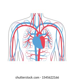
 20 5 Circulatory Pathways Anatomy And Physiology
Infraclavicular Approach In 2018 Ultrasound Access To The
 Understanding The Central Venous Catheter Azura Vascular Care
 Azygous Vein Anatomy Britannica
 Right Gonadal Vein The Anatomy Of The Veins Visual Guide
 Brachiocephalic Vein Wikipedia
![]() Thorax Anatomy Wall Cavity Organs Neurovasculature
Thorax Anatomy Wall Cavity Organs Neurovasculature
 Vascular Access For Hemodialysis
 Superior Vena Cava Syndrome Cancer Net
 Venous Access
 Thoracic Wall Atlas Of Anatomy
 Surgical Anatomy Of The Chest Wall Springerlink
 Central Venous System Anatomy Central Venous Anatomy Choice
 Cvc Central Venous Catheter Dialysis Acces Stock Vector
 Central Lines
Belum ada Komentar untuk "Chest Venous Anatomy"
Posting Komentar