Meniscus Anatomy Definition
It usually refers to either of two specific parts of cartilage of the knee. Mostly converted to bone in adults.
 Quantitative And Qualitative Assessment Of The Posterior
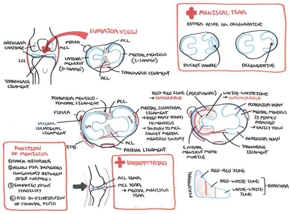
It is less mobile than the lateral meniscus because it is firmly attached to the tibial collateral ligament.
Meniscus anatomy definition. The word meniscus refers to a crescent shaped structure. Both are cartilaginous tissues that provide structural integrity to the knee when it undergoes tension and torsion. Mostly converted to bone in adults.
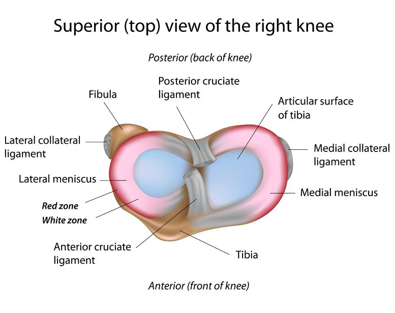
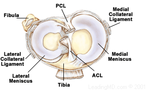
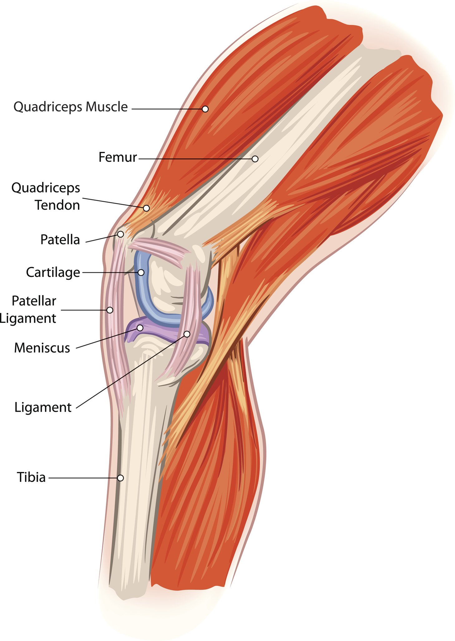
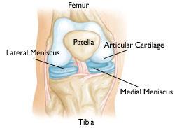
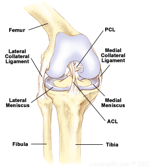
Meniscus anatomy a disk of cartilage that serves as a cushion between the ends of bones that meet at a joint semilunar cartilage cartilage gristle tough elastic tissue. The knee joint contains the meniscus structure comprised of both a medial and a lateral component situated between the corresponding femoral condyle and tibial plateau figure 1. A piece of cartilage shaped like a crescent and located at the junction of two bones in a joint.
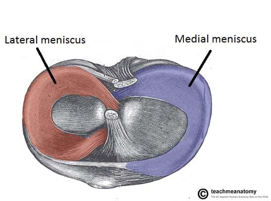
A disk of cartilage between the articulating ends of the bones in a joint. The lateral and medial menisci. The medial condyles are areas of these bones located on the inner sides of the knees.
Torn meniscus the meniscus is a c shaped piece of tough rubbery cartilage that acts as a shock absorber between your shinbone and thighbone. Each is a glossy white complex tissue comprised of cells specialized extracellular matrix ecm molecules and region specific innervation and vascularization. The meniscus acts to absorb shock.
A concavo convex or convexo concave lens. Meniscus anatomy a disk of cartilage that serves as a cushion between the ends of bones that meet at a joint semilunar cartilage cartilage gristle tough elastic tissue. The menisci are also known as semi lunar cartilages referring to their half moon c.
Medial meniscus of the knee. The medial meniscus of the knee is a thickened crescent shaped cartilage pad between the two joints formed by the femur the thigh bone and the tibia the shin bone. A crescent or a crescent shaped body.
Meniscus anatomy they are attached to the small depressions fossae between the condyles of the tibia intercondyloid fossa and towards the center they are unattached and their shape narrows to a thin shelf. A lens with a crescent shaped section. The medial meniscus is often injured when the knee is twisted or sprained with sudden force.
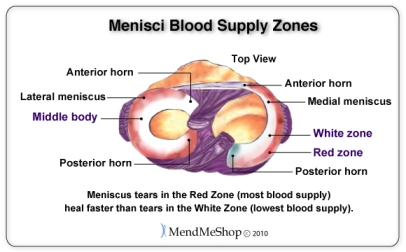
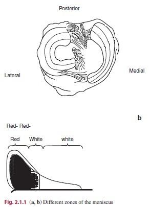
The blood flow of the meniscus is from the periphery outside to the central meniscus. The meniscus acts as a smooth surface for the joint to move on. The convex or concave upper surface of a column of liquid the curvature of which is caused by surface tension.
It can be torn if you suddenly twist your knee while bearing weight on it.
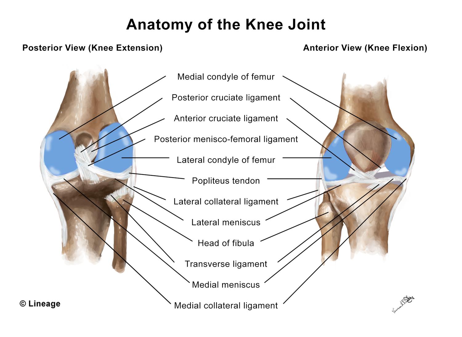
 Anatomy Of The Knee Joint
 Torn Meniscus Symptoms And Diagnosis Jeffrey H Berg M D
 Discoid Meniscus Physiopedia
:max_bytes(150000):strip_icc()/vector-illustration-of-a-meniscus-tear-and-surgery-871162428-03ac23d73f854954a8082f2ae3ce9219.jpg) Meniscus Vs Cartilage Tear Of The Knee
Meniscus Vs Cartilage Tear Of The Knee
 Should I Have Surgery For My Meniscus Tear Caring Medical
 Lateral Meniscus Physiopedia
 Meniscal Tears Causes Symptoms Diagnosis Treatment
 Aspetar Sports Medicine Journal Meniscal Pathology In The
 Meniscal Tears Classification Surgical Repair
 Discoid Meniscus Orthoinfo Aaos
 Tear Of Meniscus Wikipedia
 The Many Types Of Meniscus Tears Caring Medical
 Lateral Meniscus Physiopedia
 Torn Meniscus Symptoms Treatment Mri Test Recovery Time
 Pdf The Human Meniscus A Review Of Anatomy Function
 Torn Meniscus Cedars Sinai
 Meniscus Dictionary Definition Meniscus Defined
 Lateral Meniscus Tear
 Pdf Quantitative And Qualitative Assessment Of The
 Meniscus Tear Orthopedics Medbullets Step 2 3
 Meniscal Transplant Surgery Orthoinfo Aaos
 Meniscal Repair Physiopedia
Torn Meniscus Johns Hopkins Medicine
 Torn Meniscus Picture Image On Medicinenet Com
 Meniscus Injuries
 Meniscus Anatomy Wikipedia
 Meniscal Injury Armando Hasudungan
 Discoid Meniscus Wikipedia
 Understanding Posterior Meniscal Roots Lesions From Basic
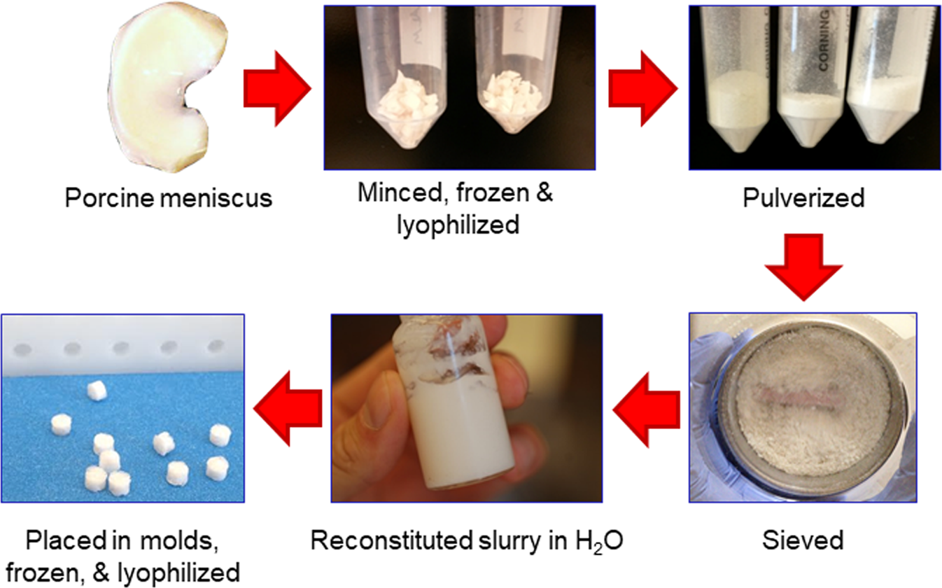 Meniscus Derived Matrix Scaffolds Promote The Integrative
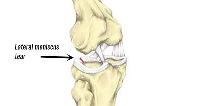 Lateral Meniscus Tear Symptoms Causes Treatment
 Uncommon Injuries Getting To The Root Of Meniscal Tears


Belum ada Komentar untuk "Meniscus Anatomy Definition"
Posting Komentar