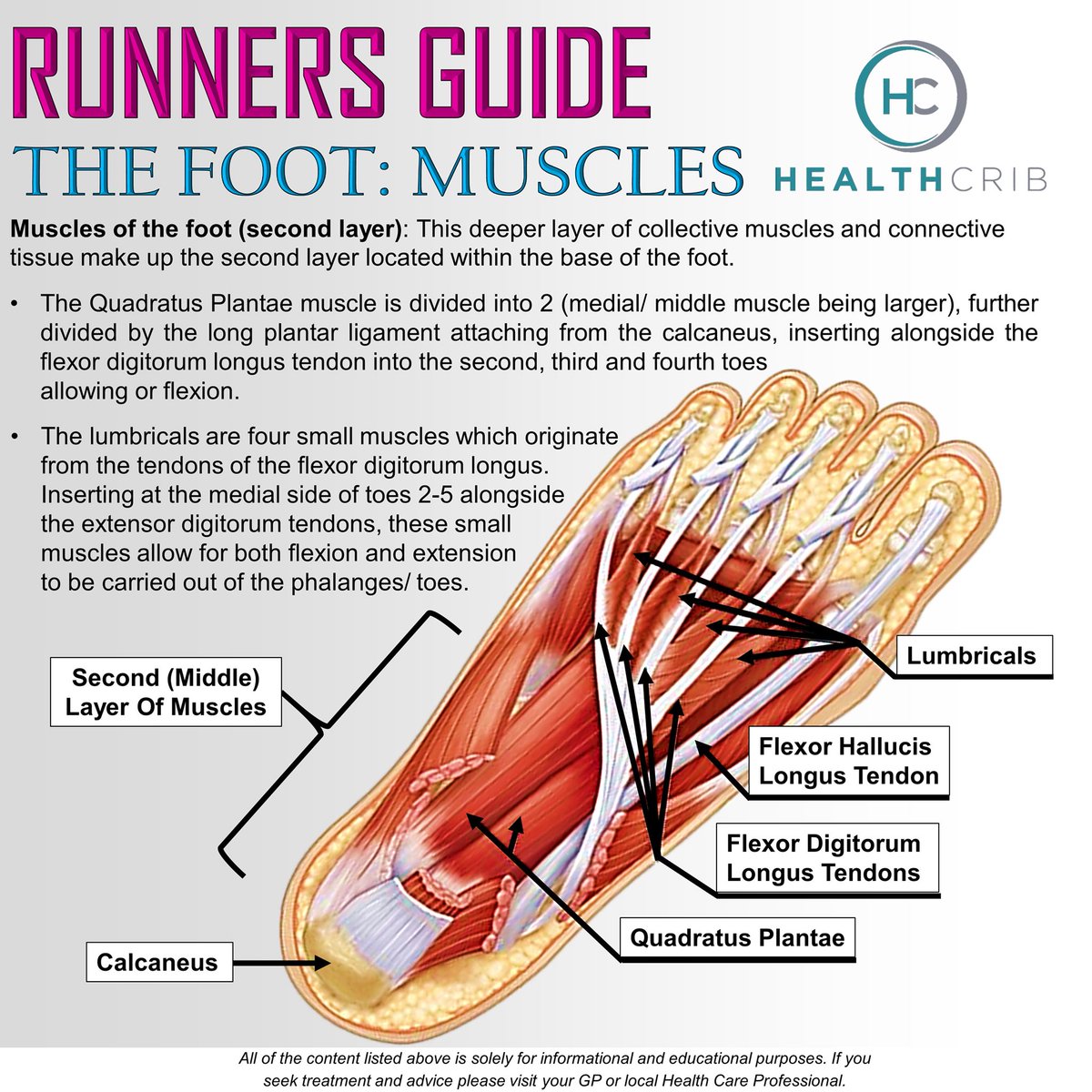
Bottom Of Foot Anatomy
The bones of the feet are. The sole is the bottom of the foot.
 Foot And Leg Anatomy The Foot Trainer
Click on one of the pictures below and point to the area of the foot or ankle where it hurts.

Bottom of foot anatomy. The midfoot is a pyramid like collection of bones that form the arches of the feet. Tarsals five irregularly shaped bones of the midfoot that form the foots arch. Picture of foot anatomy detail.
At the same time the foot must be strong to support more than 100000 pounds of pressure for every mile walked. The talus bone supports the leg bones tibia and fibula forming the ankle. First click on a view of the foot select an area of the foot below to view conditions.
There are also fat pads in the foot to help with weight bearing and absorbing impact. The hindfoot forms the heel and ankle. The foot muscles along with a tissue known as plantar fascia provide secondary support.
Foot definition the foot is a part of vertebrate anatomy which serves the purpose of supporting the animals weight and allowing for locomotion on land. When you experience discomfort or pain thats an indication that something is wrong. In h the foot is a part of vertebrate anatomy which serves the purpose of supporting the animals weight and allowing for locomotion on land.
Below the juncture of these bones are the arches of the foot which are three curves at the bottom of the foot that makes walking easier and less taxing for the body. Foot anatomy reference author. Again thank you from the bottom of my heart for taking the time to answer my questionyour an angel.
These include the three cuneiform bones the cuboid bone and the navicular bone. The foot is an extremely complex anatomic structure made up of 26 bones and 33 joints that must work together with 19 muscles and 107 ligaments to execute highly precise movements. Diagram of normal foot and ankle anatomy.
Use it to pin point where you are having foot or ankle pain. Marc mitnick dpm. Talus the bone on top of the foot that forms a joint with the two bones of the lower leg.
Calcaneus the largest bone of the foot which lies beneath the talus to form the heel bone. These arches the medial arch lateral arch and fundamental longitudinal arch are created by the angles of the bones and strengthened by the tendons. The human foot has 42 muscles 26 bones 33 joints and at least 50 ligaments and tendons made of strong fibrous tissues to keep all the moving parts together plus 250000 sweat glands.
In humans the sole of the foot is anatomically referred to as the plantar aspect. Click to see some of the diagnoses that cause foot symptoms in that area. The foot is an evolutionary marvel capable of handling hundreds of tons of force your weight in motion every day.
 Muscles Of The Lower Leg And Foot Human Anatomy And
 Foot Anatomy Bones Ligaments Muscles Tendons Arches
 Ball Of Foot Pain Relief Management Dr Scholl S
 Abductor Hallucis Muscle Wikipedia
 Ball Of Foot Pain Do The Bottoms Of Your Feet Toes Hurt
 Plantar Fasciitis Medlineplus Medical Encyclopedia
 Picture Of Foot Muscles And Tendons For Manulations
 Head Hands Feet Reflect Body Arms Leg General
 Plantar Fasciitis Info Florida Orthopaedic Institute
 Foot Pain Causes Toe Ball Arch Heel Pain With Treatment
 Layers Of The Plantar Foot Foot Ankle Orthobullets
 Foot Bones Anatomy Conditions And More
 Bottom Of Foot Anatomy Fa02 Ankle Anatomy Foot Anatomy
 Exercise Advice For Foot Pain The Chartered Society Of
 Metatarsalgia Symptoms And Causes Mayo Clinic
 Foot Pain Causes Treatment And When To See A Doctor
 Healthcrib On Twitter The Foot Muscles Distal Bottom
 Rheumatoid Arthritis In Feet Symptoms Treatments And More
 Anatomy Of The Plantar Foot Myfootshop Com
 The Foot Advanced Anatomy 2nd Ed
 Military Disability Ratings For Conditions Of The Feet Legs
 Ball Of Foot Pain Do The Bottoms Of Your Feet Toes Hurt
 Affordable Methods For Relieving Plantar Fasciitis
 Anatomical Overlays Bottom Of The Foot These Images Will


Belum ada Komentar untuk "Bottom Of Foot Anatomy"
Posting Komentar