Fetal Anatomy
The second trimester extends from 13 weeks and 0 days to 27 weeks and 6 days of gestation although the majority of these studies are performed between 18 and 23 weeks. The fetal position should be determined as precisely as possible before an interpretation of fetal anatomy is begun because the position of a structure will often influence our interpretation.
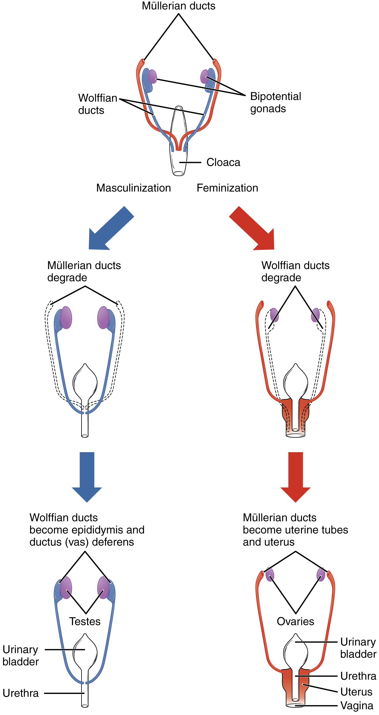 Fetal Development Anatomy And Physiology
Anatomy scan of the fetal head at 20 weeks of pregnancy in a fetus affected by spina bifida.
Fetal anatomy. By the 20th week of pregnancy the baby can weigh up to 11 ounces and measure 10 inches outstretched. Those who want to can find out the sex of the baby if desired. The general fetal orientation is first assessed ie longitudinal oblique or transverse.
The fetal circulatory system becomes much more specialized and efficient than its embryonic counterpart. Fetal ultrasound images can help your health care provider evaluate your babys growth and development and monitor your pregnancy. Most anatomy scans are performed in the second trimester of pregnancy typically at 20 weeks but they can be done anytime between 18 weeks and 22 weeks.
When a level 2 ultrasound is done. A fetal anatomy survey cannot detect all fetal problems and the accuracy of the scan depends on the position of the fetus the gestation and maternal build. A fetal ultrasound sonogram is an imaging technique that uses sound waves to produce images of a fetus in the uterus.
If you have a condition that needs to be monitored such as carrying multiples you may have more than one detailed ultrasound. During this period male and female gonads differentiate. In some cases fetal ultrasound is used to evaluate possible problems or help confirm a diagnosis.
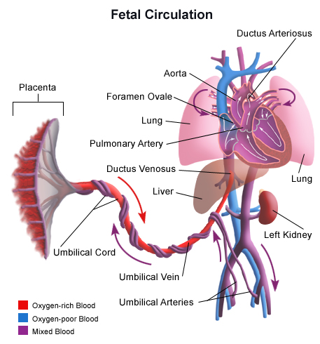
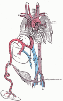
For larger women with increased body fat the diagnostic accuracy is reduced. This sonogram is used to determine fetal anomalies the babys size and weight and also to measure growth to ensure that the fetus is developing properly. Fetal circulation unlike postnatal circulation involves the umbilical cord and placental blood vessels which carry fetal blood between the fetus and the placenta.
The anatomy scan is a level 2 ultrasound which is typically performed on pregnant women between 18 and 22 weeks. When the pregnancy hits the 20th week of gestation an anatomy ultrasound is often ordered. In the axial scan the characteristic lemon sign and banana sign are seen.
It is usually established in the fetal period of development and is designed to serve prenatal nutritional needs as well as permit the switch to a neonatal circulatory pattern at birth. The second trimester scan is a routine ultrasound examination in many countries that is primarily used to assess fetal anatomy and detect the presence of any fetal anomalies. The fetal period lasts from the ninth week of development until birth.
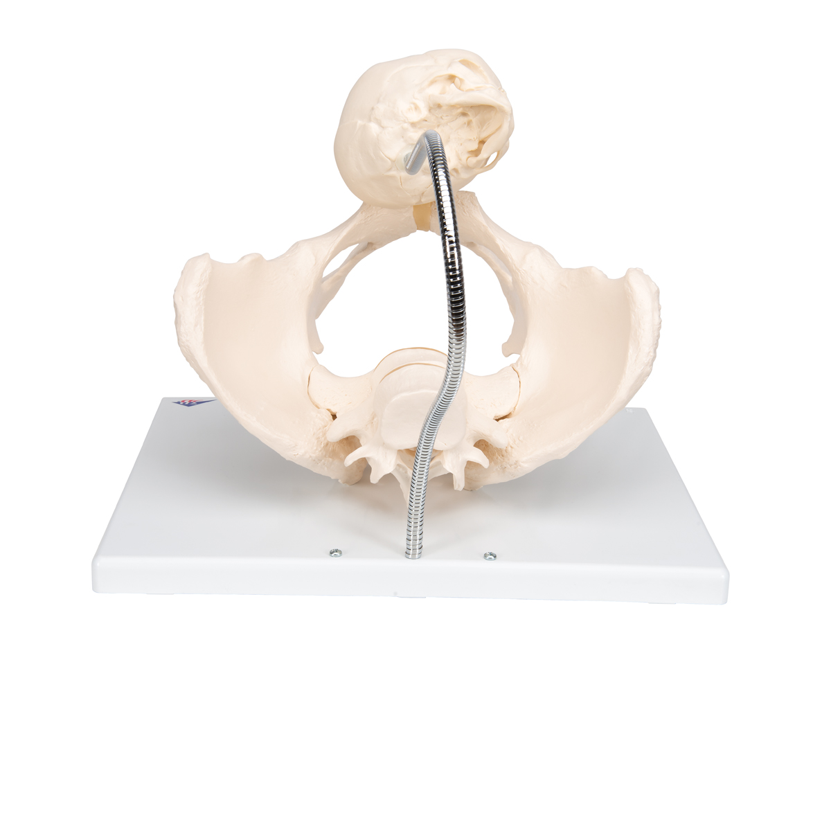
 Childbirth Demonstration Pelvis Skeleton Model With Fetal
 Spina Bifida Myelomeningocele Pavilion For Women
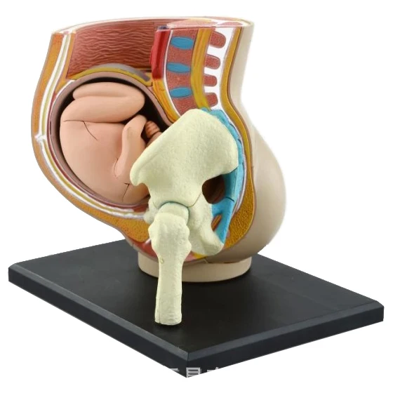 Us 25 9 3d Women Pregnancy Uterine Fetal Anatomy Assembled Model Human Body Medical Teaching Aids In Medical Science From Office School Supplies
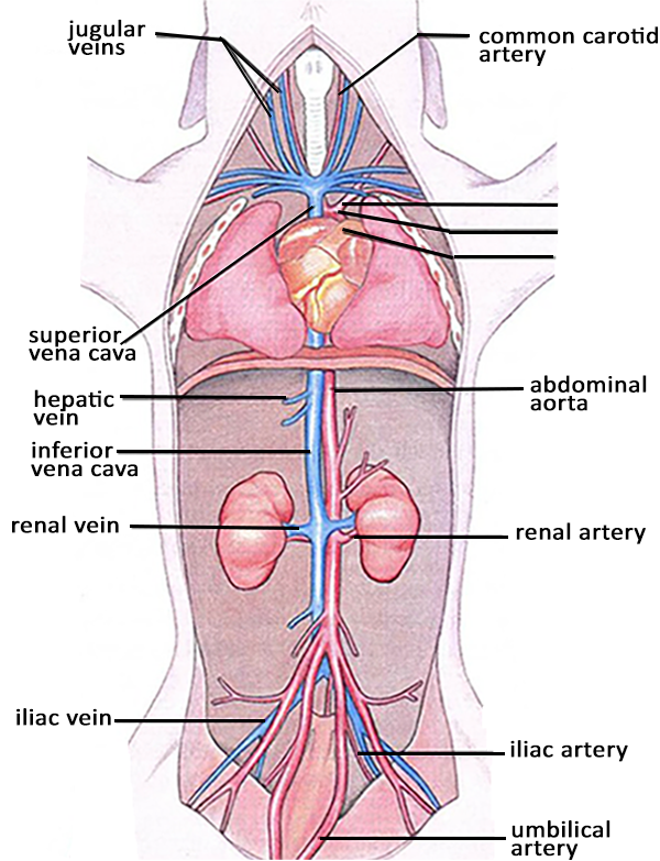 The Ultimate Fetal Pig Dissection Review
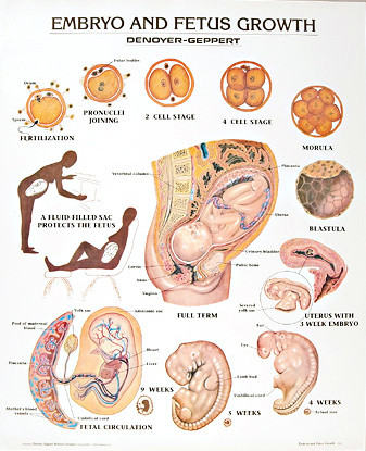 Embryo And Fetus Growth Anatomy Poster

 Pin On Ob Labor Delivery Nursing
 Medical Textbook In The Net Anatomy Of The Neonates
 Human Anatomy And Physiology Laboratory Manual Fetal Pig
 Fetal Circulation Shunts Placenta Oxygenated Rich Blood
 Archive Image From Page 582 Of The Cyclopaedia Of Anatomy And
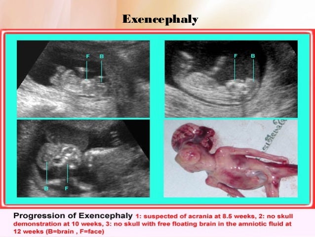 Routine Fetal Anatomy Scan At 18 23 Weeks
 Fetal Pig Anatomy Brian Mccauley
Fetal Biometry Challenge Us Isbi 2012
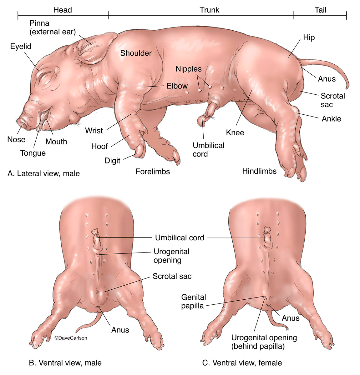 Fetal Pig Surface Anatomy Carlson Stock Art
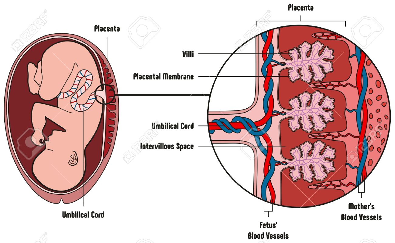 Human Fetus Placenta Anatomy Diagram With All Part Including
 Normal Fetal Brain Anatomy At 11 13 Weeks 2d Scan
 Anatomy And Physiology In Context Reading Assignment
 6 Fetal Heart Anatomy And Electrical Activation Sequence
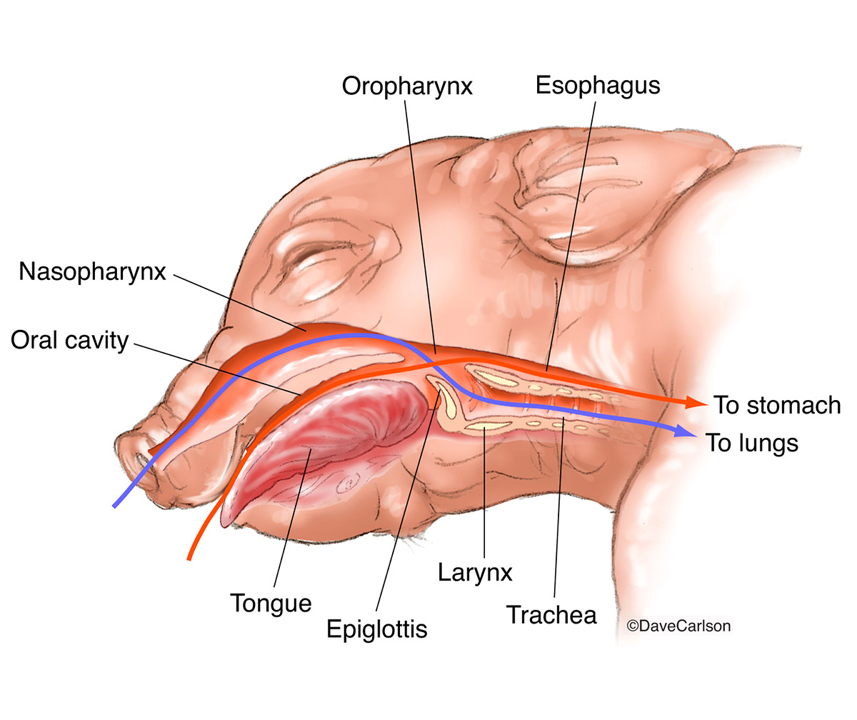 Fetal Pig Anatomy Pharynx Larynx Carlson Stock Art
 Fetal Descent Stations Birth Presentation Medical
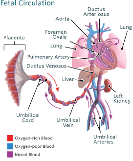 Blood Circulation In The Fetus And Newborn Children S
 External Anatomy Of The Fetal Pig Figure 27 1 Diagram
 Normal Fetal Anatomy Medical Illustration Human Anatomy
 Routine Fetal Anatomy Scan At 18 23 Weeks
 A Fetal Abdominal Anatomy B True Fasp C False Fasp
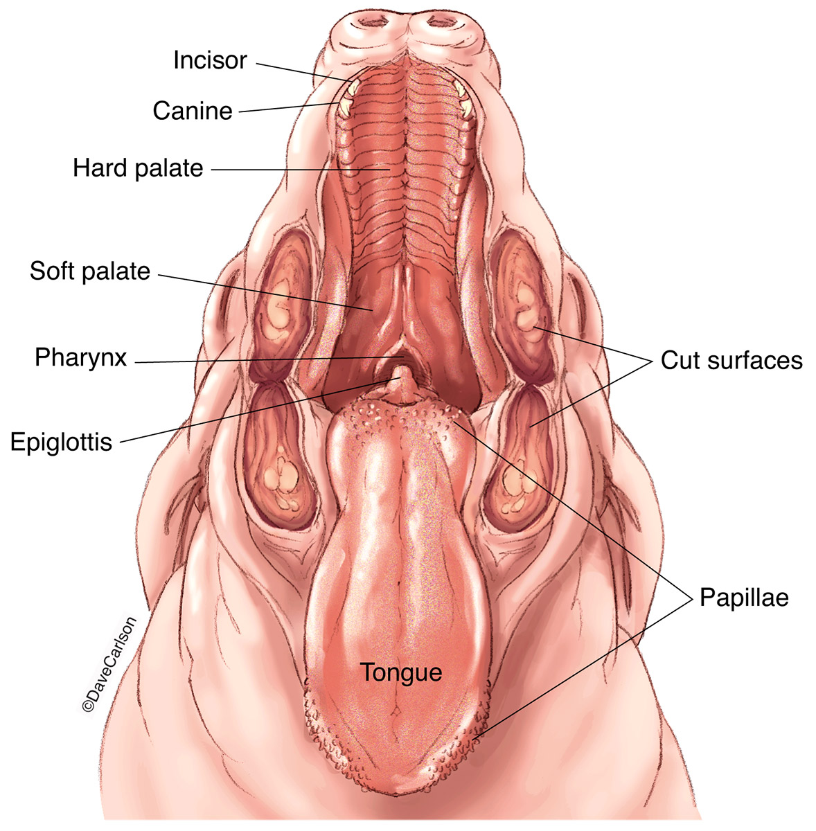 Fetal Pig Anatomy Oral Cavity Carlson Stock Art
 Fetal Heart An Overview Sciencedirect Topics


Belum ada Komentar untuk "Fetal Anatomy"
Posting Komentar