Eye Socket Anatomy
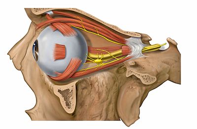
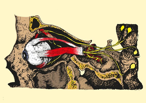
Six extraocular muscles in the orbit are attached to the eye. 101 us fl oz.
 Orbit And Eye
The eye is the organ responsible for vision.
Eye socket anatomy. A review for ocularists introduction and rationale understanding the basic anatomy of the human eye is a requirement for all health care providers but it is even more significant to eye care practition ers including ocularists. Orbit can refer to the bony socket or it can also be used to imply the contents. The white portion of the eyeball is called the sclera.
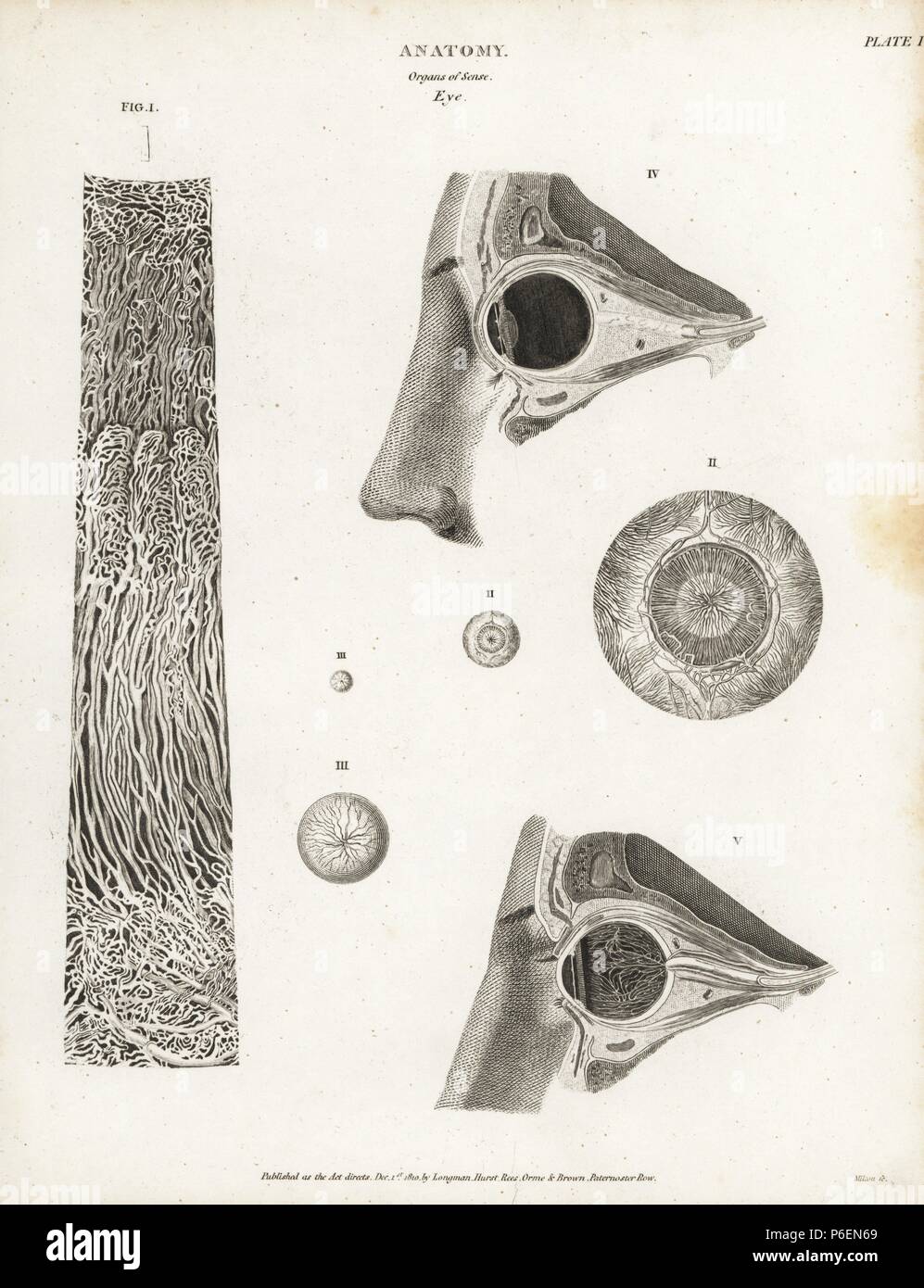
In the adult human the volume of the orbit is 30 millilitres 106 imp fl oz. The diagrams below show cross sections of the human eyeball. Anatomy of the eye.
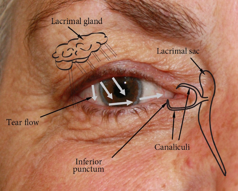
Nerve signals that contain visual information are transmitted through the optic nerve to the brain. At the inner side of the lids there is a tear duct. The eyeball is covered by a top and bottom lid.
It moves the eye upward. The extraocular muscles are attached to the white part of the eye called the sclera. The eye socket refers to the hole in the skull where the eyeball sits.
Tearfilm socket a pictorial anatomy of the human eyeanophthalmic socket. These muscles move the eye up and down and side to side and rotate the eye. They enclose the eyeball and its associated structures.
The superior rectus is an extraocular muscle that attaches to the top of the eye. Pink eye may cause eye socket pain. The bony orbits or eye sockets are bilateral and symmetrical cavities in the head.
Human eye specialized sense organ in humans that is capable of receiving visual images which are relayed to the brain. External extraocular anatomy extraocular muscles. Vision is our window to the outside world.
Infections can cause eye socket pain. The eye sits in a protective bony socket called the orbit. The anatomy of the eye includes auxillary structures such as the bony eye socket and extraocular muscles as well as the structures of the eye itself such as the lens and the retina.
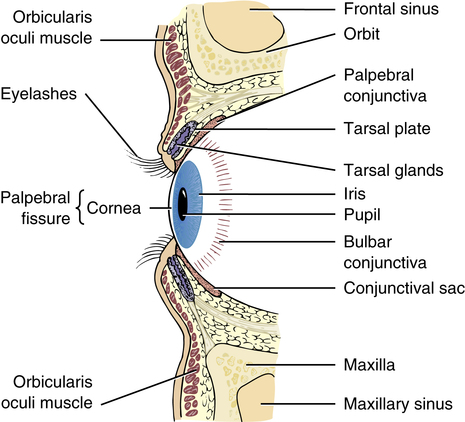
The eye is surrounded by the orbital bones and is cushioned by pads of fat within the orbital socket. Extraocular muscles help move the eye in different directions. The anatomy of the human eye includes the cornea retina lens pupil optic nerve and more.
The type of eye anatomy that ocularists know how. This is a strong layer of tissue that covers nearly the entire surface of the eyeball. This article explores the anatomy of the eye looking at the different structures of the human eye and their function.
In this article we shall look at the borders contents and clinical correlations of the bony orbit. Anatomy of the eye. These muscles work to move the eye up down side to side and rotate the eye.
Orbit anatomy in anatomy the orbit is the cavity or socket of the skull in which the eye and its appendages are situated. The top edge of this hole has a ridge thats called the brow ridge. There are six muscles that are present in the orbit eye socket that attach to the eye to move it.
How to draw eyes structure. They enclose the eyeball and its associated structures.
 Title Human Anatomy The Eye Socket Ball Seeing With Human And Psychic Eyes Like Hilma Af Klint
 Anatomy Of The Human Eye Showing Socket Surface Of The

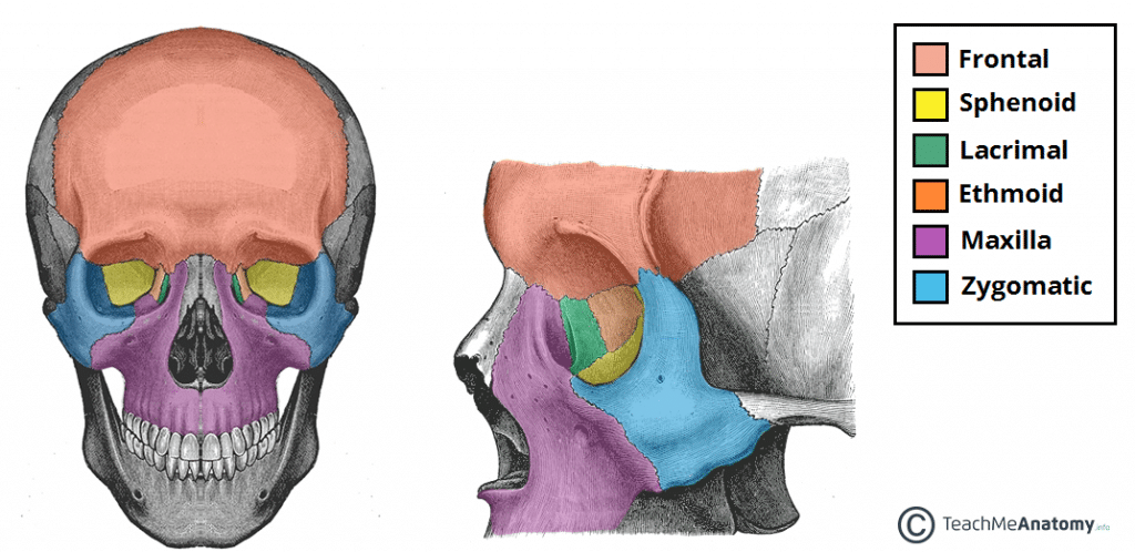
 The Skull Anatomy And Physiology I
 Skull Bones Of The Orbit Human Anatomy Kenhub
 Amazon Com Gothic Mug Skull With Dry Red Rose In Teeth
 Amazon Com Gothic Decor Ceramic Piggy Bank Skull With Dry
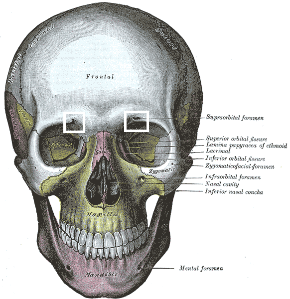 Supraorbital Foramen Wikipedia
 Eye And Nerves Medical Exhibit Medivisuals

 The Bony Orbit Borders Contents Fractures Teachmeanatomy
 The Eye Musculoskeletal Key


 Eye Socket Anatomy Medical Illustration
 Amazon Com Custom Double Sided Seasonal Garden Flag Gothic
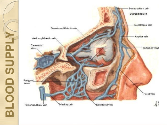
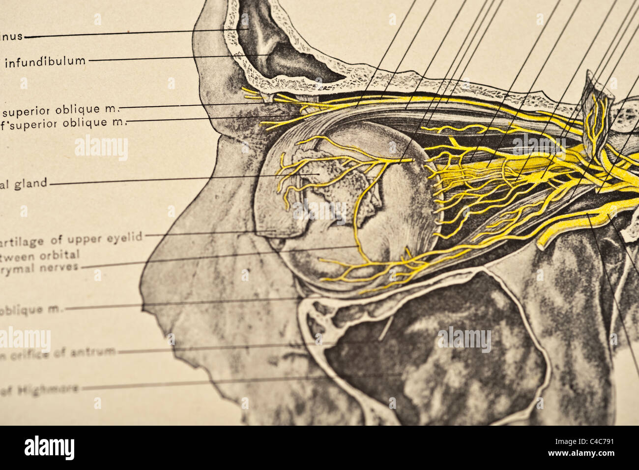 Eye Socket With Optic Nerves Circa 1904 Stock Photo
 Eye Position In The Eye Socket Eyeball Anatomy Eye Study
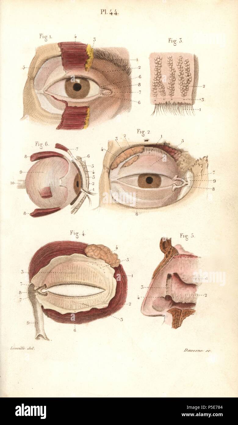 Sections Of The Eye Socket And Nasal Cavity Handcolored
 Orbital Tumor Eye Socket Cancer Anatomy
 101 Amazing Eye Facts Lenstore Co Uk
 Eye Socket Anatomy Illustration Stock Image C046 1439
 Eye Socket Canvas Prints Fine Art America

Extraocular Muscles Wikipedia
 Image Result For Eye Socket Cutaway Human Drawing Eye
 An Easy Guide To Your Eye S Anatomy Lenstore Co Uk
Belum ada Komentar untuk "Eye Socket Anatomy"
Posting Komentar