Fossa In Anatomy
A trench or channel. The cubital fossa is triangular in shape and thus has three borders.
 Nasal Fossa An Overview Sciencedirect Topics
Plural fossae ˈfsiː or ˈfsa.

Fossa in anatomy. In anatomy a fossa ˈfs. Arm and cubital fossa. Superior border hypothetical line between the epicondyles of the humerus.
A slender long tailed carnivorous mammal cryptoprocta ferox of the family eupleridae of madagascar that has retractile claws usually reddish brown or sometimes black short thick fur and anal scent glands. Medial border lateral border of the pronator teres muscle. In anatomy a hollow or depressed area.
When it is lateral to the humerus it pierces the lateral intermuscular septum as it moves into the forearm anterior to the lateral epicondyle between the brachialis and brachioradialis. Fossa a concavity in a surface especially an anatomical depression pit. Fossa ovalis is one of the most important parts of the cardiovascular system of human since the fetal development until the death.
The fossa evolved on the catless island of madagascar where it became the ecological equivalent of a cat. Condylar fossa condyloid fossa either of two pits on the lateral portion of the occipital bone. Descends inferolaterally with the deep artery of the arm in the radial groove between the lateral and medial heads of the triceps.
Teachme anatomy part of the teachme series the medical information on this site is provided as an information resource only and is not to be used or relied on for any diagnostic or treatment purposes. Lateral border medial border of the brachioradialis muscle. Glenoid cavity glenoid fossa the concavity in the head of the scapula that receives the head of the humerus to form the shoulder joint.
Cerebral fossa any of the depressions on the floor of the cranial cavity. Amygdaloid fossa the depression in which the tonsil is lodged. The superomedial aspect of the popliteal fossa is bounded by the semimembranosus and.
Closure of the foramen ovale as the baby grows and starts using the lungs the pressure is created in the foramen ovale causing it to close. From the latin fossa ditch or trench is a depression or hollow usually in a bone such as the hypophyseal fossa the depression in the sphenoid bone. The popliteal fossa is 25 cm wide and mainly consists of fat tissue.
Blood vessels are located deep to the nerves within the fossa and include.
 J Neurosurgery On Twitter Neurosurgicalatlas Anatomy For
 B3w4an Infratemporal Fossa Block3 2015 Anatomy Flashcards
 Pterygopalatine Fossa Anatomy Arterial Supply Venous
 Supraclavicular Fossa Supraclavicular Lymph Nodes Anatomy
 The Popliteal Fossa Human Anatomy
 Pterygopalatine Fossa Wikipedia
 Popliteal Fossa And Knee Joint Last S Anatomy Regional
 Figure Cubital Fossa Image Courtesy S Bhimji Md
 Anatomy Atlases Illustrated Encyclopedia Of Human Anatomic
 Anatomy Infratemporal Fossa Flashcards Quizlet
 Infratemporal Fossa An Overview Sciencedirect Topics
 The Pterygopalatine Fossa Imaging Anatomy Communications
 Popliteal Fossa Anatomy And Posterior View Of The Leg
 Anatomy Of The Infratemporal Fossa Ami 2018 Meeting
 Image Result For Cranial Fossa Anatomy Anatomy Miller
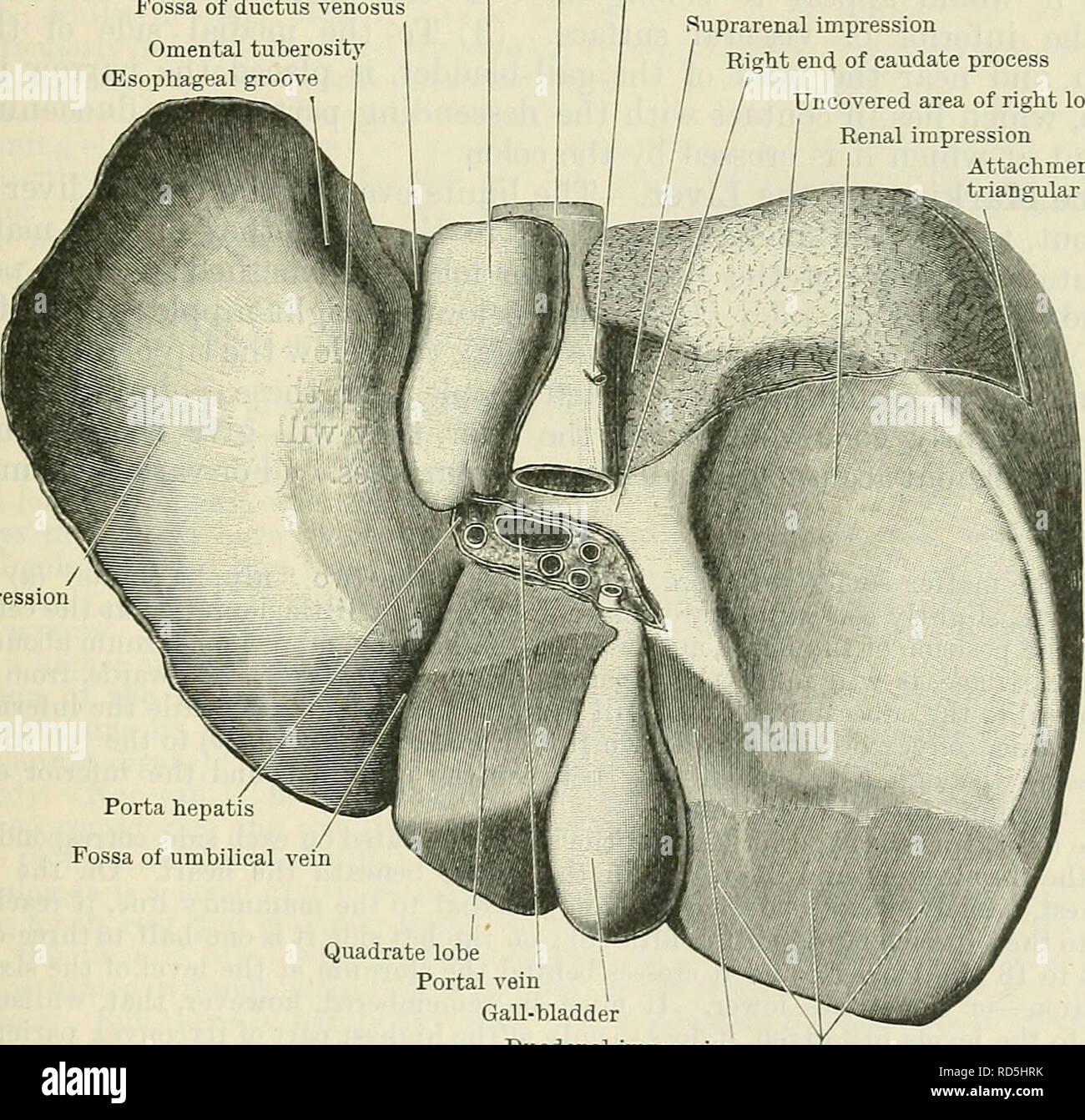
 Cunningham S Text Book Of Anatomy Anatomy The Liver 1193
 Basic Sciences Anatomy Of The Popliteal Fossa
 Posterior Cranial Fossa Boundaries Contents Teachmeanatomy
 Anatomy Of The Left Popliteal Fossa Download Scientific
 Temporal Muscle Temporal Bone Infratemporal Fossa Anatomy
 What Is The Difference Between Ridge Process Fossa And
 The Scapula Surfaces Fractures Winging Teachmeanatomy
 Temporal Fossa Anatomy Borders And Contents Kenhub
 The Intercondylar Fossa Of The Femur Definition Function
 Easy Notes On Cubital Fossa Learn In Just 4 Minutes
 Anatomy Of The Posterior Fossa Clinical Gate
 Rectum Digestion Gastrointestinal Tract Human Digestive
 Posterior Cranial Fossa With Cranial Nerves Relation
 Infratemporal Fossa Approach The Modified Zygomatico
 Fossa Printout Enchantedlearning Com
 Middle Cranial Fossa Surgery Offers A Better Chance For


Belum ada Komentar untuk "Fossa In Anatomy"
Posting Komentar