Dog Leg Anatomy
Understanding and knowing your dogs leg anatomy will help learn the possible weaknesses injuries and the best ways how to treat them. Understanding the dog leg anatomy is also important as this is an area that is very much prone to injury.
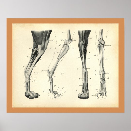 Dog Leg Bones Muscle Veterinary Anatomy Print
The bones and ligaments can easily be cracked stretched or twisted when impact is applied through running jumping or by virtue of an accident or jolting impact as listed below.
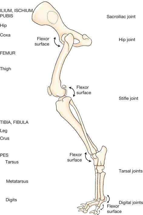
Dog leg anatomy. Only one third is carried on their hind legs. The rear legs of the dog begin with the femur bone which extends to a pair of bones known as the tibia and the fibula. Dog leg anatomy just like humans have arms and legs dogs have forelegs and hind legs.
The elbow is the first joint in the dogs leg located just below the chest on the back of the foreleg. Two thirds of a dogs body weight is carried on their front legs. These dog leg injuries include bone fractures bone cracks ligament tears ligament damage cuts bruises and joint pain.
The forelegs or front legs carry two thirds of its body weight. The dog is carried around by the forelegs and the hind legs. Directly below the shoulder of the foreleg is the humerus bone which ends at the elbow the first joint located just below the chest on the back of the foreleg.
The anatomy of a dogs hind leg and foreleg differs just as a human arm and leg differ according to for dummies. Like your arms its comprised of the ulna and radius. The upper arm on the foreleg is right below the shoulder and is comprised of the humerus bone.
In reality the anatomy of a dogs leg is very complex. The forelegs and hind legs of a dog are as different as human arms and legs. The long bone that runs after the elbow on the foreleg is the forearm.
The forearm may have feathering on the back. Much as the hind legs have got larger muscles which make them stronger they only carry around one third of its body weight. However the muscles on their hind legs are larger and therefore stronger.
These further extend to the heel bone known as tarsus the paw bone known as metatarsus and the toe bone phalange. Dog leg problems can be classified to further understand how they should be dealt with and treated. Dogs legs are comprised of bones muscles ligaments and tendons.
It ends at the elbow the elbow is the first joint in the dogs leg located just below the chest on the back of the foreleg.
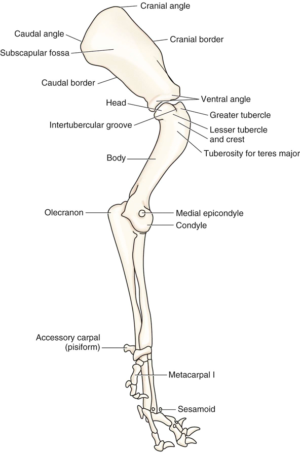 Canine Anatomy Veterian Key
 Dog Front Legs Anatomy Bones
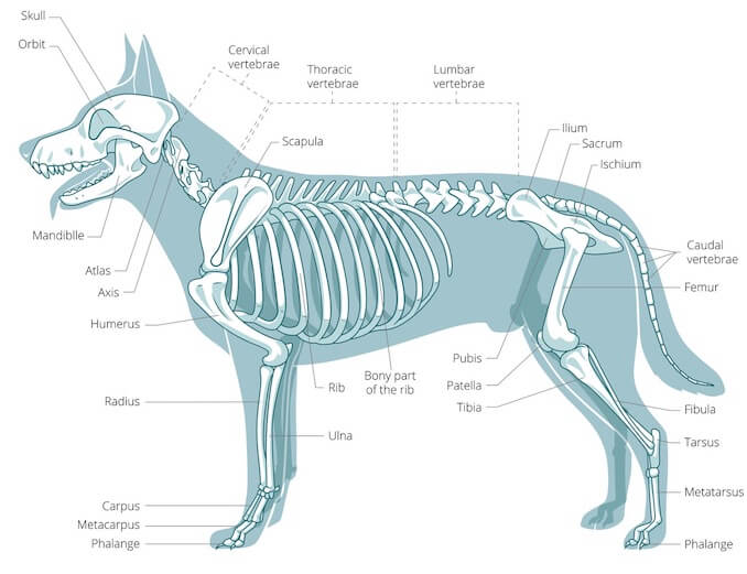
 Dog Leg Skeletal Anatomy Animal Leg Bones Dog Anatomy

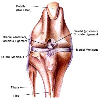 Clicking Sound Cranial Cruciate Ligament Ccl Or Knee
 Components Of The Musculoskeletal System In Dogs Dog
 Harnesses Homeskooling 4 Dogs
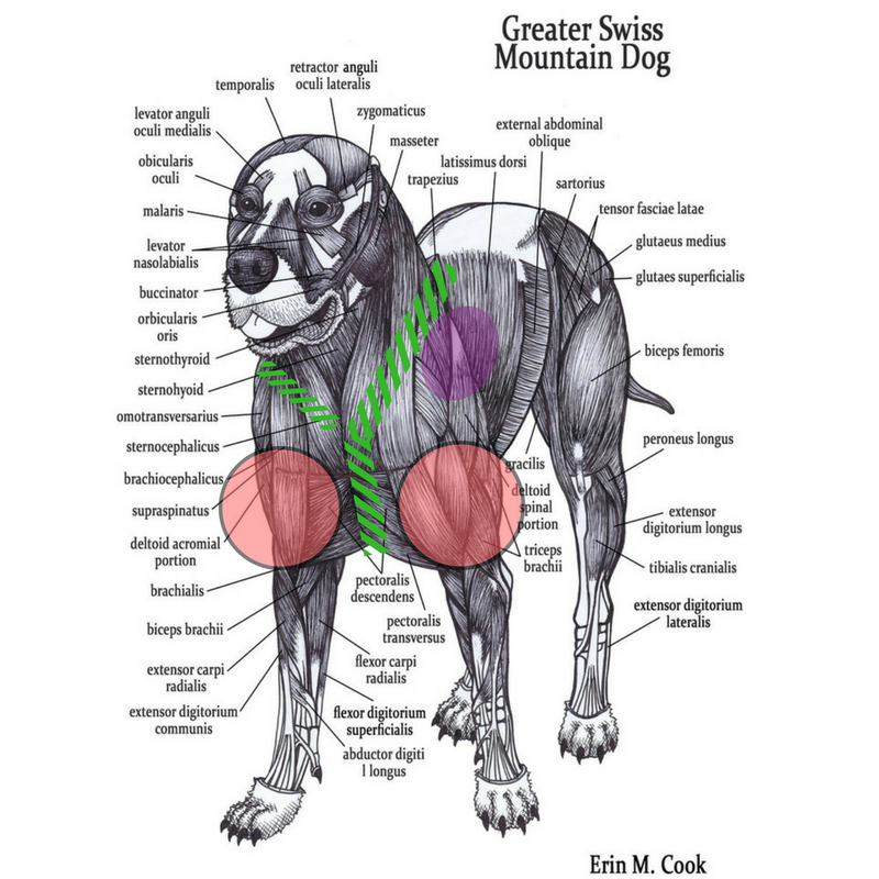
 A Visual Guide To Dog Anatomy Muscle Organ Skeletal
 Canine Osteology Illustrations
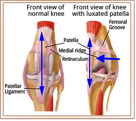 Patellar Luxation Metropolitan Veterinary Associates

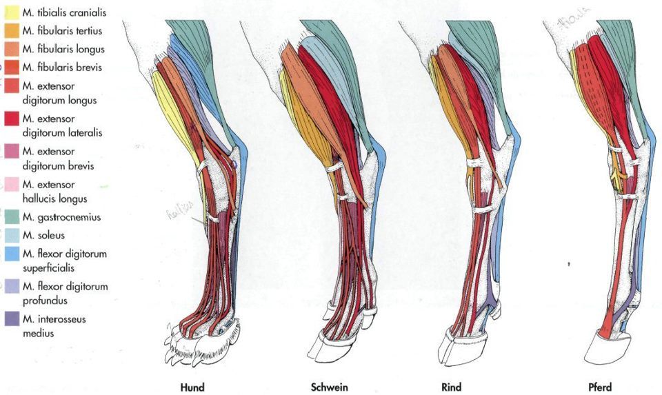 My Vet Life Comparative Leg Anatomy Dog Pig Cow Horse
 Dog Leg Anatomy Explained Injury Types And Treatments
 Canine Anatomy Veterian Key
 Dog Anatomy Hind Legs Dog Anatomy Anatomy For Artists
 2019 Ultimate Veterinary Guide To Dog Anatomy With Images
 Canius Lupis Dog Anatomy
 Yoga Anatomy Using Muscle Awareness To Lower Your Heels In
 The Project Gutenberg Ebook Of The Artistic Anatomy Of
 Acl Repair Family Vetcare
 Syntissue Leg Syndaver
 Dog Anatomy Front Legs Lion Anatomy Animal Anatomy Dog
 Alternatives To Surgery For Ligament Injuries In Dogs
 Dog Hind Legs Anatomy Bones
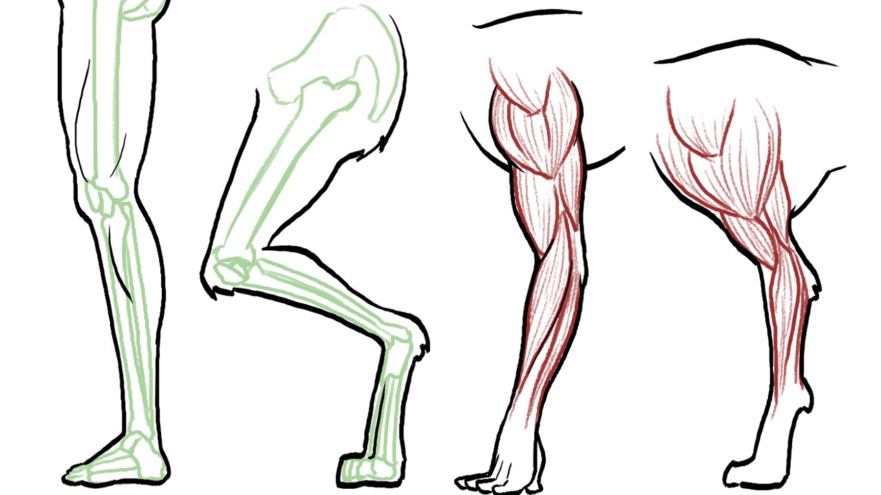
How To Draw Animal Legs Dogs Cats Horses Bears Etc
 Canine Anatomy Veterian Key
 Anatomy Of The Dogs Hl Stifle Joint Veterinary Medicine
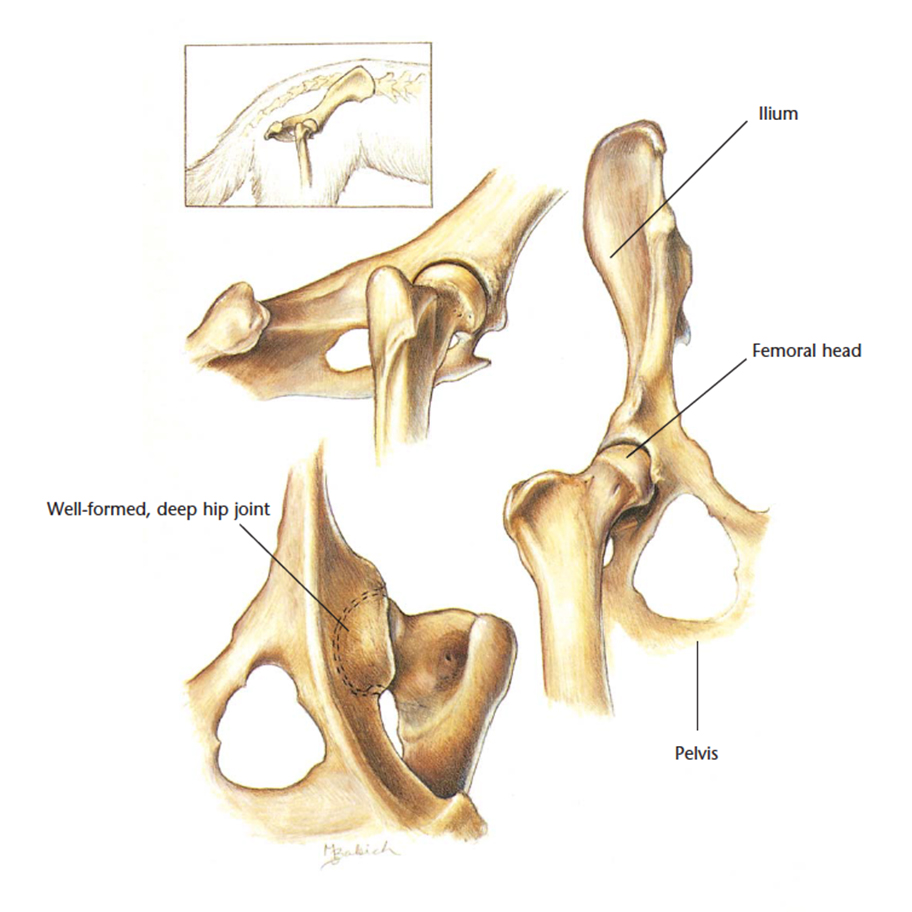
 Amazon Com Bbyt Dog Canine Hip Joint Skeleton Model
 Dog Body Parts Stock Vector Illustration Of Isolated 88316412
 Labeled Atlas Of Anatomy Illustrations Of The Dog
 Dog Leg Anatomy Explained Injury Types And Treatments
 Muscle Bone Structure Charts Dog Anatomy Dog Leg Grey
Belum ada Komentar untuk "Dog Leg Anatomy"
Posting Komentar