Iac Anatomy
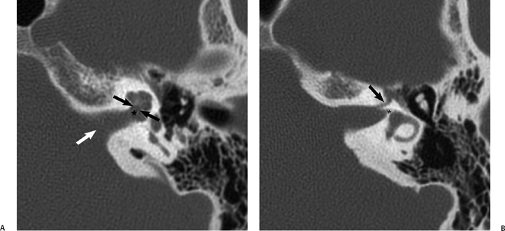 The Vestibulocochlear Nerve With An Emphasis On The Normal
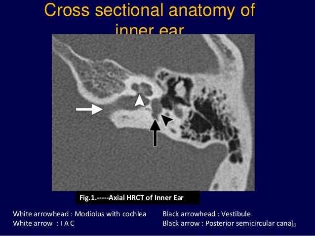
 Internal Auditory Meatus Wikipedia
The Inner Ear Imaging Anatomy With 3t Mri New Sequences A
 Magnetic Resonance Imaging Of Inner Ear

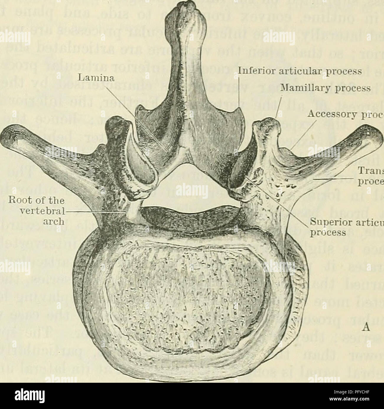 Cunningham S Text Book Of Anatomy Anatomy Lumbar Veetebeie
 Key Anatomical Landmarks For Middle Fossa Surgery A
Anatomy Of The Fundus Of The Internal Acoustic Meatus
 Three Dimensional Imaging Of The Human Internal Acoustic
 Roentgen Ray Reader Contents Of The Internal Auditory Canal
 Normal Mri Internal Auditory Canal Radiology Case
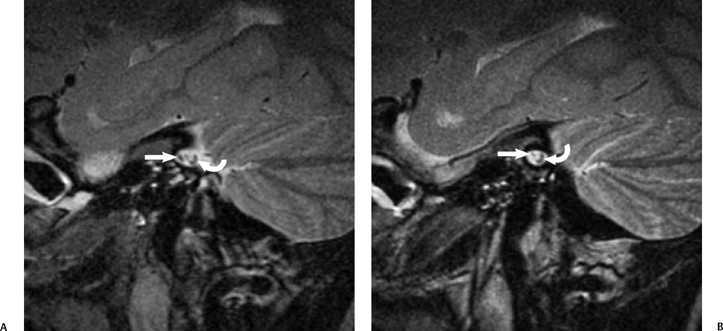 The Vestibulocochlear Nerve With An Emphasis On The Normal
 Figure 8 From Imaging Of Sensorineural Hearing Loss A
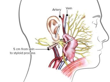 Facial Nerve Anatomy Overview Embryology Of The Facial
 A C Axial Temporal Hrct Images Of Normal Inner Ear Anatomy
Ct Images Of Normal Right Inner Ear Anatomy A F Axial
 Three Dimensional Imaging Of The Human Internal Acoustic
 7up Coke Down Mnemonic Radiology Case Radiopaedia Org
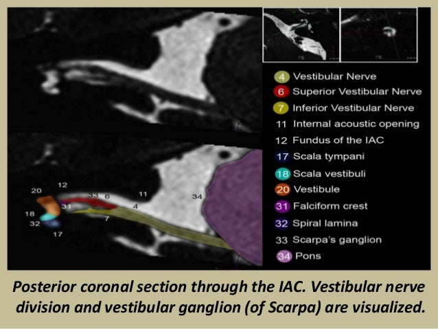 Presentation1 Pptx Radiological Anatomy Of The Petrous Bone
Surgical Exposure Of The Internal Auditory Canal Through The
 Figure Measurements Of Internal Auditory Canal Iac A
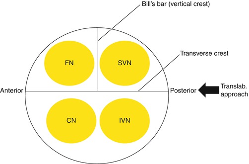 Internal Auditory Canal Iac Springerlink
 Dissection Of Extended Middle Fossa And Anterior
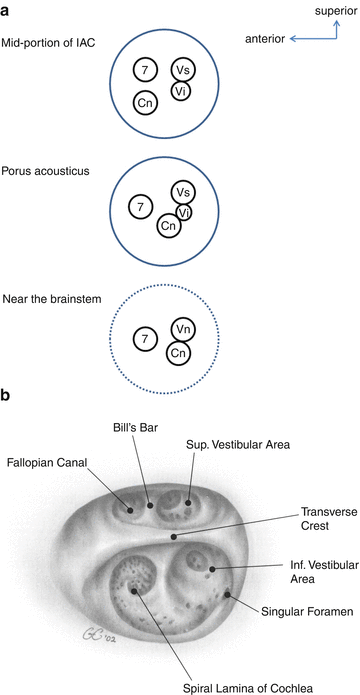 Balance Anatomy Vestibular Nerve Springerlink
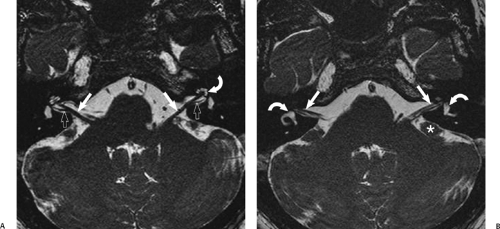 The Vestibulocochlear Nerve With An Emphasis On The Normal
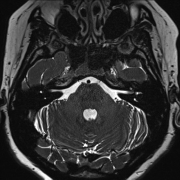 Normal Mri Internal Auditory Canal Radiology Case
Jugular Bulb And Skull Base Pathologies Proposal For A
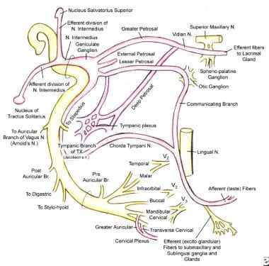 Facial Nerve Anatomy Overview Embryology Of The Facial
Imaging Findings Of Cochlear Nerve Deficiency American

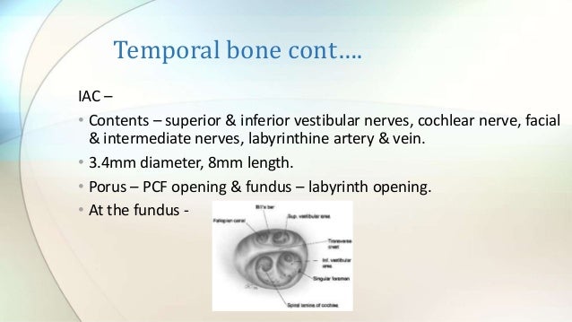


Belum ada Komentar untuk "Iac Anatomy"
Posting Komentar