Knee Anatomy Mri
Anatomy of the knee can be complicated and hard to understand. Spin echo t1 or proton density with fat saturation sequences.
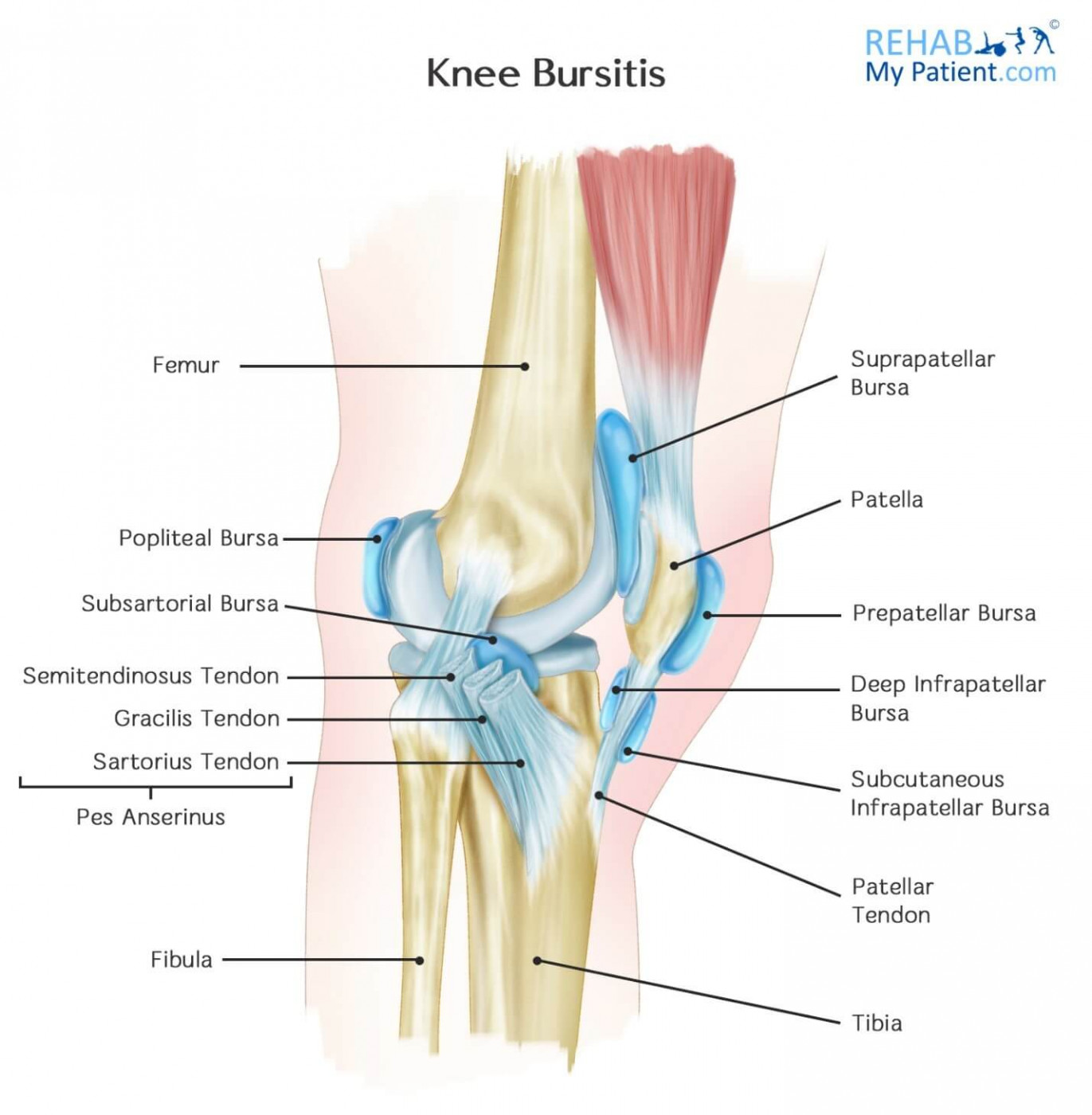 Knee Bursitis Rehab My Patient
After all an entire year of fellowship training is dedicated to musculoskeletal imaging.

Knee anatomy mri. Use the mouse to scroll. Meniscus lateral and medial cruciate ligaments vastus lateralis intermedius medialis tibial and fibular collateral ligaments. Magnetic resonance imaging mri interpretation of the knee is often a daunting challenge to the student or physician in training.
The weightings menu makes it possible to choose the type of mri sequence to be viewed. This mri knee sagittal cross sectional anatomy tool is absolutely free to use. Click on a link to get t1 coronal view t2 fatsat axial view t2 fatsat coronal view t2 fatsat sagittal view.
Atlas of knee mri anatomy. Use the mouse scroll wheel to move the images up and down alternatively use the tiny arrows on both side of the image to move the images. Anatomy of the knee on a coronal slice mri.
This webpage presents the anatomical structures found on knee mri. Colorado knee specialist dr. Robert laprade discusses how to read an mri of a normal knee.
Through the use of magnetic resonance imaging clinicians can diagnose ligament and meniscal injuries along with identifying cartilage defects bone fractures and bruises.
 Mri Anatomy Of The Knee And Shoulder Lieberman S
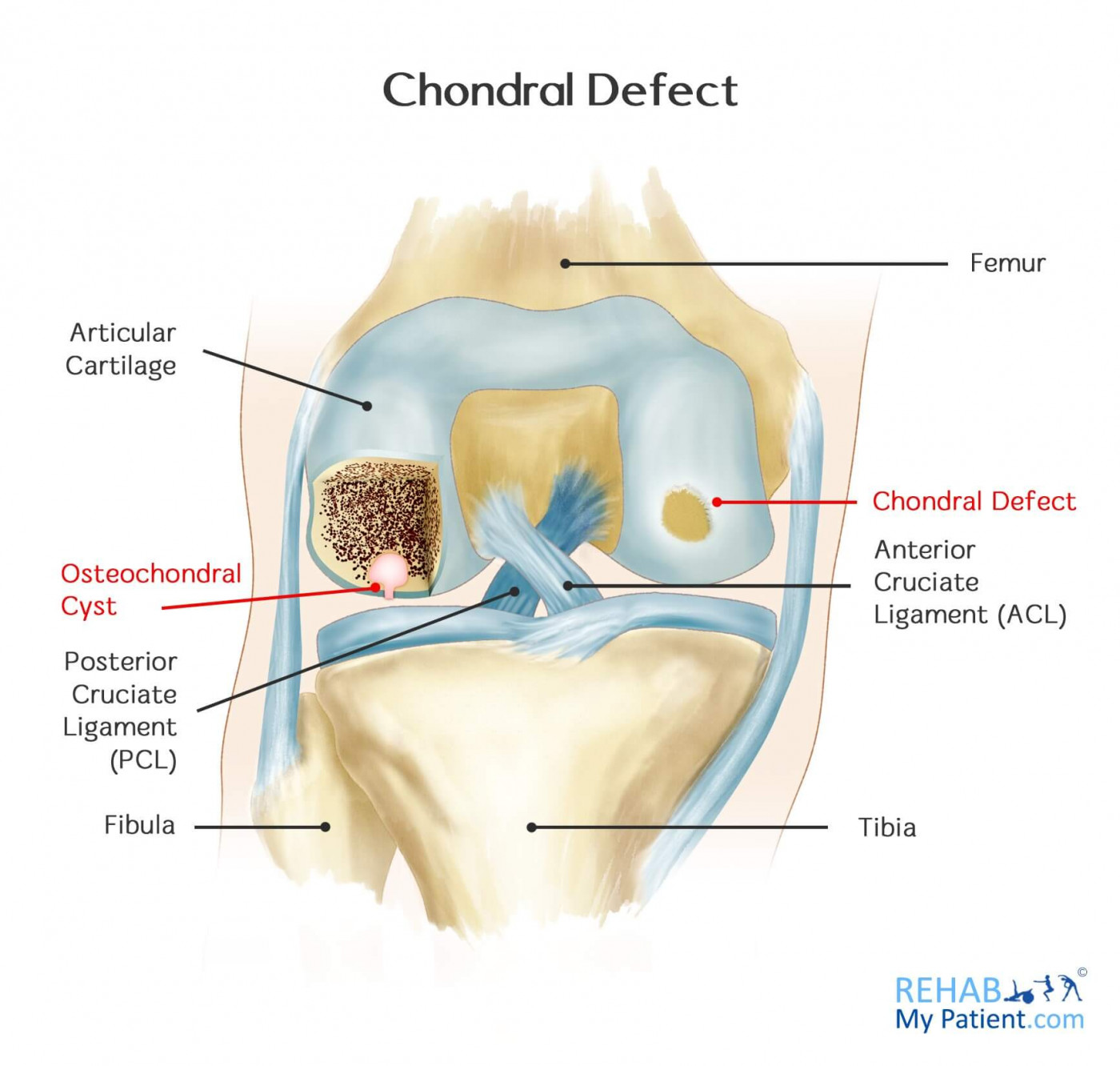 Chondral Defect Rehab My Patient
 Magnetic Resonance Imaging Of Hoffa S Fat Pad And Relevance
 Ppt Posterolateral Corner Of The Knee Mri Anatomy
 Knee Anatomy Mri Knee Coronal Anatomy Free Cross
 Mri Knee Anatomy Knee Sagittal Anatomy Free Cross
 Knee Joint Picture Image On Medicinenet Com
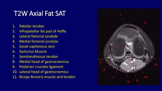 Mri Anatomy Of Knee Dr Muhammad Bin Zulfiqar
 Mri Images Of A Rat Wistar And Mouse C57bl 6 Knee Joint
 Mri Knee Anatomy Knee Sagittal Anatomy Free Cross
 Magnetic Resonance Imaging Mri And Jia

 Figure 14 From Normal Mr Imaging Anatomy Of The Knee
 Essr 2015 P 0055 Popliteal Fossa Masses A Pictorial
 Anatomy Quiz Mri Knee Radiology Case Radiopaedia Org
 Peroneal Nerve Normal Anatomy And Pathologic Findings On
 The Radiology Assistant Knee Non Meniscal Pathology
 Mri Knee Meniscus Tear Scan Stock Photo Image Of Scan
Imaging Evaluation Of The Multiligament Injured Knee
 Multi Ligament Knee Injuries Sterling Ridge Orthopaedics


Belum ada Komentar untuk "Knee Anatomy Mri"
Posting Komentar