Vein Of Galen Anatomy
Most conditions associated with the great cerebral vein are due to congenital defects. Vein of galen aneurysmal malformations vgam are the most common form of symptomatic cerebrovascular malformation in neonates and infants.
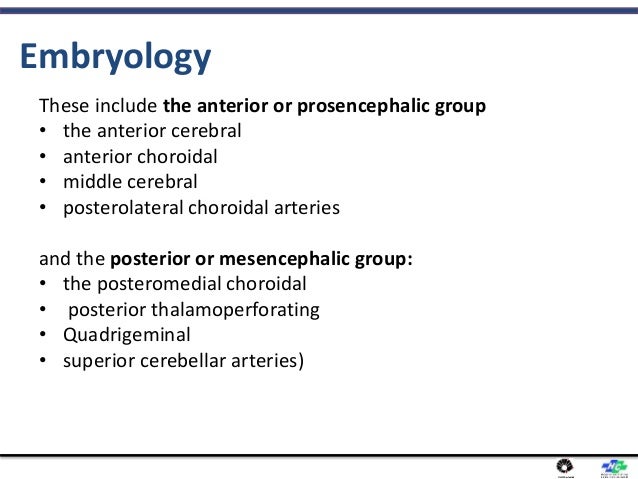
 Cerebral Venous Development In Relation To Developmental
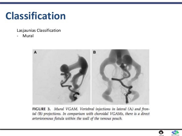
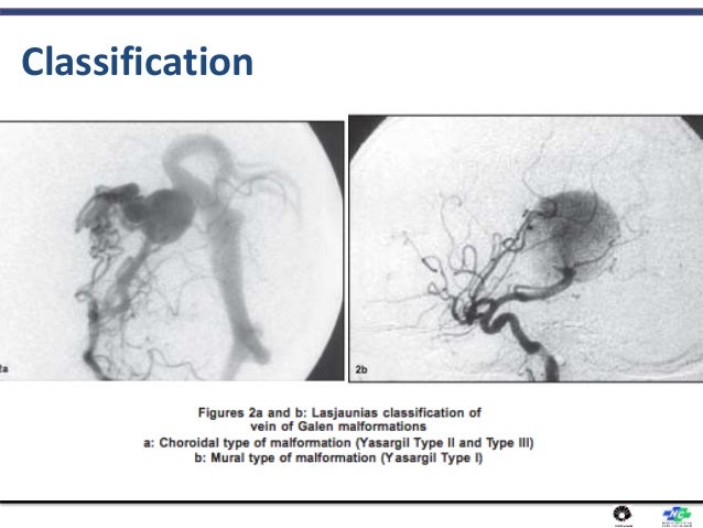
Vein of galen malformation.
Vein of galen anatomy. Vein of galen aneurysmal dilatation. It lies in the quadrigeminal cistern. In this type of lesion the vein of galen is fully developed and drains not only the lesion but also the adjacent brain parenchyma.
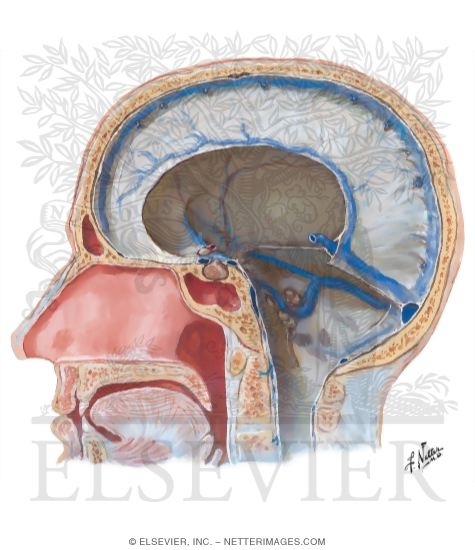
Most of these malformations present in early childhood often causing congestive heart failure in the neonate. The presence and locations of angiomas are very variable and do not follow any predictable pattern. The vein of galen also known as the great cerebral vein or great vein of galen is a short trunk formed by the union of the two internal cerebral veins and basal veins of rosenthal.
It curves backward and upward around the posterior border of the splenium of the corpus callosum. The incidence of the vein of galen malformation is unknown. This dilatation of the vein of galen is variable and depends on the outflow obstruction due to stenoses or thrombosis where the vein joins the straight sinus.
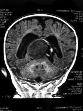
A malformed great cerebral vein will be noticeably enlarged. The vein of galen can be visualized using ultrasound or doppler. Vein of galen aneurysmal malformations vgam are rare congenital vascular malformations characterized by shunting of arterial flow into an enlarged cerebral vein dorsal to the tectum.
Fetuses with prenatally diagnosed vgam have. Vein of galen malformation background. The vein of galen is located under the cerebral hemispheres and drains.
Ultrasound is a particularly useful tool for vein of galen malformations because so many cases occur in infancy and ultrasound can make diagnoses prenatally.
 Great Cerebral Vein An Overview Sciencedirect Topics
 Vein Of Galen Malformation
 Figure 1 From Endovascular Management Of Vein Of Galen
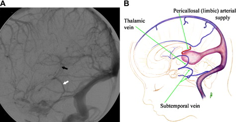
 Anatomy Of The Deep Venous System In Vein Of Galen
 Vein Of Galen Malformation
 Vein Of Galen Malformation
 Vein Of Galen Aneurysmal Malformations Critical Analysis Of
 Hemodynamic Analysis Of An Adult Vein Of Galen Aneurysm
 Great Cerebral Vein An Overview Sciencedirect Topics
 Vein Of Galen Malformation
 Great Cerebral Vein An Overview Sciencedirect Topics
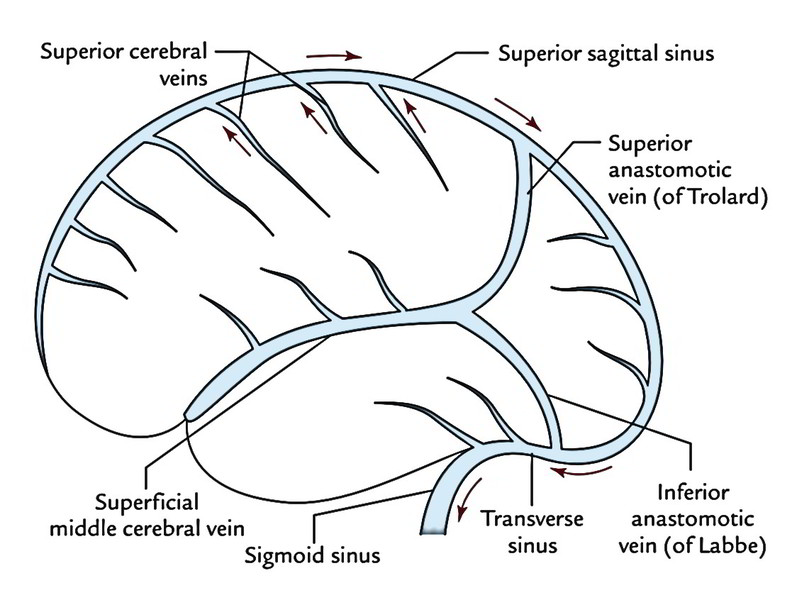 Easy Notes On Venous Drainage Of The Brain Learn In Just
 Great Vein Of Galen Discovered By Greek Physician Galen
 Vein Of Galen Malformation Mri Xray Medical
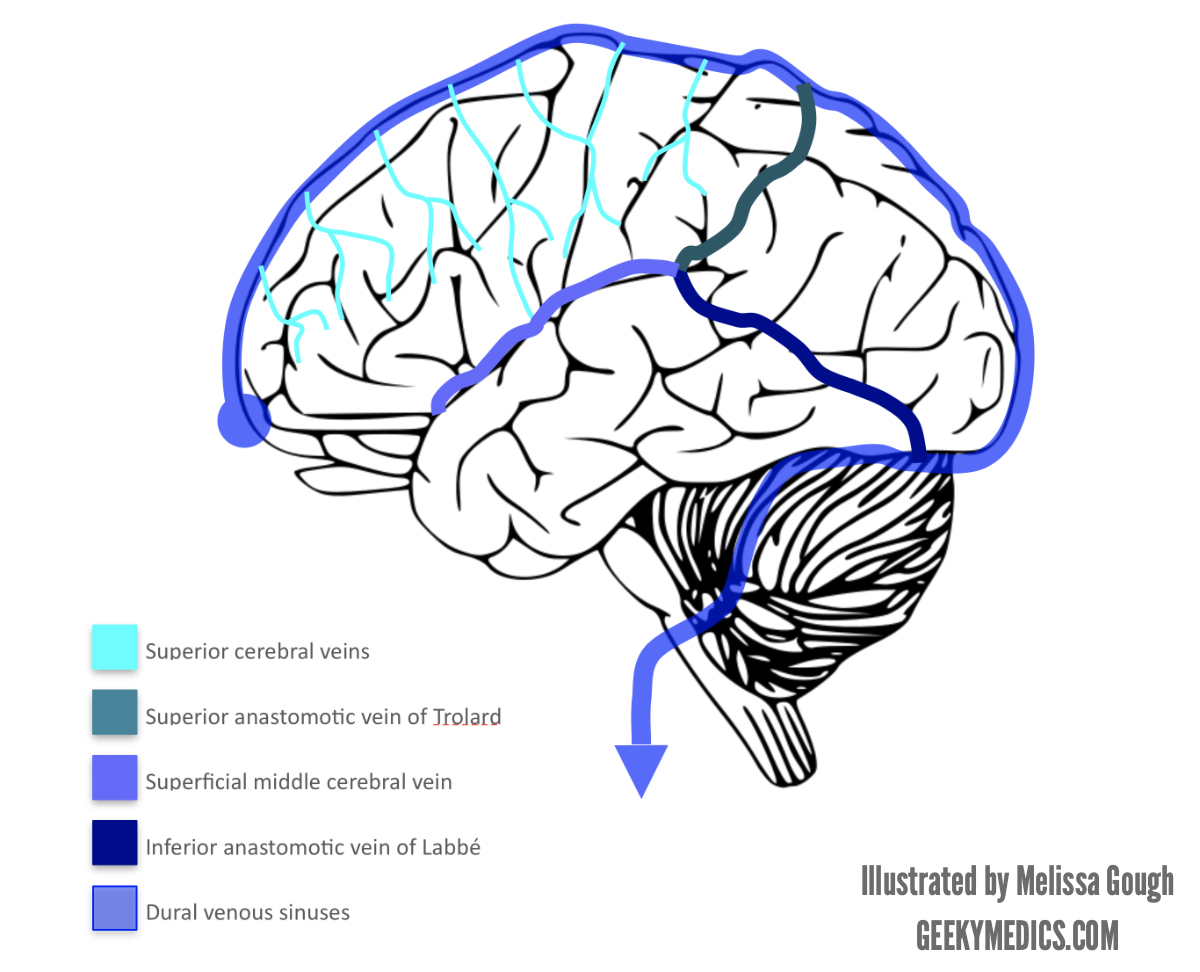 Venous Drainage Of The Brain Anatomy Geeky Medics
 Vein Of Galen Aneurysmal Malformations Radiology Key
 Endovascular Treatment Of Vein Of Galen Malformations A
 Vein Of Galen Malformation
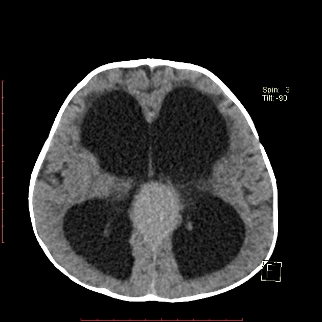 Vein Of Galen Malformation Radiology Case Radiopaedia Org
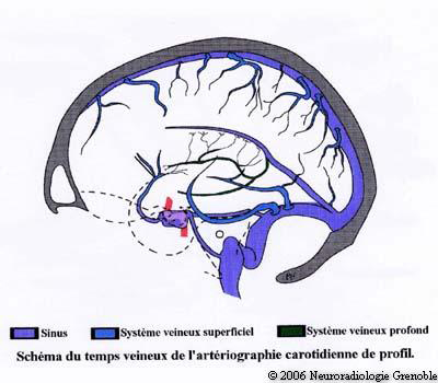 Cerebral Veins
 Anatomy Of The Deep Venous System In Vein Of Galen
 Pediagenosis
 Dural Venous Sinuses
 Vein Of Galen Malformation
 Vein Of Galen Malformation Vogm Diagnosis Treatment
 Figure 5 From Vein Of Galen Aneurysmal Malformations
 Great Cerebral Vein An Overview Sciencedirect Topics
 Vein Of Galen Malformation Workup Imaging Studies Other


Belum ada Komentar untuk "Vein Of Galen Anatomy"
Posting Komentar