Zygoma Anatomy
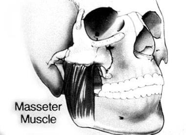
The masseter muscle important in chewing arises from the lower edge of the arch. Four processes the frontosphenoidal orbital maxillary and temporal.
 Management Of Zygomatic Fractures Pocket Dentistry
The zygomatic bone is small and quadrangular and is situated at the upper and lateral part of the face.
Zygoma anatomy. It forms the prominence of the cheek part of the lateral wall and floor of the orbit and parts of the temporal and infratemporal fossæ fig. The most common condition associated with the zygomatic bone is. The zygomatic bone functions as a structure which joins the bones.
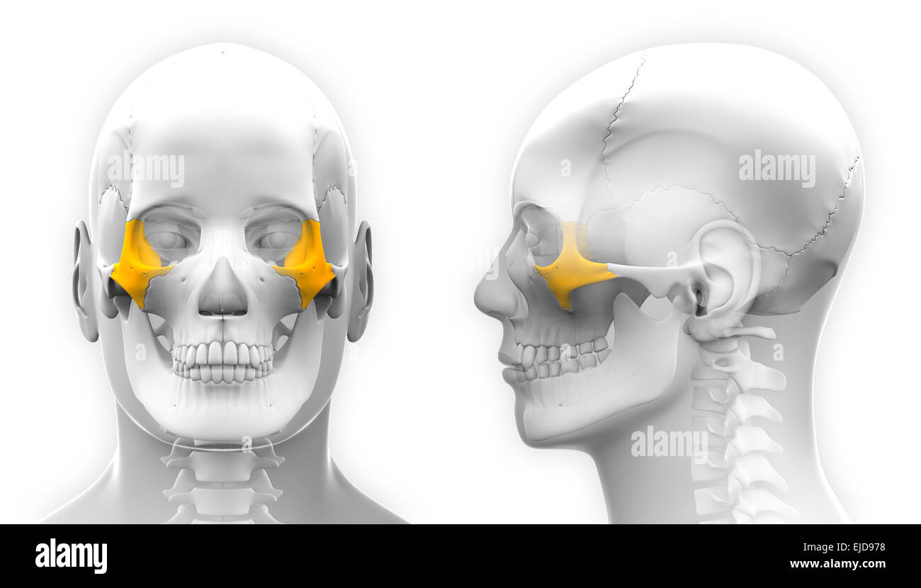
Several bones and joints surround the zygoma including the. The zygoma also known as zygomatic bone or malar bone is an important facial bone which forms the prominence of the cheek. Facial bone located below each eye socket anatomy.
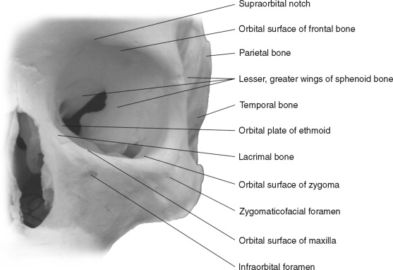
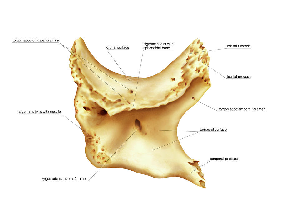
It presents a malar and a temporal surface. Zygomatic process of the maxillary bone articulated by the. Gross anatomy zygoma has three surfaces five borders and two processes.
Introduction to temporal bone anatomy. Zygomatic bone also called cheekbone or malar bone diamond shaped bone below and lateral to the orbit or eye socket at the widest part of the cheek. Each zygomatic bone articulates with the temporal bone frontal bone maxilla and sphenoid bones.
They are also commonly referred to a as the cheekbones or malar bones l mala the cheek. The zygomatic bone is somewhat rectangular with portions that extend out near. Zygomatic arch bridge of bone extending from the temporal bone at the side of the head around to the maxilla upper jawbone in front and including the zygomatic cheek bone as a major portion.
Zygomatic process of the temporal bone linked by the temporozygomatic suture. Frontal bone via the frontozygomatic suture which creates the rounded form of the bony orbit. It is roughly quadrangular in shape.
In the human skull the zygomatic bone cheekbone or malar bone is a paired irregular bone which articulates with the maxilla the temporal bone the sphenoid bone and the frontal bone. It is situated at the upper and lateral part of the face and forms the prominence of the cheek part of the lateral wall and floor of the orbit and parts of the temporal fossa and the infratemporal fossa. The zygomatic bones gr zygoma yoke are two facial bones that form the cheeks and the lateral walls of the orbits.
Another major chewing muscle the temporalis passes through the arch. It adjoins the frontal bone at the outer edge of the orbit and the sphenoid and maxilla within the orbit.


 Zygomatic Complex Facial Fractures Background Epidemiology
 Management Of Zygomatic Complex Fractures
 Extent Of The Prezygomatic Space The Space Overlies The
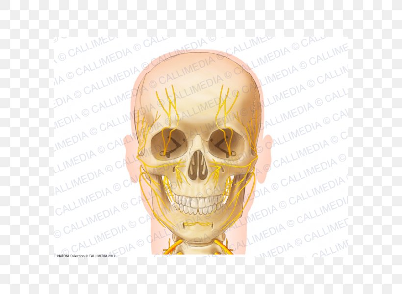 Skull Anatomy Nerve Zygomatic Bone Neck Png 600x600px
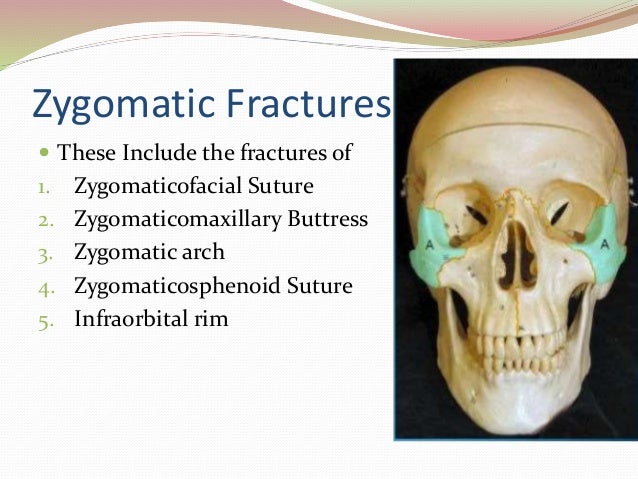
 32 Fractures Of The Zygoma Short Notes In Plastic Surgery
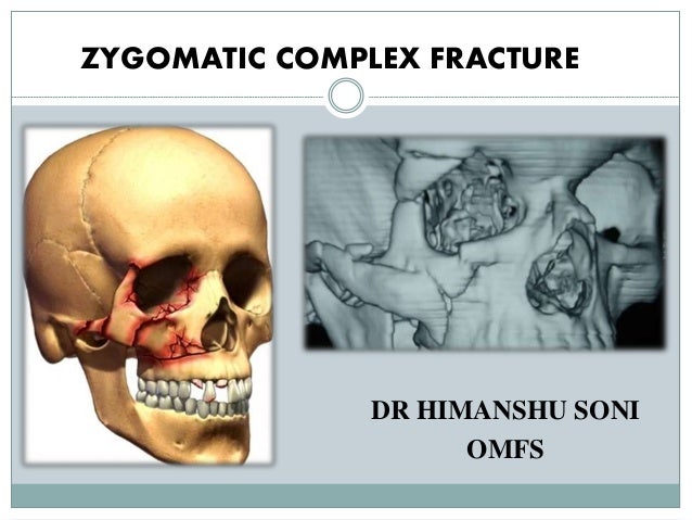 Zygomatic Complex Fracture Zmc
 Bones Of The Skull Zygomatic Bone Zygoma
![]() Zygomatic Bone Anatomy And Pathology Kenhub
Zygomatic Bone Anatomy And Pathology Kenhub
 Midface Reduction Fixation Orif 3 Point Fixation
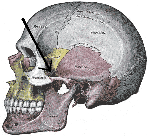 Zygomatic Arch Wikipedia
Zygomatic Bone Stock Photos Zygomatic Bone Stock Images
 Rt 233 Skull Radiography Introducing Zygomatic Arches Ppt
 Zygomatic Bone
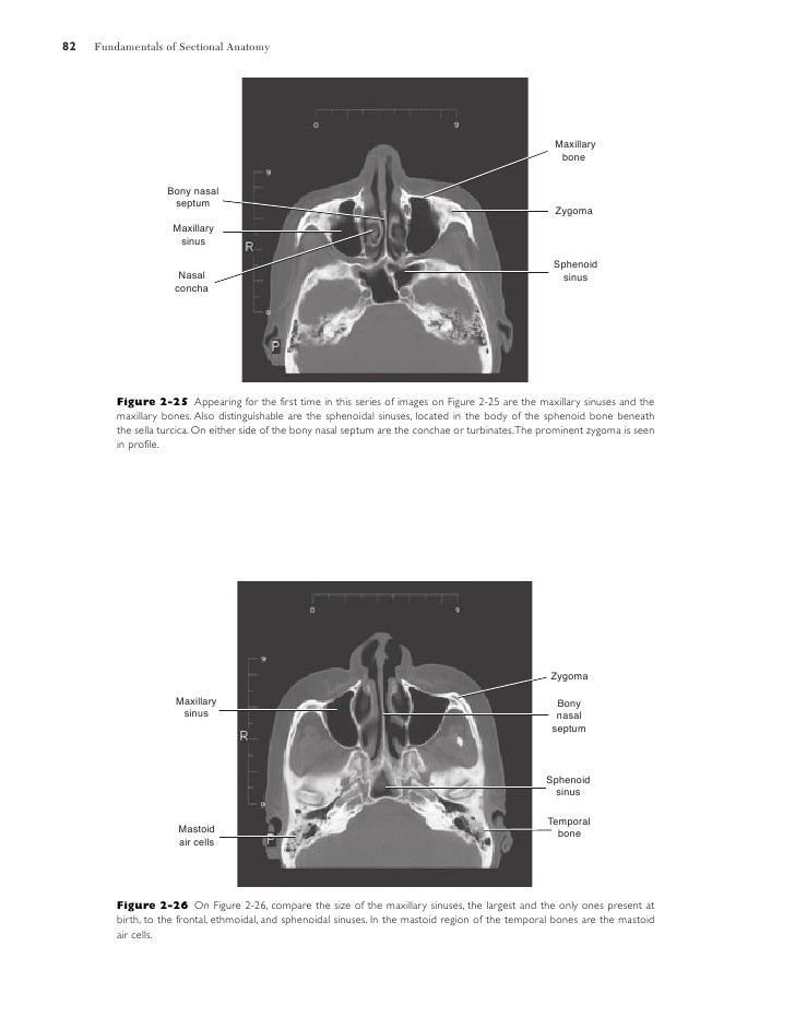 Sectional Anatomy
 Figure 3 From Zygomatic Implants Placed Using The Zygomatic
![]() Cunningham S Text Book Of Anatomy Anatomy Beanches Of The
Cunningham S Text Book Of Anatomy Anatomy Beanches Of The
 Zygomatic Process An Overview Sciencedirect Topics
 Zygomatic Bone
 Zygomatic Process Wikipedia
 Facial Nerve Paralysis Authors Added Material Ao

 Facial Bone Anatomy Overview Mandible Maxilla
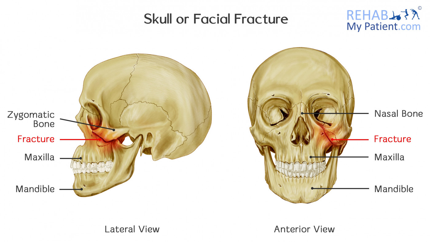 Skull Or Facial Fracture Rehab My Patient
Belum ada Komentar untuk "Zygoma Anatomy"
Posting Komentar