Hamstrings Muscles Anatomy
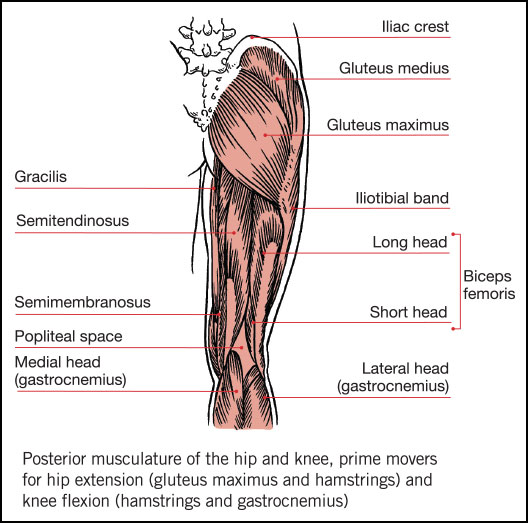
Muscles should be inserted over the knee joint in the tibia or in the fibula. As group these muscles act to extend at the hip and flex at the knee.
 Anatomy 101 Understanding Your Hamstrings Hamstring
The common criteria of any hamstring muscles are.

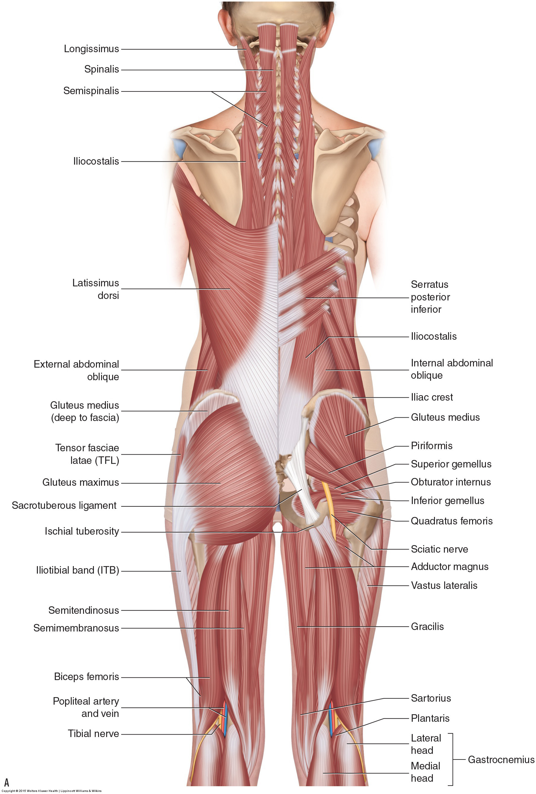
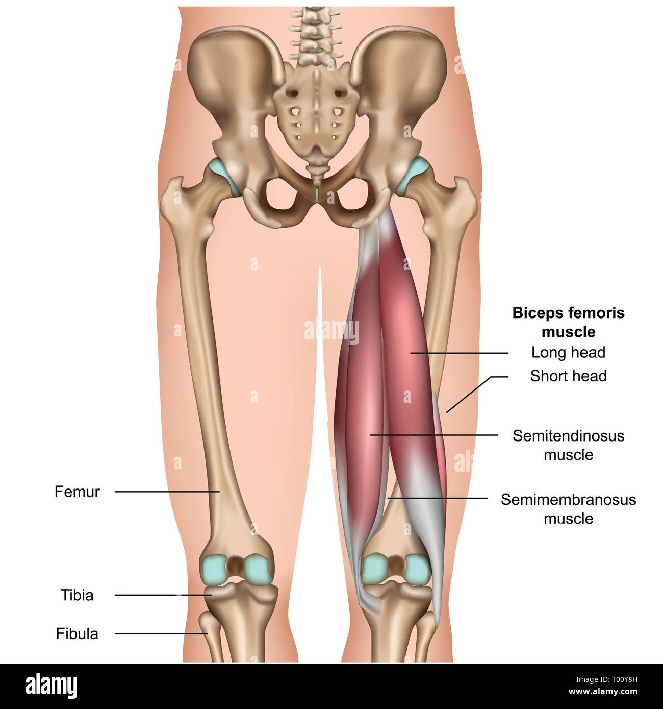
Hamstrings muscles anatomy. At the back of the upper leg behind the thigh quadriceps there are three muscles known as the hamstrings. Hamstring strains tend to be either the result of sudden stopping and starting during a sport sprinting for example or extreme stretching as might occur in gymnastics dance or yoga. Anatomy of the hamstring muscles.
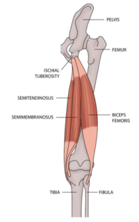
They consist of the biceps femoris semitendinosus and semimembranosus which form prominent tendons medially and laterally at the back of the knee. The hamstrings are comprised of three separate muscles. The hamstrings are a group of muscles and their tendons at the rear of the upper leg.
These muscles start at the bottom of your pelvis extending down the back of your thigh and along either side of your knee to your lower leg bones. They include the biceps femoris semitendinosus and semimembranosus. The hamstrings in the back of the thigh consist of three separate muscles.
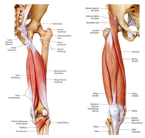
Muscles should originate from ischial tuberosity. These muscles originate just underneath the gluteus maximus on the pelvic bone and attach on the tibia. They are the antagonistic muscles to the quads.
Muscle will participate in flexion of the knee. Definition of the hamstring muscles. The hamstrings flex the knee joint adduct the leg and extend the thigh to the backside of the body.
When any one of the three hamstring muscles is stretched beyond its limit hamstring strain can occur. The muscles in the posterior compartment of the thigh are collectively known as the hamstrings. Usually a bit of midmuscle discomfort on the back of the thigh wont cause problems.
All three attach by long tendons crossing the back of the knee to the lower leg. The anatomy of the hamstring muscles. The biceps femoris semitendinosus and semimembranosus.
Muscles will be innervated by the tibial branch of the sciatic nerve. The hamstrings refer to 3 long posterior leg muscles the biceps femoris semitendinosus and semimembranosus. There are two hamstrings on the medial inner side of the back of the thigh and one on the lateral outer side.
They are used in walking and running. The biceps femoris long and short head the semitendinosus.
 Hamstring Muscle Anatomy 3d Medical Vector Illustration On White
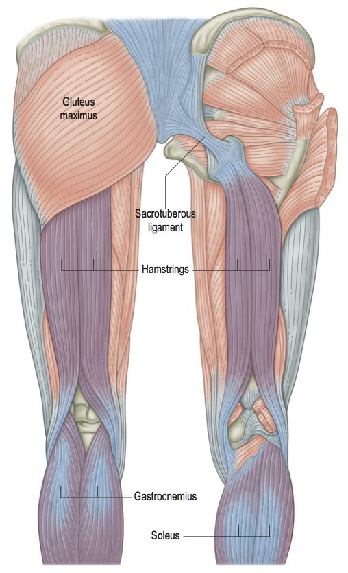
 Hamstrings 201 The Advanced Course Fleet Feet Columbus
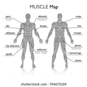
 Muscles Of The Hips And Thighs Human Anatomy And
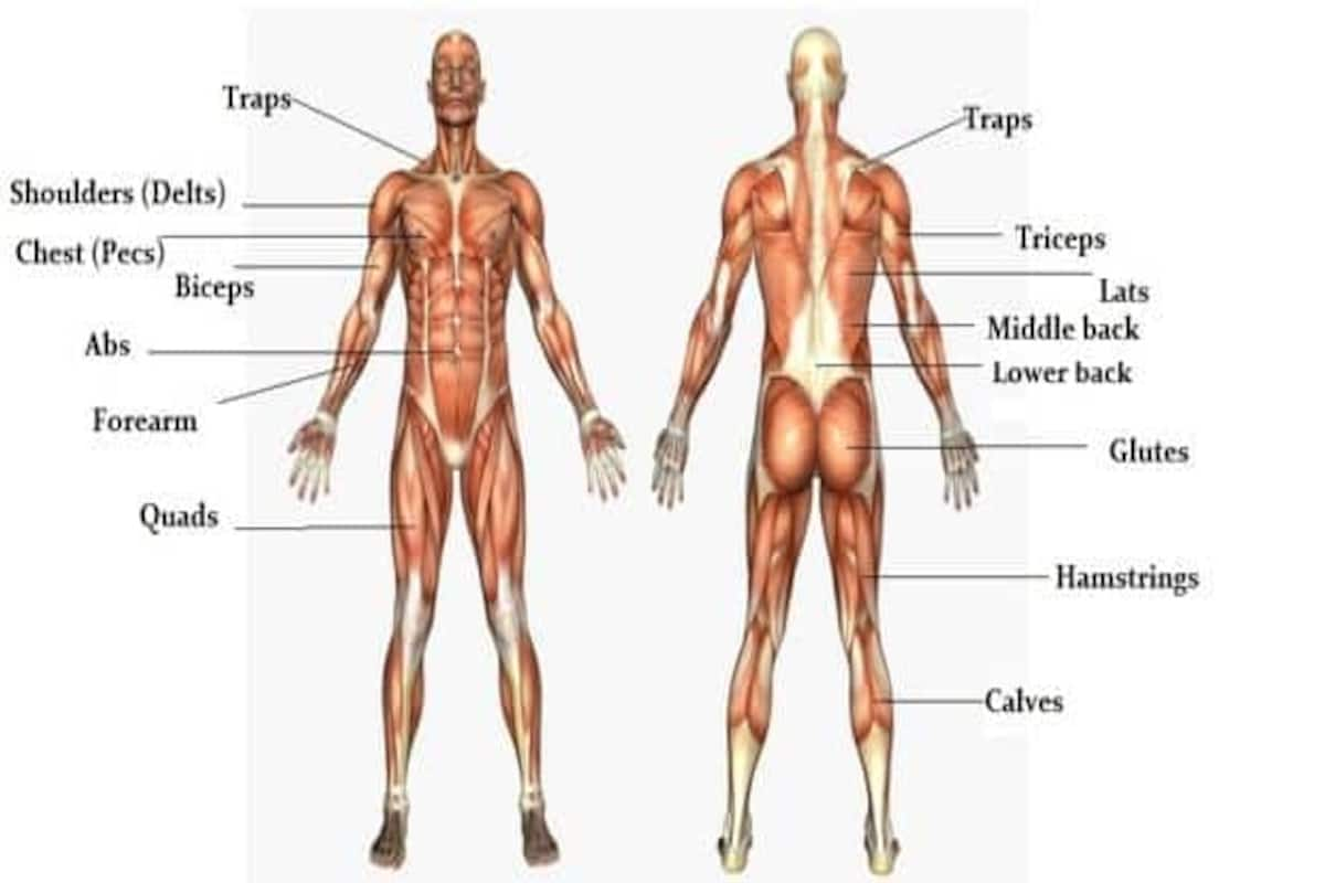 The Massive Muscle Anatomy And Body Building Guide You
 Bilder Stockfoton Och Vektorer Med Hamstring Muscle Anatomy
 Why Do So Many Of Us Have Tight Hamstrings Doctor Yogi
 Hamstrings Surgery Recovery There Is No Such Thing As The
 Hamstring Tendonitis Or Hamstring Syndrome Zion Physical
 Pure Fascial Release Vs Myofascial Release Anatomy Trains Blog
:max_bytes(150000):strip_icc()/Depositphotos_19871399_original-56a05f523df78cafdaa14cd1.jpg) Hamstring Muscles
Hamstring Muscles
 Hamstring Injuries Of The Knee Sports Medicine Specialist
 Hamstring Wikipedia
 What Is A Hamstring Strain And What Are The Causes Of A
 The Hamstrings And The Calves Corewalking
 Why Stretching Your Hamstrings Won T Help Your Back Pain In
 Hamstring Muscle Anatomy Images Stock Photos Vectors

 Hamstring Tendonitis Or Hamstring Syndrome Zion Physical
 Hamstrings Anatomy And Fitness Training Ace Prosource
 Pdf Hamstrings Muscle Anatomy And Function And
 The Hamstrings Yoga Anatomy
 Intramuscular Hamstring Tendon Injury Prognosis Surgical
 Hamstrings Articles Of Interest Biceps Head Anatomy
 Hamstring Muscle Anatomy 3d Medical Vector Illustration On
The Other Curl Get The Most Out Of Your Hamstring Training
 Hamstring Injuries Victorian Wolves Supporters Club
Belum ada Komentar untuk "Hamstrings Muscles Anatomy"
Posting Komentar