Hand Mri Anatomy
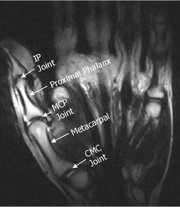
Stanford bone tumor bayesian network issssr msk lectures for residents ocad msk cases from around the world stanford msk mri atlas has served almost 800000 pages to users in over 100 countries. Anatomy the pip joint is a hinged joint with a bicondylar anatomy that allows a wide range of flexion and extension movements 14.
Anatomy of the hand and the wrist on mri.

Hand mri anatomy. Use the mouse to scroll or the arrows. Mor s n d thom asj ef r onu iv ty h p l mri of the wrist occult fracture ganglion cyst tumor ligament tear avascular necrosis arthritis tendon pathology nerve impingement infection occult fracture not visible on initial radiographs follow up xray ct mri. The main stabilizers of the joint are the surrounding soft tissues espe cially the collateral ligaments and the volar plate fig 1 15.
The basic mri anatomy of the tendon units of the finger and thumb are discussed. With high resolution 3 t mri the complex anatomy of the fingers can be imaged in exquisite detail to provide an accurate diagnosis of clinically important ligament and tendon injuries. This webpage presents the anatomical structures found on wrist mri.
The extensor mechanism flexor ten dons and retinacular ligaments play a. Atlas of wrist mri anatomy. Medical imaging anatomy atlas.
This section of the website will explain how to plan for an mri hand scan protocols for mri hand how to position for mri hand and indications for mri hand. Cross sectional anatomy of the hand on mr imaging wrist metacarpus fingers the hand mr. Normal tendons and ligaments of the hands typically demonstrate low signal intensity on mr imaging.
Traumatic injuries of the hand and fingers. The fine soft tissue contrast resolution of mri allows assessment of the tendons ligaments and intricate pulley systems of the fingers. Select a zone.
Mr imaging of the wrist and hand w illiam b. Click on a link to get t1 axial view t1 coronal view. Anatomy of the hand on mr imaging.
It is mri that has emerged as a powerful tool in the identification and characterization of lesions of the extensor and flexor tendons of the hand and finger as a result of its excellent soft tissue contrast and multiplanar imaging capabilities.
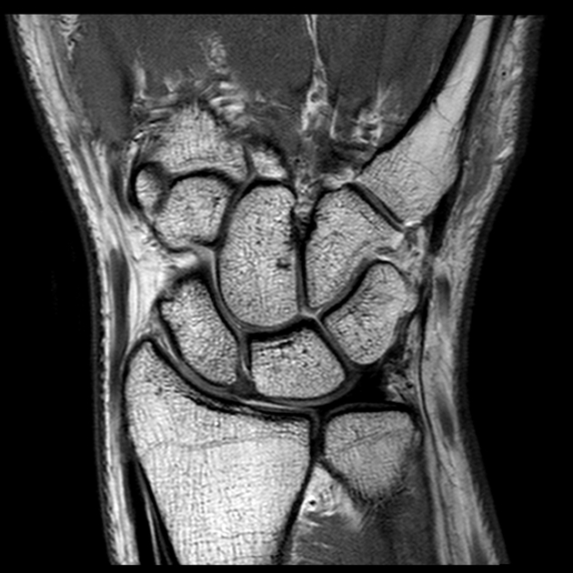
 Mri Wrist Coronal Anatomy Wrist Tendon And Ligaments
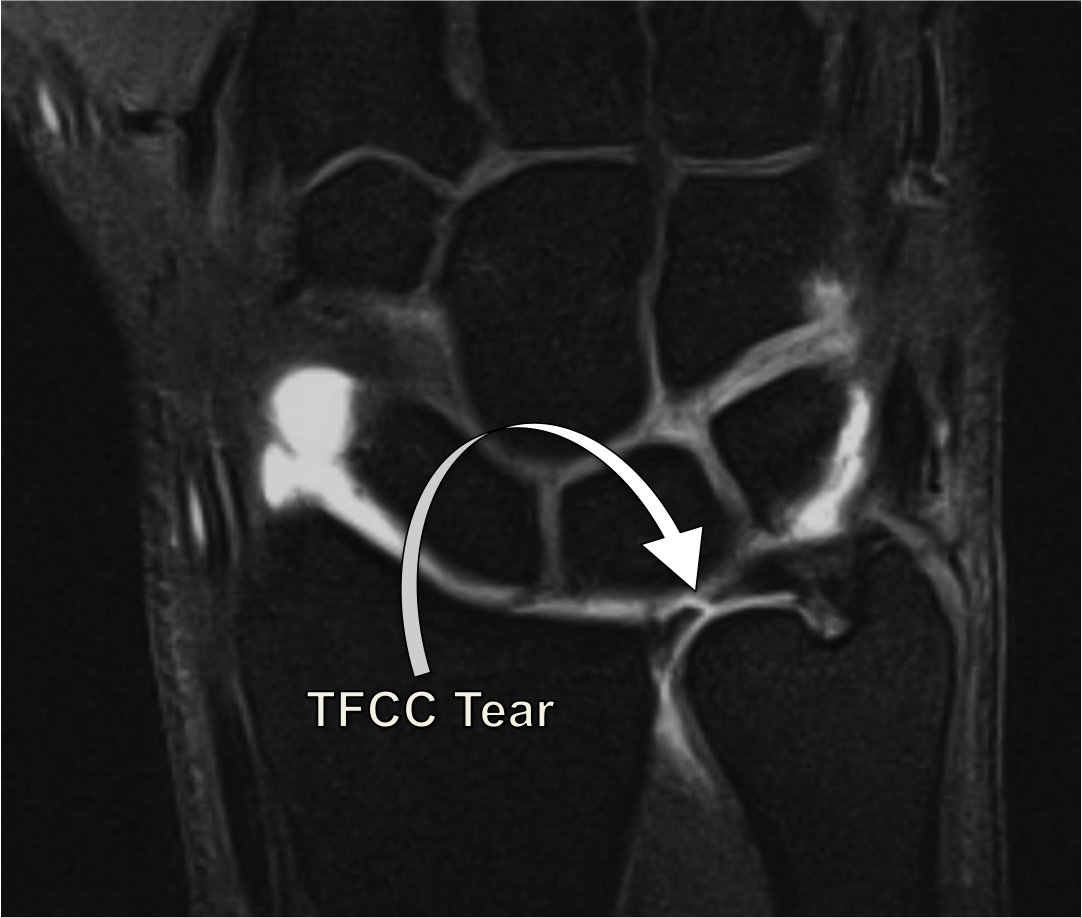
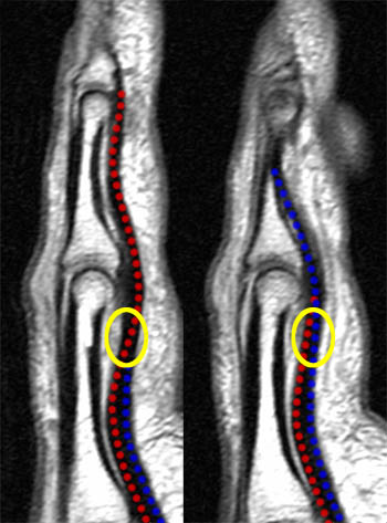
 Stener Lesion Skier S Thumb On Mri
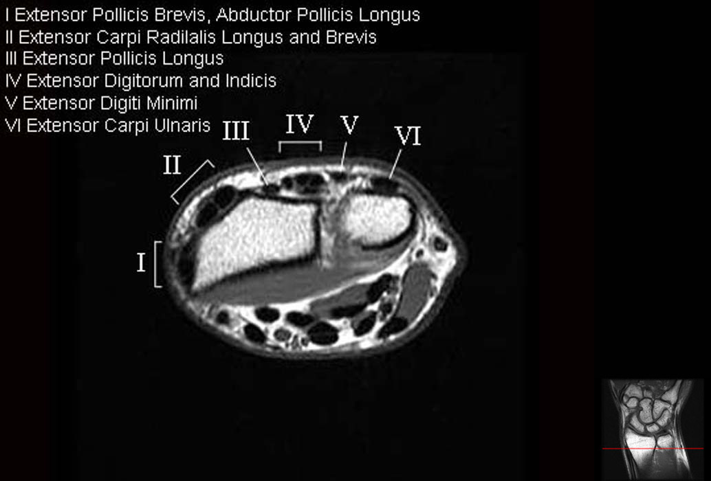
 Wrist Tendons Topographic Anatomy Radiology Case
 Musculoskeletal Mri

 Mri Wrist Coronal Anatomy Wrist Tendon And Ligaments
 Magnetic Resonance Imaging Of The Digital Nerves Of The Hand
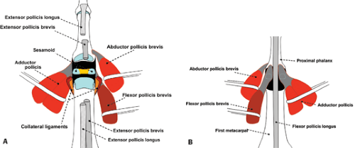
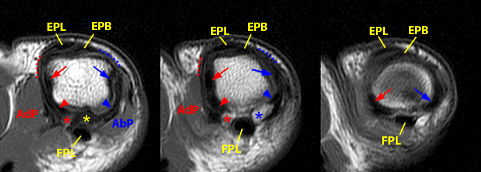
Mri Of The Thumb Anatomy And Spectrum Of Findings In
 Wrist Anatomy Mri Wrist Axial Anatomy Free Cross
 Wrist Mri Approach To Msk Mri Series
Mri Of The Thumb Anatomy And Spectrum Of Findings In
 Arun S Mri Protocols Thumb Mri Imaging Planes
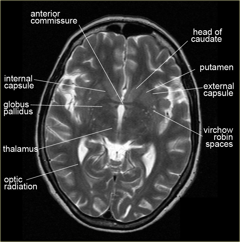 The Radiology Assistant Brain Anatomy
 I Love Physical Therapy Atlas Of Knee Mri Anatomy
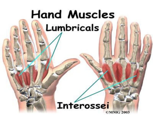 Muscular Anatomy Of Upper Limb Mri Anatomy

 Mri Of The Thumb Anatomy And Spectrum Of Findings In
 Mri Wrist Anatomy
 Magnetic Resonance Imaging Of The Digital Nerves Of The Hand
 Mri Of Finger Tendons Radiology Key
 Arm Forearm And Hand Mri Of Anatomy
 The Hand Mr Medical Imaging Anatomy Atlas
Mri Glove Captures Clear Images Of Hand Anatomy The Hindu
 Normal Wrist Mri Radiology Case Radiopaedia Org
 Arm Forearm And Hand Mri Of Anatomy
A Radiologist S Guide To Wrist Alignment
 Wrist Mri
 Mri Technique Startradiology
 Lister S Tubercle Wikipedia
 Knee Imaging Knee Sports Orthobullets
 Arm Forearm And Hand Mri Of Anatomy

Belum ada Komentar untuk "Hand Mri Anatomy"
Posting Komentar