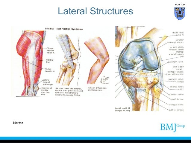
Lateral Knee Anatomy
In posterolateral corner injuries the lateral compartment has lost all or part of its stability and cannot maintain normal anatomic positioning when stressed. The medial and lateral menisci are fibrocartilage structures in the knee that serve two functions.
 X Knee Startradiology
Numerous bursae or fluid filled sacs help the knee move smoothly.
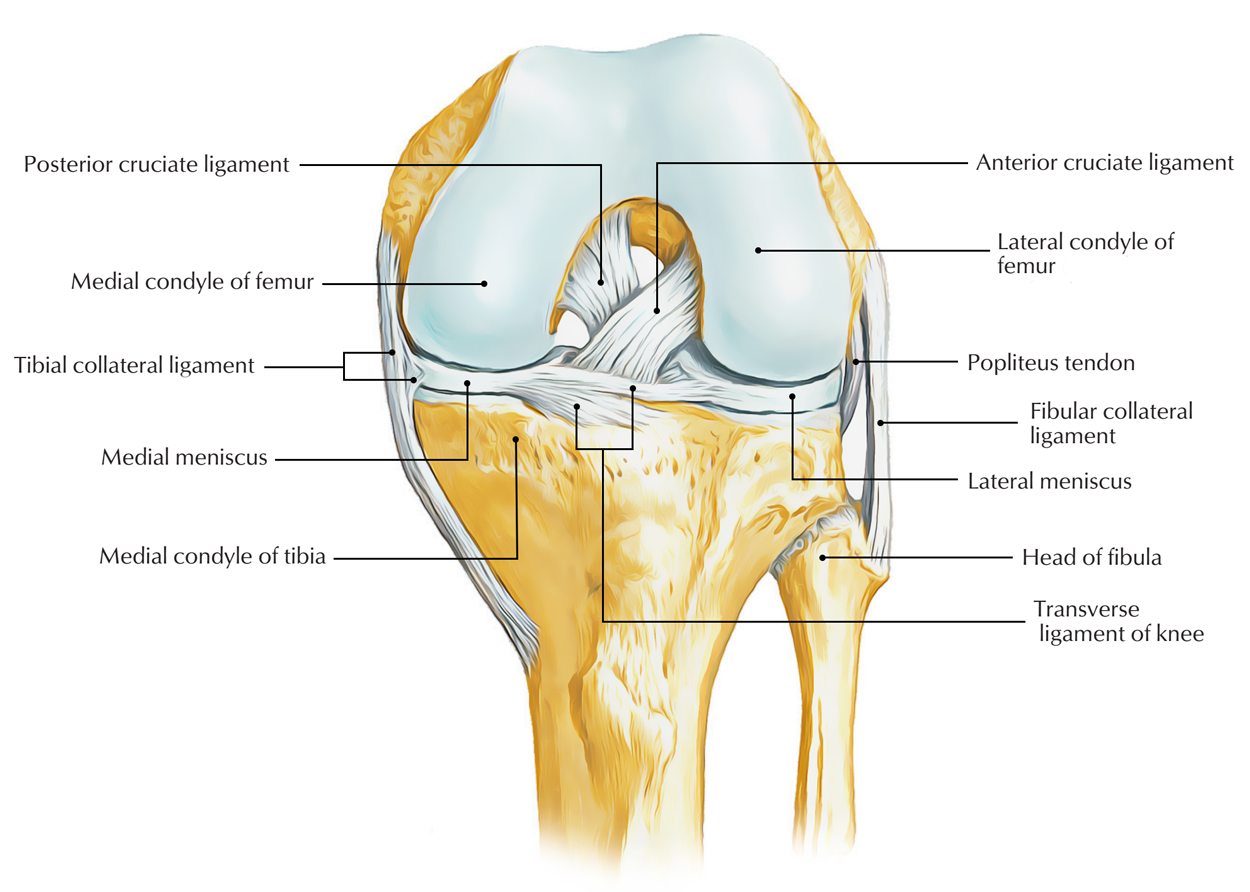
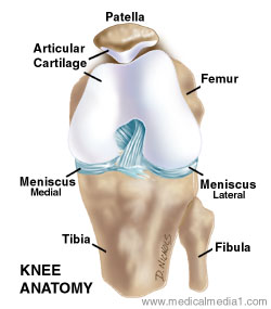
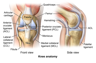
Lateral knee anatomy. To act as shock absorbers by increasing surface area to further dissipate forces. To deepen the articular surface of the tibia thus increasing stability of the joint. Two c shaped pieces of cartilage called the medial and lateral menisci act as shock absorbers between the femur and tibia.
As the foot makes contact with the ground the compartments of the knee should remain tight and stabilize the joint through the impact and movements of walking. It may come on gradually over time or may develop suddenly after an injury. It may or may not be connected to a specific activity.
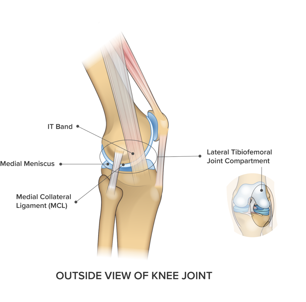
A varus thrust gait occurs as the foot strikes and the lateral compartment opens due to the forces applied on the joint. Outer knee pain may be a general ache or specific sharp pain and movement may be restricted. A lack of familiarity leads to hesitancy when performing approaches in these areas of the knee.
As a result the symptoms are varied too. 16 the peroneal nerve also courses through the posterolateral aspect of the knee. A lack of familiarity leads to hesitancy when performing approaches in these areas of the knee.
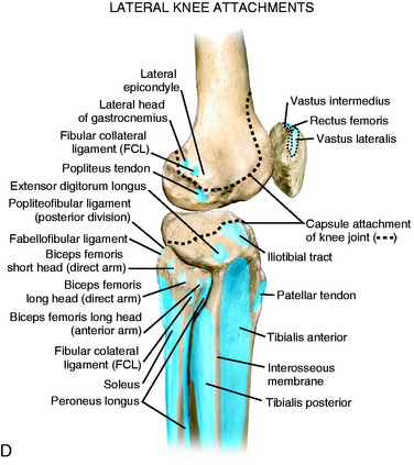
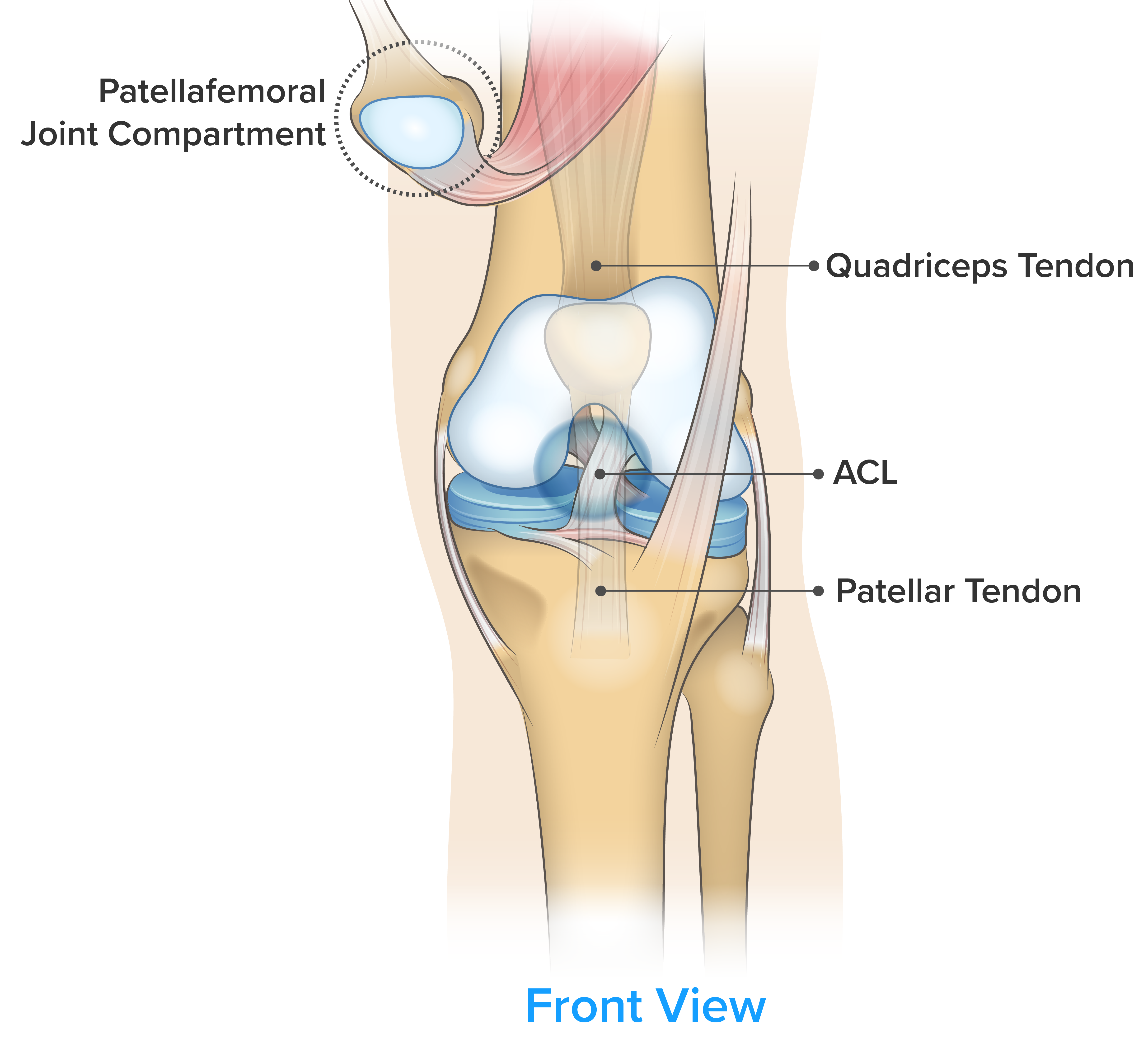
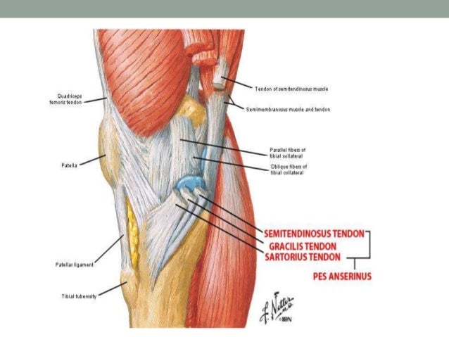
Anatomy of the lateral knee the lateral knee is comprised of 28 unique static and dynamic stabilizers. The posterior and lateral anatomy of the knee joint presents a challenge to even the most experienced knee surgeon. The 3 primary stabilizers that are com monly reconstructed surgically include the fcl pfl and plt fig.
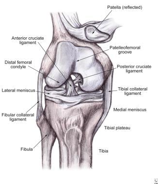
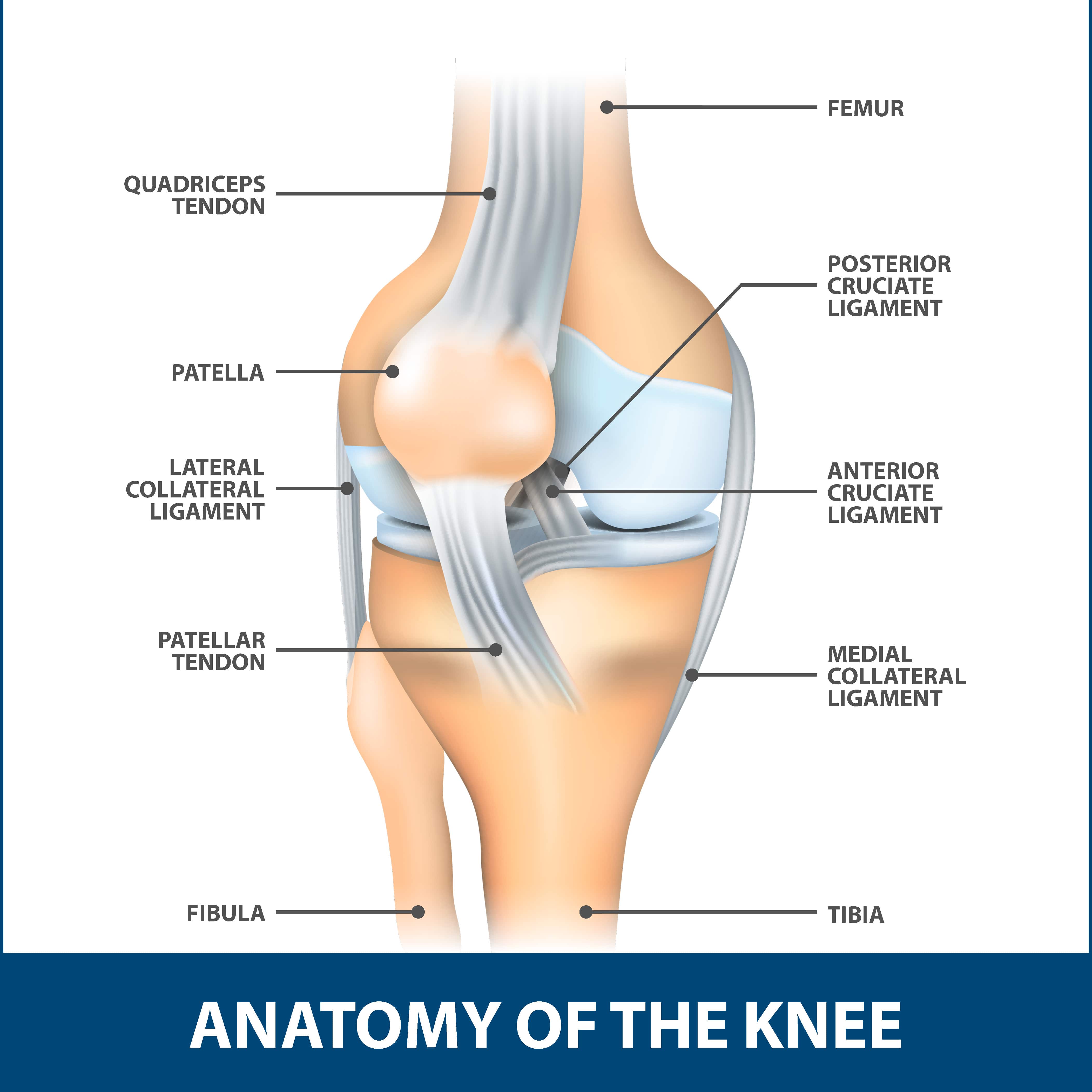
In knee joint anatomy knee ligaments are the main stabilising structures of the knee preventing excessive movements and instability. Ligaments are tough fibrous connective tissues which link bone to bone made of collagen. This arch type ligament is composed of a medial arm and a lateral arm.
Knowledge of the bony topography will result in a greater number of anatomic ligament reconstructions. Knowledge of the bony topography will result in a greater number of anatomic ligament reconstructions. At the posterior lateral corner of the knee located distally to the posterior horn fascicles is the arcuate ligament.
Lateral knee pain is pain that occurs on the outside of the knee. The posterior and lateral anatomy of the knee joint presents a challenge to even the most experienced knee surgeon.
 A Guide To Your Knees Well Guides The New York Times
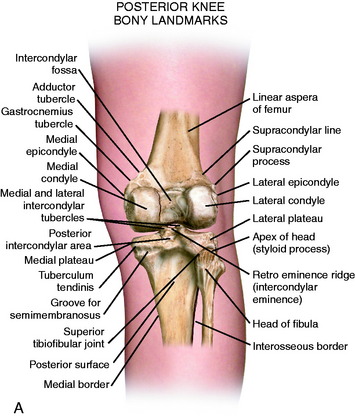
 Lateral Posterior And Cruciate Knee Anatomy Clinical Gate
 Anatomy Of Knee
 Redding Hospital Knee Anatomy
 Knee Injuries Musculoskeletal Key
 X Knee Startradiology
 Lateral Posterior And Cruciate Knee Anatomy Clinical Gate
 Complete Guide To Knee Pain Spring Loaded Technology
 Soft Tissue Knee Injury Practice Essentials Background
 Knee Wikipedia
 Vector Illustration Of A Healthy Human Knee Joint And
 Anatomy Of The Knee Joint Paley Orthopedic Spine Institute
 Medial Knee Injuries Wikipedia
 Anatomy And Examination Of The Knee

 Anatomy Of The Knee Joint 1 Lateral Collateral Ligament
 Lateral Medial And Posterior Knee Pain Brukner Khan S
 Lateral Knee Injury Don T Get Sidelined By Iliotibial Band
 4 Common Causes Of Knee Pain
/188058334-crop-56aae7425f9b58b7d0091480.jpg) What Is Causing Your Knee Pain
What Is Causing Your Knee Pain
 Lateral Collateral Ligament Florida Orthopaedic Institute
 Knee Pain On The Outside Of Your Joint Five Reasons Why
 The Knee Anatomy Injuries Treatment And Rehabilitation
Easy Notes On Ligaments Of The Knee Joint Learn In Just 3
Belum ada Komentar untuk "Lateral Knee Anatomy"
Posting Komentar