Acetabular Anatomy
Although simplicity is suggested in its most basic understanding as a containing device for the femoral head the acetabulum in reality is a complex and elegantly designed structure. The hip joint or acetabulum is responsible for many movements including walking.

Bursae in the hip.

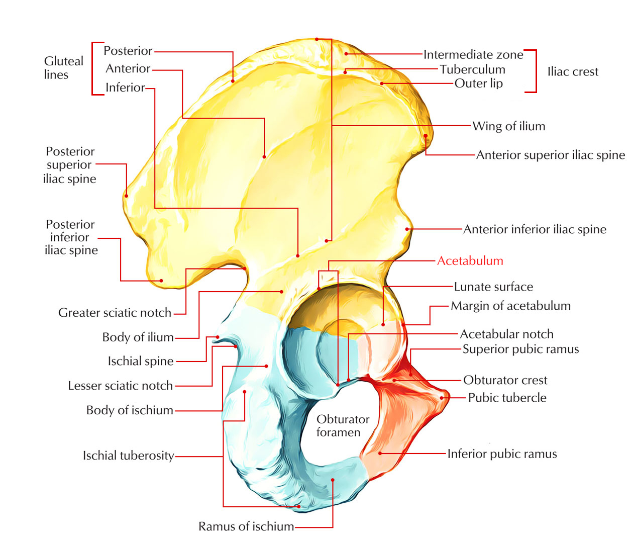
Acetabular anatomy. Pectoral girdle and pelvic girdle contributes a part of the acetabulum the deep cavity into which the head of the thighbone or femur is fitted. Gross anatomy the acetabular labrum is a c shaped fibrocartilaginous structure with an opening anteroinferiorly at the site of the acetabular notch. Bursae plural for bursa are flat fluid filled sacs.
Together with the labrum it is responsible for both the high stability of the hip joint and the mechanics and lubrication of the articular surface. Anatomy of human skeleton in human skeleton. Here it is bridged by the transverse ligament thus forming the acetabular foramen beneath it.
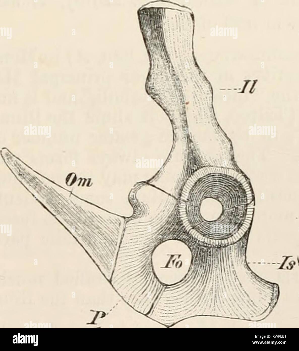
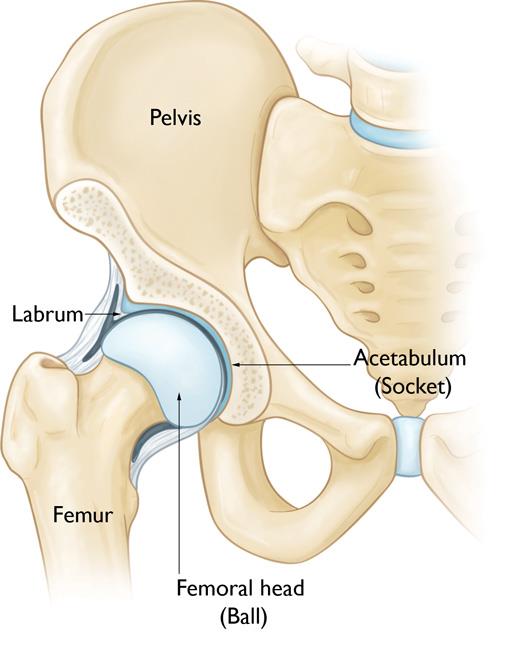
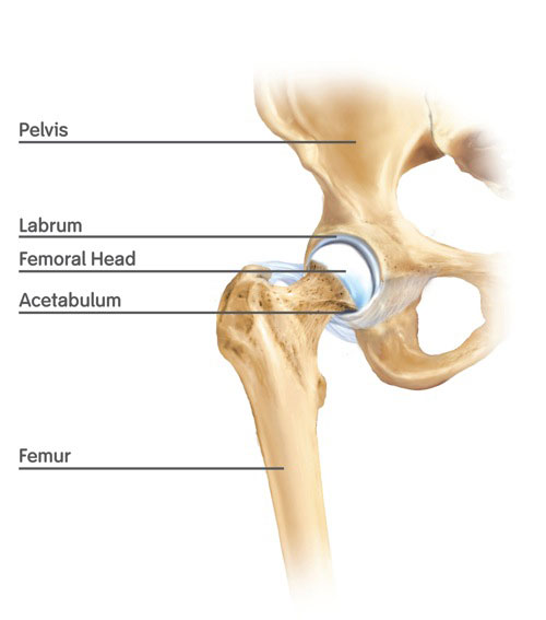
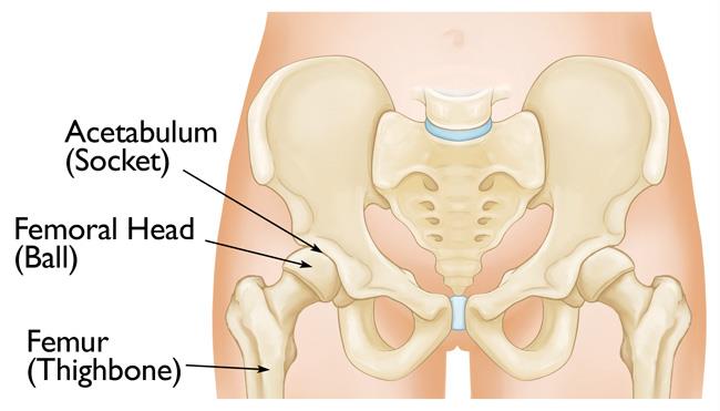
The upper leg is called the femur bone and at the very top of that bone there is a ball like structure called the femoral head the closed fist so in short the acetabulum is the cup shaped portion of the hip bone that receives the femoral head of the femur bone and together these two bony structures form the hip joint. Acetabula is the large cup shaped cavity on the anterolateral aspect of the pelvis that articulates with the femoral head to form the hip joint. All three bones of the pelvis the ilium ischium and pubis together form the acetabulum.
Ligaments of the hip are extremely tough and strong. The labrum forms a gasket around the socket creating a tight seal and helping to provide stability to the joint. The acetabulum is ringed by strong fibrocartilage called the labrum.
The acetabulum is the socket of the ball and socket hip joint. The head of the femur meets with the pelvis at the acetabulum forming the hip joint. The acetabulum æ s ə ˈ t æ b j ʊ l ə m cotyloid cavity is a concave surface of a pelvis.
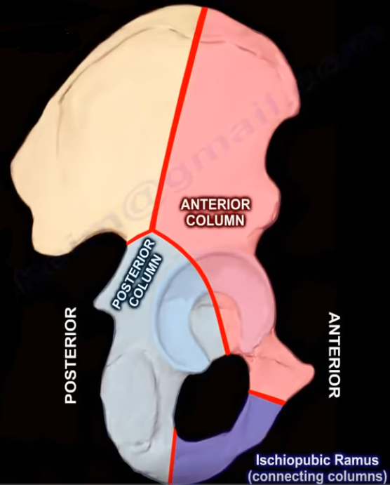
The three bones are initially separated by a y shaped triradiate cartilage that begins to fuse after puberty. Hip anatomy the acetabular joint the hip joint. Ligaments of the hip joint.
Elsewhere it is attached to the margins of the acetabulum. The largest window trial that bottomed out in the acetabulum was secured with its face parallel to the face of the acetabulum making the anteversion and abduction of the window trial equal to that of the native acetabulum. Anatomy of the acetabulum.
It creates a smooth low friction surface that helps the bones glide easily across each other during movement.
Hip Labral Tear Cleveland Clinic
 Is Yoga Tearing Labrums Yoga Anatomy
 Elements Of The Comparative Anatomy Elements Of The
Lower Limb Final Acb 3110 0001 Principles Of Human Anatomy
 Acetabulum Liner Hip Knee Book
 Acetabulum Fracture Imaging Practice Essentials
Hip Anatomy Yoga Understanding The Hips For Yoga Jason
 Acetabular Fracture Radiographic Evaluation Everything You Need To Know Dr Nabil Ebraheim
 Hip Joint Illustrates Acetabular Foramen Made Up Of
 Anatomy Hip Joint And Gluteal Muscles Flashcards Quizlet
 26 Acetabular Fractures Anatomy Evaluation And
 Femoroacetabular Impingement Orthoinfo Aaos
 Anatomy Of The Acetabulum Nabil Ebraheim Medium
Search Right Acetabular Anatomy
![]() Acetabulum Definition Anatomy Fracture Study Com
Acetabulum Definition Anatomy Fracture Study Com
Transient Osteoporosis Of The Hip Orthoinfo Aaos
 Normal Hip Anatomy Anatomy Of The Hip Nashville Tn
 Acetabulum Earth S Lab
 Acetabulum Anatomy Britannica
 A The Acetabular Anatomy Is Assessed By Three Measurements
 Acetabular Fractures Orthoinfo Aaos
 Transverse Acetabular Ligament Wikipedia
 Anatomy Of The Acetabulum Nabil Ebraheim Medium
 Acetabulum Radiology Reference Article Radiopaedia Org
 Hip Anatomy Recon Orthobullets
 Acetabulum Anatomy
 Acetabular Fractures Presentation And Treatment Bone And Spine
Belum ada Komentar untuk "Acetabular Anatomy"
Posting Komentar