Mandible Bone Anatomy
Intra and extracapsular condylar fractures are the most frequent mandibular fractures. Movement of the lower jaw opens and closes the mouth and also allows for the chewing of food.
 Mandible
Introduction to mandible bone anatomy.

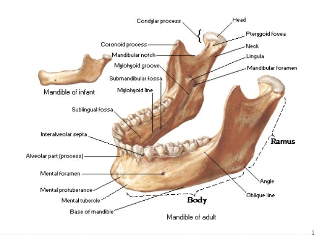
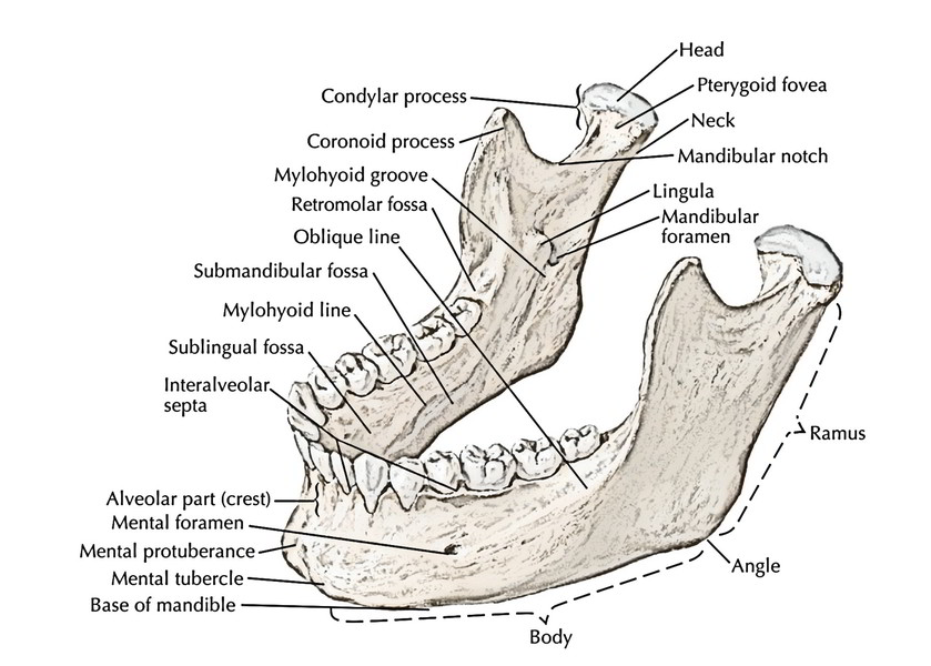
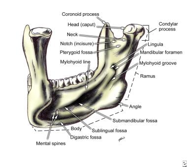
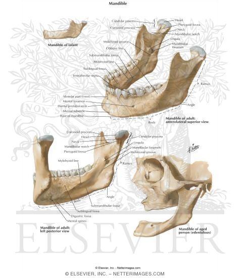
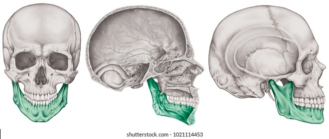
Mandible bone anatomy. Here the most common bony disturbances have been noted. It consists of right and left halves that fuse together early in life. It is formed by intramembranous ossification.
The two processes meet medially in. Other mandibular fracture areas include the body the angle the. The mandible or lower jaw is the bone that forms the lower part of the skull and along with the maxilla upper jaw forms the mouth structure.
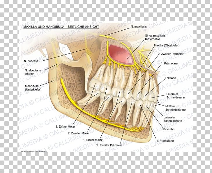
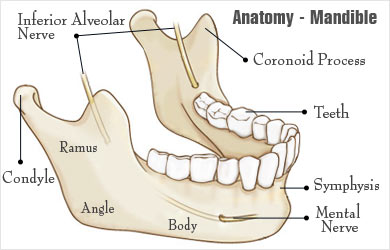
Four different muscles connect. The body of the mandible is located in the anterior part of the lower jawbone has a curved shape and can be divided in two parts. Alveolar bone resorption occurs when the teeth are lost.
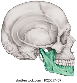
The body of the mandible has two surfaces external internal and two borders superior or alveolar and inferior. The mandible located inferiorly in the facial skeleton is the largest and strongest bone of the face. The mandible the largest and strongest bone of the face serves for the reception of the lower teeth.
The mandible l mandere to chew is the facial bone that forms the lower jaw and contains the lower teeth. It consists of a curved horizontal portion the body and two perpendicular portions the rami which unite with the ends of the body nearly at right angles. Forms part of lateral aspect of bridge of nose.
It also articulates on either side with the temporal bone forming the temporomandibular joint. Forms the anterior part of the hard palate. The maxillary bones important structures.
It is the only mobile bone of the facial skeleton and since it houses the lower teeth its motion is essential for mastication. The base of the mandible and the alveolar part of the mandible. The mandible is a u shaped bone.
The anterior portion of the mandible called the body is horseshoe shaped and runs horizontally. The mandible is composed of 2 hemimandibles joined at the midline by a vertical symphysis. It forms the lower jaw and acts as a receptacle for the lower teeth.
The lower set of teeth in the mouth is rooted in the lower jaw. There is a lack. Helps form the zygomatic arches.
 Anatomy Of The Jaw Mandible Doctor Stock
 Amazon Com Ronten Human Skull Model Life Size Replica
 Easy Notes On Mandible Learn In Just 4 Minutes Earth S Lab
 Mandible Lower Jaw Or Jawbone
 Teeth And Jaw Bone Anatomy Print
 Pin By Renee Mccarty On Diagnostic Imaging Dental Anatomy
The Skull Anatomy And Physiology Openstax
 Bones Of The Head And Neck Interactive Anatomy Guide
 Facial Bone Anatomy Overview Mandible Maxilla
 671 02094380
 Vector Art Mandible Fractures Is The Largest Strongest
 Mandible
 Mandible Bone Images Stock Photos Vectors Shutterstock
 The Mandibular Bone Of A 13 Kg Six Month Old Male Clawn
 Mandible Download Free 3d Model By University Of Dundee
 Skull Anatomy Pictures And Information
 Front Anatomical View Of Human Skull Bone With Mandible And The
 Mandibular Nerve Maxilla Mandible Alaleuanluu Png Clipart
Mandible Bone Anatomy
 Mandible Structure And Bony Landmarks Preview Human Anatomy Kenhub
 Skull Tutorial 4 Mandible Anatomy Tutorial
 1000 Mandible Stock Images Photos Vectors Shutterstock
Mandibular Fossa
 Normal Anatomy Of The Jaw This Lateral View Of The Skull

 The Mandible Lower Jaw Human Anatomy

 Maxilla Mandible Anatomy Bone Human Tooth Anatomi Png
 The Bones Of The Skull Human Anatomy And Physiology Lab
 Broken Jaw Mandibular Fracture Types Causes Symptoms

 Only Movable Bone In The Face Largest And Strongest Facial
Belum ada Komentar untuk "Mandible Bone Anatomy"
Posting Komentar