Mouse Heart Anatomy
Left lateral aspect of skull. To proceed click here.
![]() Free Photos Mouse Heart Anatomy Avopix Com
Free Photos Mouse Heart Anatomy Avopix Com
The latter is formed from the proximal part of the left cranial caval vein lccv webb et al.

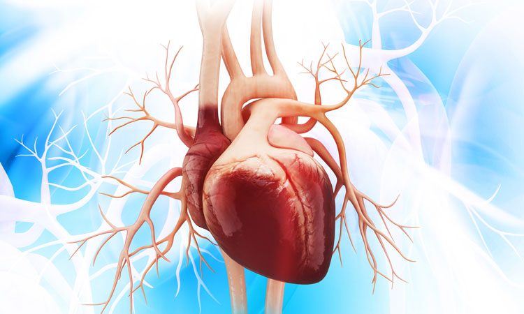
Mouse heart anatomy. Cardiomyocytes differentiate from precursor cells in the primitive streak and move anterior laterally to form bilateral paired cardiogenic plates myocardial primordial in the mouse embryo at e75. Information is provided about the anatomical features and landmarks for conducting a physical examination. The anatomy of the postnatal heart in mouse and human the basic anatomical features of the postnatal heart in the human and mouse are very similar fig.
Early mouse heart development the heart is the first organ to develop and function in the embryo. Mus musculus lac grey strain. Dorsal aspect of skull.
Two atria separated by an interatrial septum ias and two ventricles separated by an interventricular septum ivs. Thus in both species the heart has four chambers. A color atlas and text provides detailed comparative anatomical information for those who work with mice and rats in animal research.
Skeleton of lac grey mouse. They empty to the right atrium or to the coronary sinus. Heart in situin situ.
Order your anatomy atlas from the aalas store. 1996 1998. Quicktime mouse radiographic atlas of skeletal anatomy.
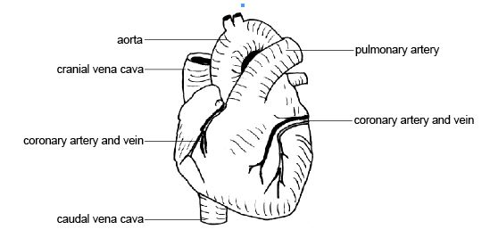
The warriors series meets redwall in mouseheart the first book in an epic animal adventure series by lisa fiedler set in the subway tunnels of brooklyn. Abbreviated title page foreword introduction externals 4. Coronary artery anatomy in the mouse is comparable to that of other mammals with early branching of a large septal coronary artery also seen in hamsters and rabbits from the left coronary system.
The following link will take you to a series of radiographic images with color overlays and labels. In mice the cardiac veins run on the surface of the heart within the subepicardium draining the myocardium of the left and the right ventricles as well as the left atrium. The anatomy of the laboratory mouse margaret j.
Comparative anatomy of the mouse and rat. The diameter of the mouse coronary arteries at their ostia averages 016 mm.
 Anatomy Of The Laboratory Mouse In Vivo Imaging Atlas On A
 Heart Stock Image N200 0076 Science Photo Library
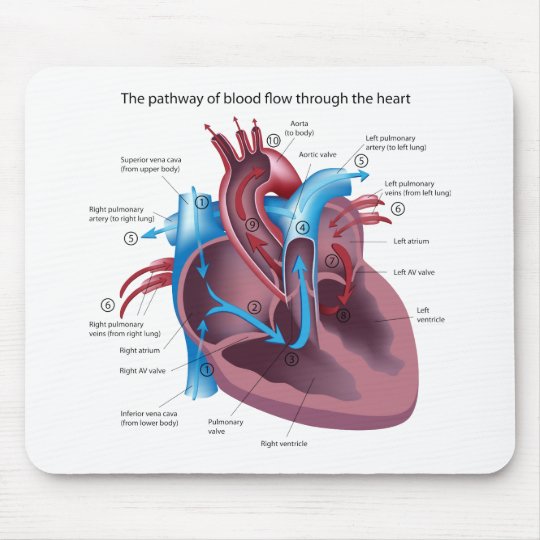 Heart Anatomy Mousepad
 Cardiac Regeneration Strategies Staying Young At Heart
 Kinase Identified As Potential Target For Heart Failure
 Anatomy And Physiology Of Animals Cardiovascular System The
 Anatomical Heart Mouse Pad Horror Mouse Pad Anatomy
 Black And White Tattoos Tattoo Heart Octopus Sketch Snake
 Amazon Com Boszina Mouse Pads Chamber Human Heart Anatomy
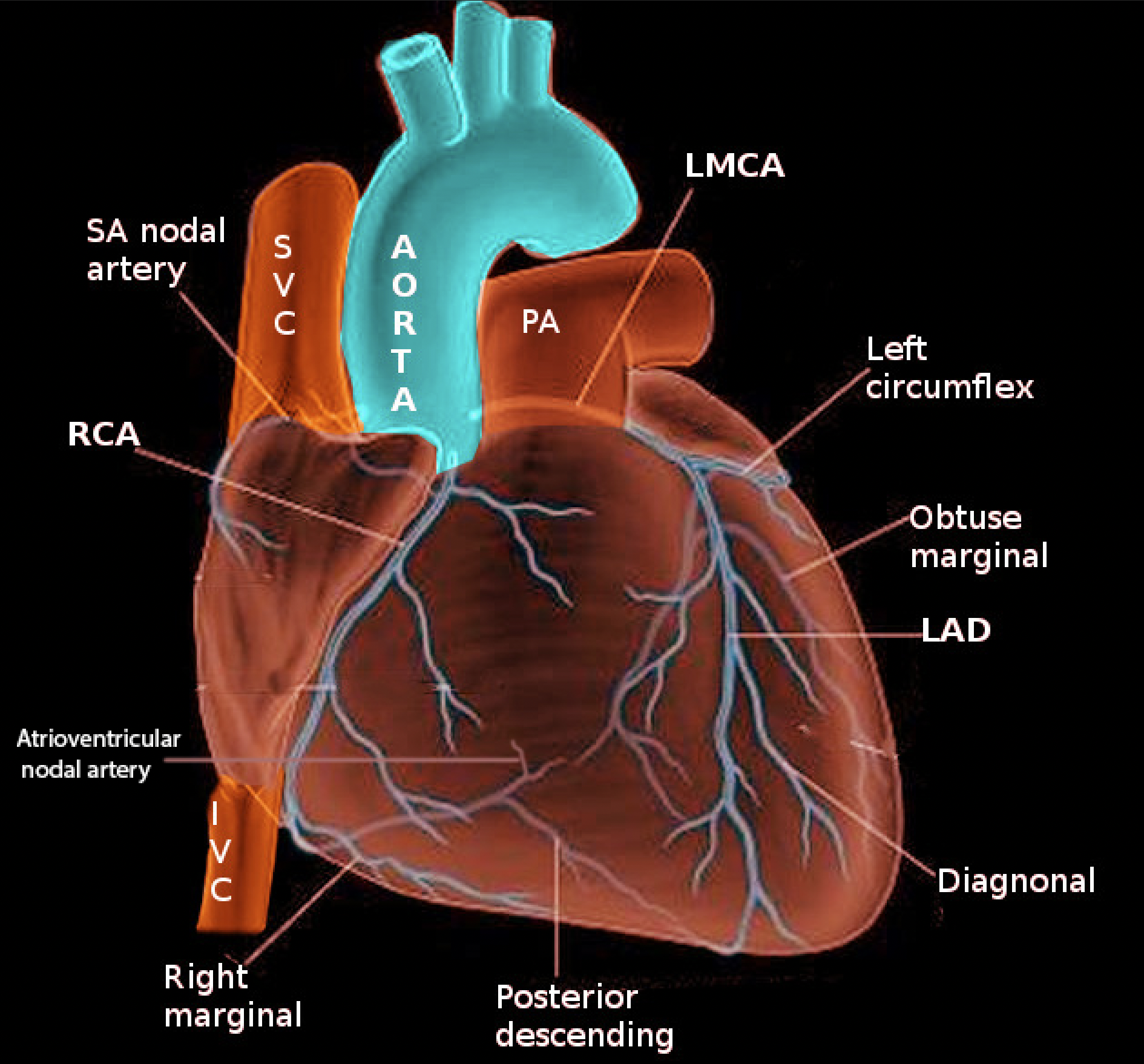
 Abstract 130 The Diversity Of Coronary Artery And
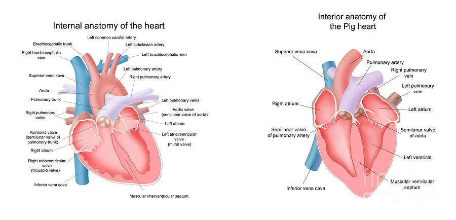 Pig And Human Heart Illustrations
 From Fruit Fly Wings To Heart Failure Why Not Ch
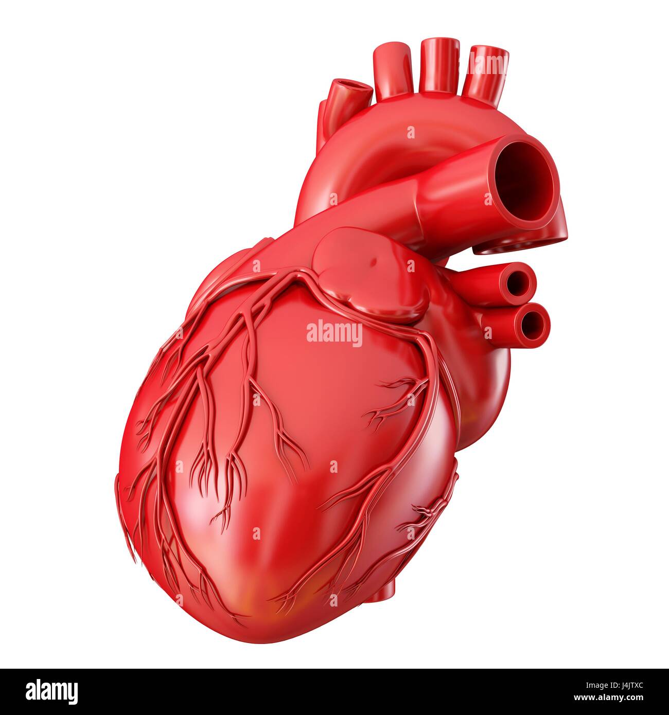 Illustration Of Human Heart Anatomy Stock Photo 140556260
![]() Mouse Heart Anatomy Free Images And Photos Avopix Com
Mouse Heart Anatomy Free Images And Photos Avopix Com
 Heart Anatomy Coffee Tea Mug White
 Experimental Myocardial Infarction Mouse Model A
 Techniques And Best Practices For Cardiomyocyte Isolation
 Heart Anatomy Science Otterbox Iphone Case Zazzle Com
 Human Heart Drawing K6138623 Fotosearch
 Mouse And Mice Models Scintica Instrumentation
![]() Anatomy And Physiology Of The Adult Mouse Heart A A
Anatomy And Physiology Of The Adult Mouse Heart A A
Belum ada Komentar untuk "Mouse Heart Anatomy"
Posting Komentar