Muscle Fiber Anatomy Definition
The primary metabolic pathway used by a muscle fiber determines whether the fiber is classified as oxidative or glycolytic. Nouna muscle cell especially one of the cylindrical multinucleate cells that make up skeletal muscles and are composed of numerous myofibrils that contract when stimulated.
Type i muscle fibres type i fibre are also known as slow twitch fibre.
Muscle fiber anatomy definition. Muscle fs fast twitch paler colored muscle fibers of larger diameter than slow twitch fibers and having less sarcoplasm and more prominent cross striping. More atp can be produced during each metabolic cycle making the fiber more resistant to fatigue. In the deep fascia around the muscles are several large arteries veins and nerves.
Fibre fiber any of several elongated threadlike cells especially a muscle fiber or a nerve fiber muscle musculus one of the contractile organs of the body. Any of the elongated cells characteristic of muscle. Muscle cell muscle fibre.
Skeletal muscle is a specialized contractile tissue found in animals which functions to move an organisms body. Muscle fiber any of the cells of skeletal or cardiac muscle tissue. Mammalian skeletal muscle fibers are characterized by d their morphological traits such as total number of fibers tnf and cross sectional area of fibers csaf and fiber e type characteristics such as contractile and metabolic f properties lee et al.
Muscle fiber an elongated contractile cell that forms the muscles of the body. Skeletal muscle fiber location and arrangement. If a fiber primarily produces atp through aerobic pathways then it is classified as oxidative.
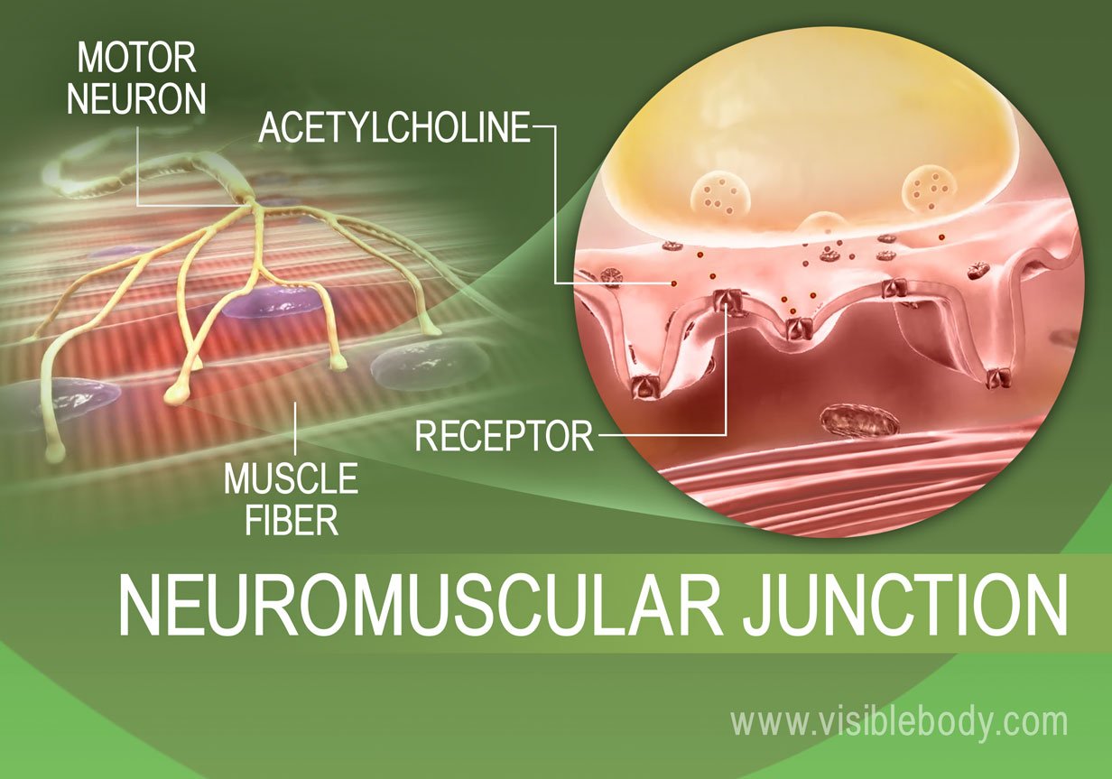
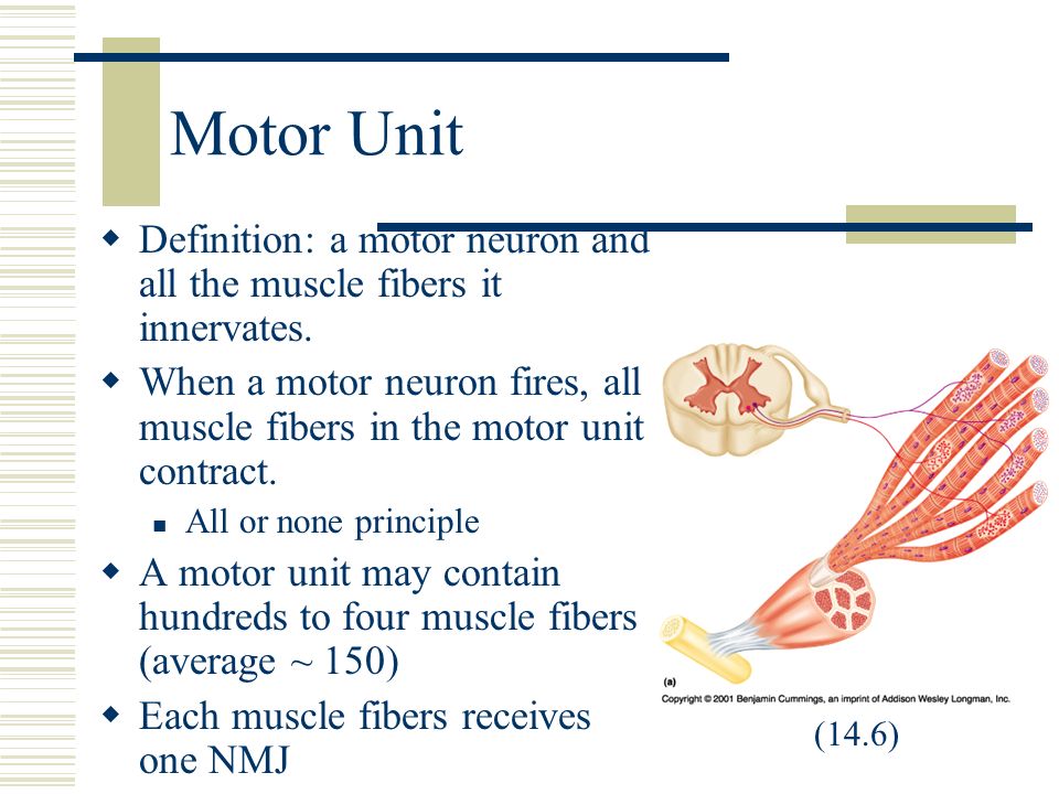
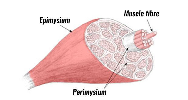
They are red in colour due to the presence of large volumes of myoglobin and so oxygen and high numbers of mitochondria. The strength of a muscle contration is determinded not only by the frequency of stimulation but also by the number and size of motor units recruited the number of motor units that are recruited is determined by the number of motor neurons that are stimulated by the central nervous system. Branches of these vessels and nerves penetrate each muscle close to its midpoint then extend into the epimysium and perimysium layers.
Used for forceful and rapid contractions over short periods of time. About half of the skeletal muscle fibers in adults are slow fibers. Medical definition of muscle fiber.
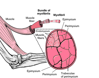
In the endomysium individual muscle fibers cells are supported by nearby capillaries and nerve cells. Skeletal muscle is comprised from a series of bundles of muscle fibers surrounded by protective membranes.
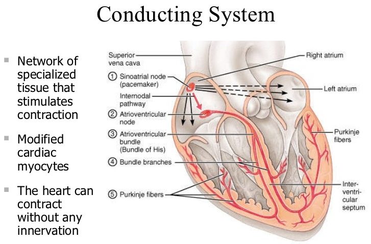 Purkinje Fibers Anatomy Location Function Anatomy Info
 Human Muscle Cell Types Interactive Anatomy Guide
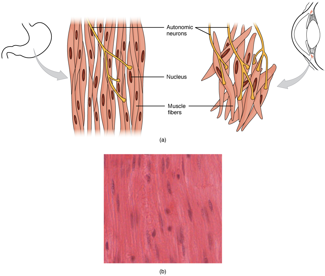 10 8 Smooth Muscle Anatomy And Physiology
 Muscle Physiology An Introduction
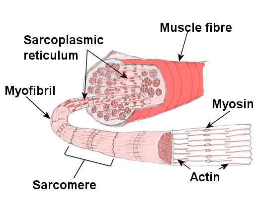 Muscle Anatomy Skeletal Muscle Structure Explained
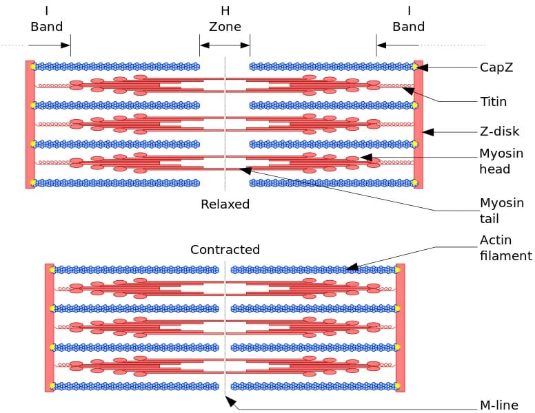 Myofibril Definition Function And Structure Biology
 Endomysium Anatomy Of Muscle Structure
 Muscle Contractions Learn Muscular Anatomy
 Chapter 10 Muscle Tissue Muscle Tissue Functions Movement
 Kondo Lab 4 Muscle Set Biology 200 With Kondo At Citrus
 Biology For Kids Muscular System

 Muscular System 3 Types Of Muscle Skeletal Muscle Aka
Microscopic Anatomy And Organization Of Skeletal Muscle
 Myofibril Definition Of Myofibril By Medical Dictionary
 A Closer Look At Skeletal Muscles Muscle Fibres Big Picture
 Skeletal Muscle Anatomy And Physiology I
Skeletal Muscle Fiber Structure And Function Open
 Muscle Anatomy Skeletal Muscle Structure Explained
 Muscular System Muscles Of The Human Body
 Muscle Structure Muscle Under The Microscope Science
 Muscle Structure Fiber And Cells


Belum ada Komentar untuk "Muscle Fiber Anatomy Definition"
Posting Komentar