Shin Anatomy Muscles
The posterior compartment holds the large muscles that we know as the calf muscles. The two muscles join together at the achilles tendon and insert on the back side of your heel bone called the calcaneus.
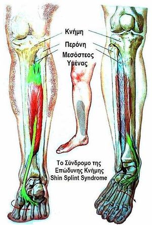
Shin splint pain most often occurs on the inside edge of your tibia shinbone.
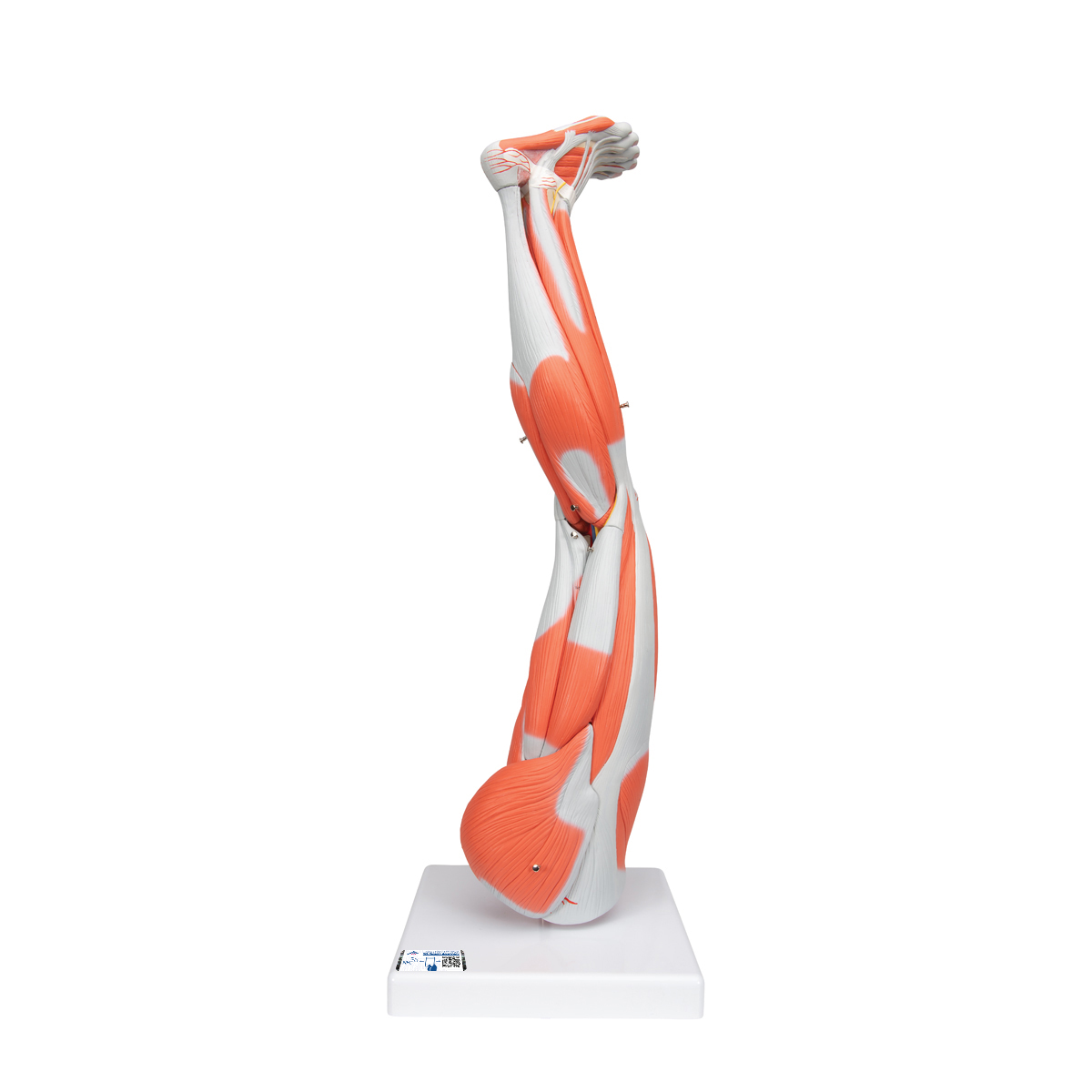
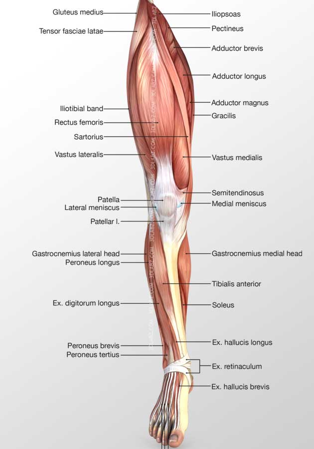
Shin anatomy muscles. The calf muscle on the back of the lower leg is actually made up of two muscles. The muscles of the lower leg the anterior compartment in the front of the shin holds the tibialis anterior. The lateral compartment is along the outside of the lower leg.
They are simple muscles to exercise either on your own or with a resistance band. The tibialis anterior is a muscle in humans that originates in the upper two thirds of the lateral outside surface of the tibia and inserts into the medial cuneiform and first metatarsal bones of the foot. Many professionals consider the two heads of the gastrocnemius calf muscle and the single soleus to be one muscle group called the triceps surae.
This muscle is mostly located near the shin. The muscles in the front allow for dorsiflexion. This motion will stretch the dorsiflexion muscles mainly the anterior tibialis extensor hallucis longus and extensor digitorum longus by slowly causing the muscles to lengthen as body weight is leaned on the ankle joint by using the floor as resistance against the top of the foot.
Shin muscles such as the tibialis anterior and extensor digitorum longus dorsiflex the foot and extend the toes. The deep posterior compartment. The gastrocnemius is the larger calf muscle forming the bulge visible beneath the skin.
The soleus muscle courses down the back of your lower leg and is located just beneath your larger gastrocnemius muscle. These muscles are found on the front and back sides of the lower leg. Shin splints medial tibial stress syndrome is an inflammation of the muscles tendons and bone tissue around your tibia.
The muscles of the calf also work subtly to stabilize the ankle joint and foot and to maintain the bodys balance. In order to stretch the anterior muscles of the lower leg crossover shin stretches work well. This involves pointing the toes upward.
How to exercise your shin muscles. Your shin muscles in the front of your lower legs are important muscles to use when running and walking. Some nerves of the sacral plexus innervate this area namely the superficial fibular nerve the deep fibular nerve and the tibial nerve.
It acts to dorsiflex and invert the foot. The anterior tibial posterior tibial and the fibular arteries supply blood to the lower leg. Pain typically occurs along the inner border of the tibia where muscles attach to the bone.
The main muscle in this area of the leg is the gastrocnemius which gives the calf a bulging muscular appearance.
 Muscular Function And Anatomy Of The Lower Leg And Foot
 Gastrocnemius Calf Muscle Anatomy
 Human Leg Muscle Anatomy 3dsmax 3d Model
 Anatomy Quiz Muscles Of The Leg
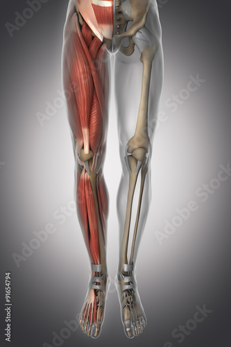 Leg Anatomy Muscle Bone Cartilage Ligament Buy This
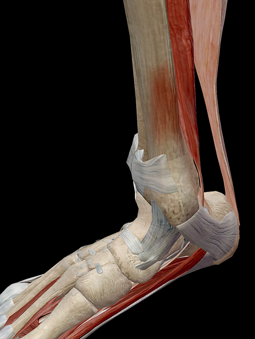 Hold On To Your Tibias The Anatomy And Causes Of Shin Splints
 Muscles Of The Lower Leg And Foot Human Anatomy And
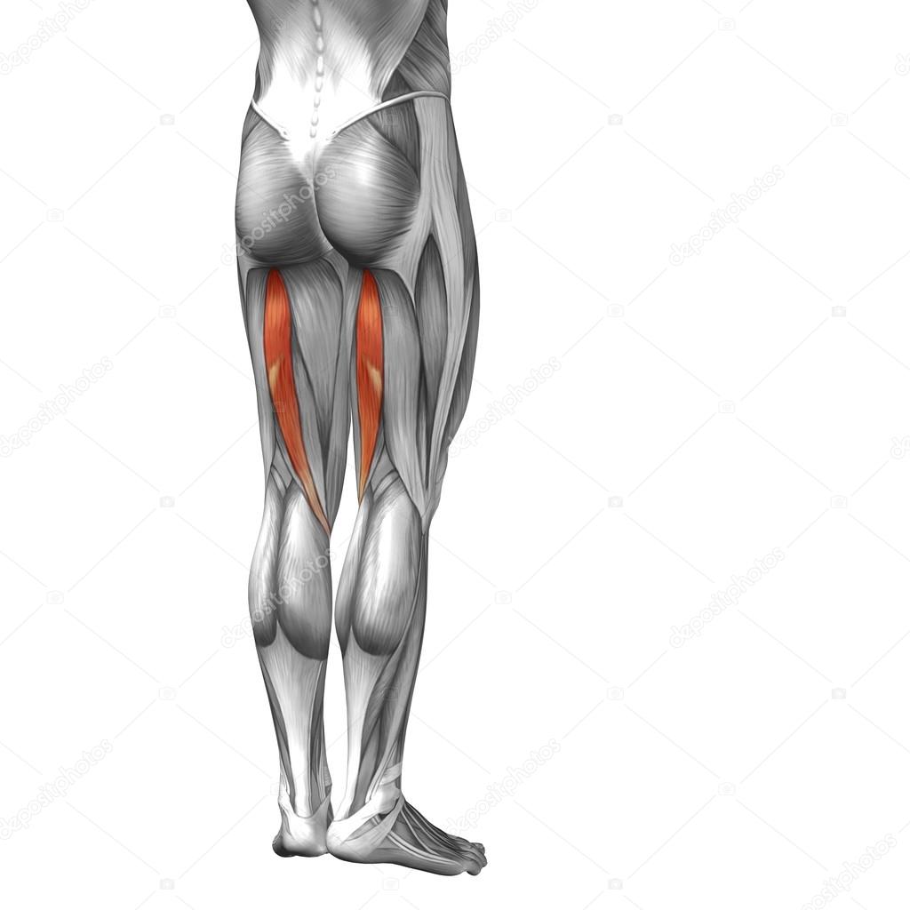 Conceptual 3d Human Upper Leg Anatomy Or Anatomical And
 Thigh Leg Muscles Human Anatomy Course
 Amazon Com Muscles Of The Leg Laminated Anatomy Chart By
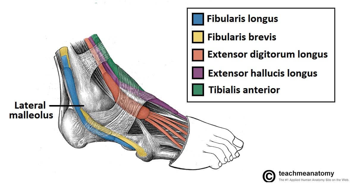 Muscles Of The Anterior Leg Attachments Actions
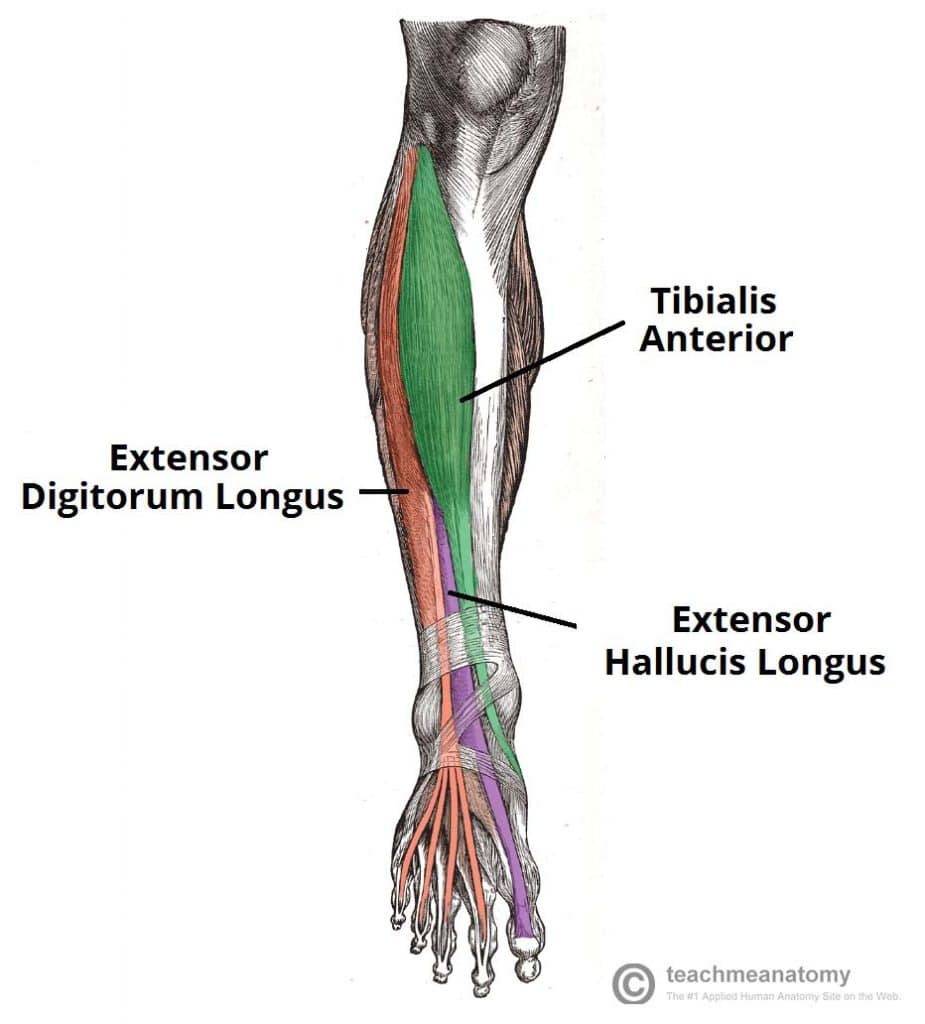 Muscles Of The Anterior Leg Attachments Actions

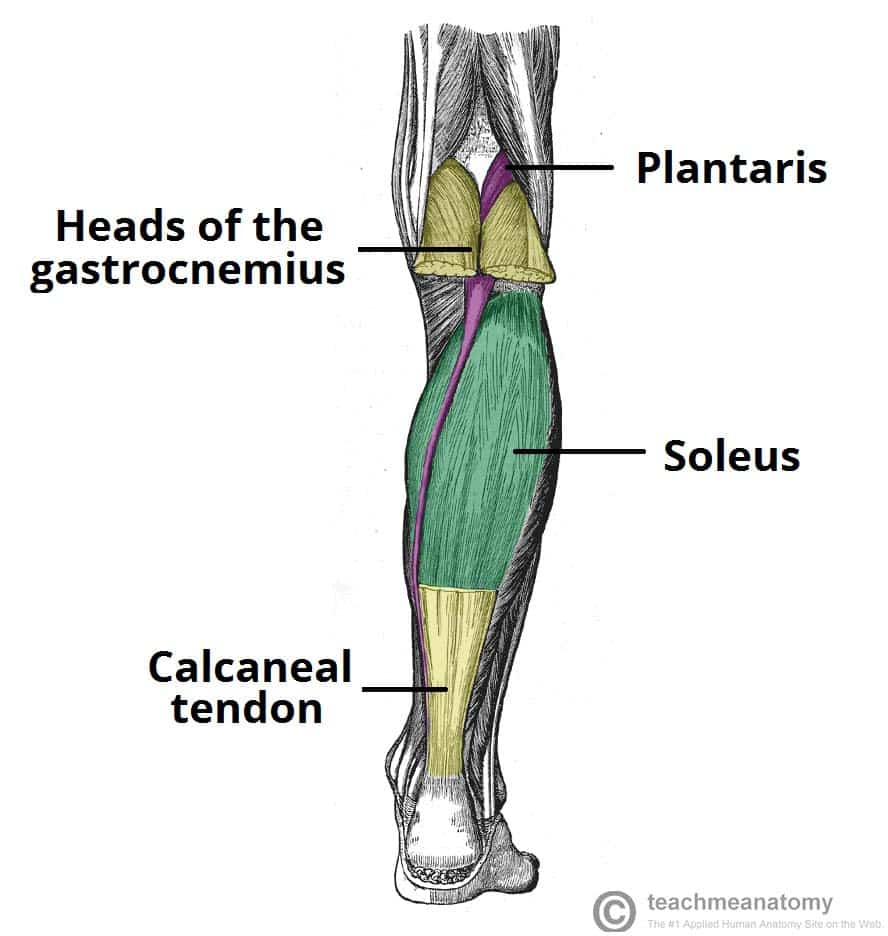 Muscles Of The Posterior Leg Attachments Actions
 Tibialis Anterior Muscle Wikipedia
 Muscles Of The Lower Leg And Foot Human Anatomy And
 How To Build A Pair Of Muscular Legs As A Natural Trainer
:background_color(FFFFFF):format(jpeg)/images/library/11153/muscles-tibia-fibula_english__2_.jpg) Leg And Knee Anatomy Bones Muscles Soft Tissues Kenhub
Leg And Knee Anatomy Bones Muscles Soft Tissues Kenhub
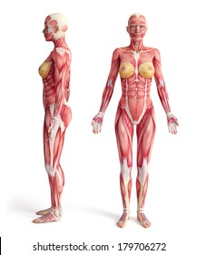 Woman Leg Anatomy Images Stock Photos Vectors Shutterstock
 Anatomical Teaching Models Plastic Human Muscle Models
 Numbered Muscle Leg Model 9 Part
 Quads Mov 1 High Bar Back Squat 5 Sets 5 4 3 2 1 Reps
 Leg Anterior Muscles 3d Illustration
 Tibialis Anterior Muscle Wikipedia
 Illustration Of Fit Strong Leg Anatomy Stock Photo
Belum ada Komentar untuk "Shin Anatomy Muscles"
Posting Komentar