Anatomy Of The Hip Joint
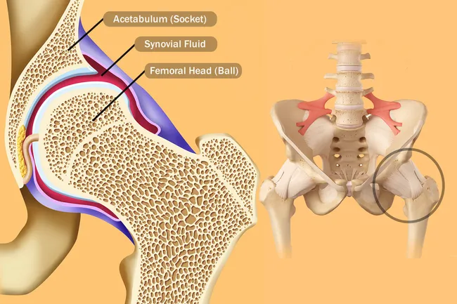
The socket is a concave depression in the lower side of the pelvis also called the acetabulum. It is a ball and socket joint at the juncture of the leg and pelvis.
 Hip Surgery Illustrations Pelvis Hip Anatomy Medical
The hip joint is one of the most important joints in the human body.
Anatomy of the hip joint. It forms a connection from the lower limb to the pelvic girdle and thus is designed for stability and weight bearing rather than a large range of movement. Standing and dynamic eg. Weight bearing stresses on the hip during walking can be 5 times a persons body weight.
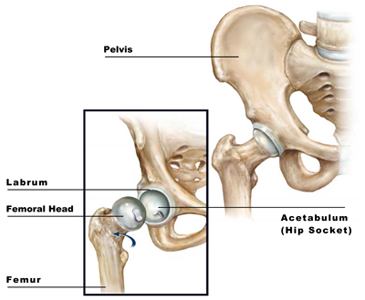
General hip anatomy the hip is a ball and socket join t similar to the joint in the shoulder. The pelvis and the femur the thighbone. Part of the reason for the hips stability is that there is a very deep socket called the acetabulum in the hip joint.
A strong capsule joint supported by ligaments and muscles also provides extra stability to the hip. The hip is the bodys second largest weight bearing joint after the knee. The hip joint is an intricate structure including hip bones hip articular cartilage muscles ligaments and tendons and synovial fluid.
It allows us to walk run and jump. It is the largest ball and socket joint in your body. The hip joint scientifically referred to as the acetabulofemoral joint art.
The ball is the rounded end of the femur also called the femoral head. A problem with any one of these parts of the hip anatomy can result in pain. It bears our bodys weight and the force of the strong muscles of the hip and leg.
The hip joint is the articulation of the pelvis with the femur which connects the axial skeleton with the lower extremity. It is a ball and socket joint at the juncture of the leg and pelvis. A healthy hip can support your weight and allow you to move without pain.
Coxae is the joint between the femur and acetabulum of the pelvis and its primary function is to support the weight of the body in both static eg. The adult os coxae or hip bone is formed by the fusion of the ilium the ischium and the pubis which occurs by the end of the teenage years. Hip anatomy function and common problems.
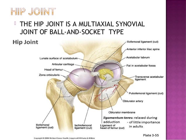
Walking or running postures. The hip joint is a ball and socket synovial joint formed by an articulation between the pelvic acetabulum and the head of the femur. Yet the hip joint is also one of our most flexible joints and allows a greater range of motion than all other joints in the body except for the shoulder.
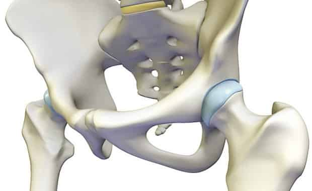
The hip joint is one of the largest joints in the body and is a major weight bearing joint. The hip joint is made up of two bones.
 Hip Anatomy Dr Sujit Kadrekar Arthroscopy And Joint
 Hip Anatomy Diagram From Bones To Joints Science Trends
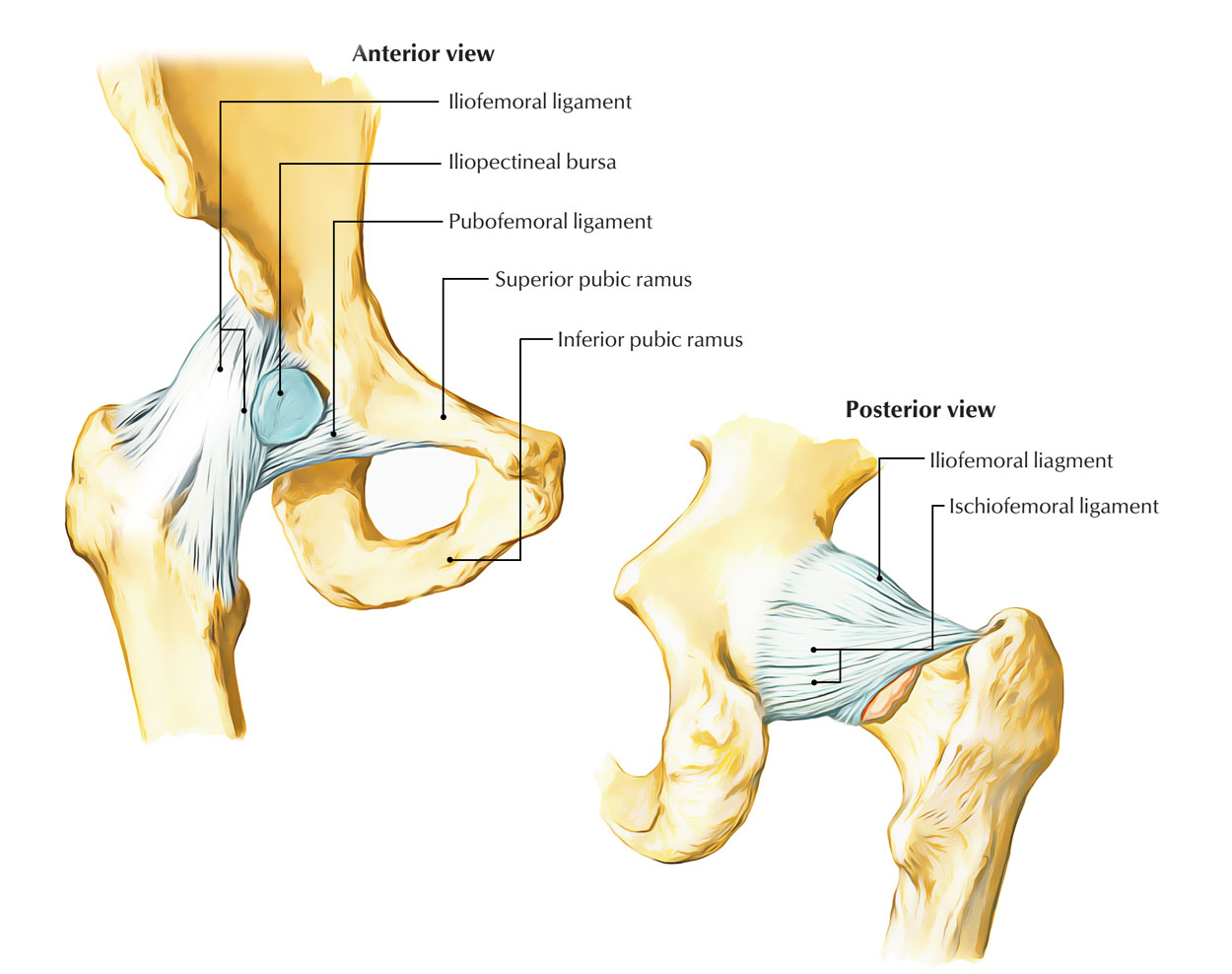
 Hip Joint Bones Ligaments Blood Supply And Innervation Anatomy Kenhub
 Hip Joint Anatomy Overview Gross Anatomy
 Ultimate Hip Joint Anatomy In One Picture Hip Anatomy
 1 Anatomy Of Hip Joint Adapted From 33 Download
 Hip Joint Treatment New York Labral Tear Treatment New
Transient Osteoporosis Of The Hip Orthoinfo Aaos
Hip Resurfacing Orthoinfo Aaos
![]() Hip And Thigh Bones Joints Muscles Kenhub
Hip And Thigh Bones Joints Muscles Kenhub


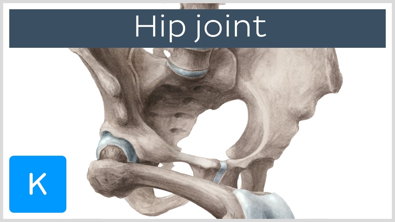
 Hip Joint 3d Anatomy Tutorial
 Hip Anatomy Pictures Function Problems Treatment
 International Hip Dysplasia Institute
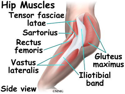
 Hip Anatomy Eorthopod Com
Easy Notes On Hip Joint Learn In Just 4 Minutes
 Anatomy Of The Hip Central Coast Orthopedic Medical Group
 Hip Joint Anatomy Hip Bones Ligaments Muscles
 Hip Joint Anatomy And Its Biomechanics
 Yoga For Hip Stability Understanding Hypermobility
 Coxal Articulation Or Hip Joint Human Anatomy
 Hip Bone Wikipedia
 Hip Joint Treatment Orange County Hip Surgeon Fountain
 Reasons Your Hips May Hurt
Belum ada Komentar untuk "Anatomy Of The Hip Joint"
Posting Komentar