Anatomy Of Ankle And Foot
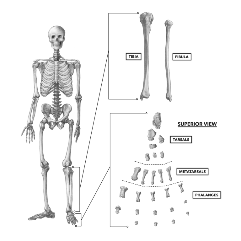
The ankle joint talocrural joint is formed where the distal end of the leg meets the foot. The foot consists of thirty three bones twenty six joints and over a hundred muscles ligaments and tendons.

The bones of the foot and ankle begin with the ankle joint itself.
Anatomy of ankle and foot. The articulating surfaces ligaments movements and any clinical correlations. The last two together are called the lower ankle joint. Descriptive topographic functional by kelikian md armen s and sarrafian md facs shahan k.
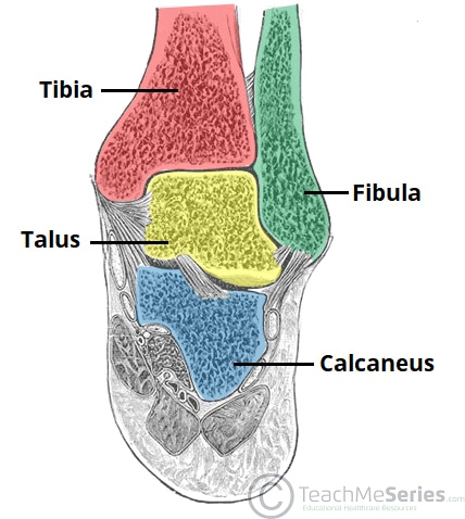
In order to understand conditions that affect the foot and ankle it is important to understand the normal anatomy of the foot and ankle. The ankle consists of three bones attached by muscles tendons and ligaments that connect the foot to the leg. The ankle joint also known as the talocrural joint allows dorsiflexion and plantar flexion of the foot.
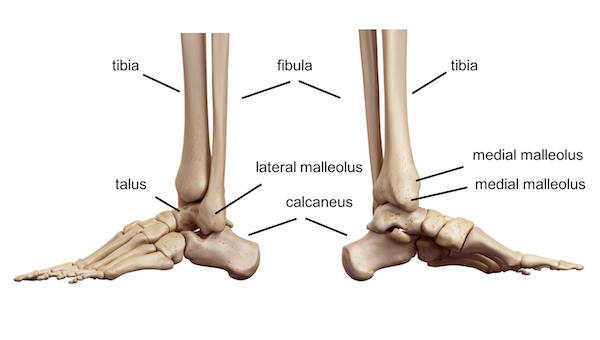
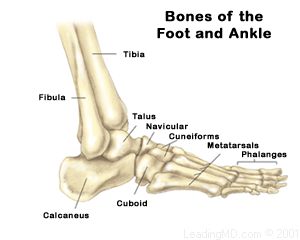
In the lower leg are two bones called the tibia shin bone and the fibula. These include the three cuneiform bones the cuboid bone and the navicular bone. Upper ankle joint tibiotarsal talocalcaneonavicular and subtalar joints.
You can also see the bands of ligaments where the metatarsals and phalanges are connected to each other. The talus bone supports the leg bones tibia and fibula forming the ankle. Foot and ankle anatomy is quite complex.
The hindfoot forms the heel and ankle. Here you can see that the ankle is also a thick web of ligaments where the tibia is connected to the bones of the ankle and the core of the foot. Foot and ankle anatomy.
It is made up of three joints. The ankle joint is formed where the talus the uppermost bone in the foot and the tibia shin meet. Sarrafians anatomy of the foot and ankle.
Footeducation is committed to helping educate patients about foot and ankle conditions by providing high quality accurate and easy to understand information. The ankle joint or talocrural joint is a synovial joint formed by the bones of the leg and the foot the tibia fibula and talus. The calcaneus heel bone is the largest bone in the foot.
In this article we shall look at the anatomy of the ankle joint. Use our anatomy tools to learn about bones joints ligaments and muscles of the foot and ankle. 50 out of 5 stars 6.
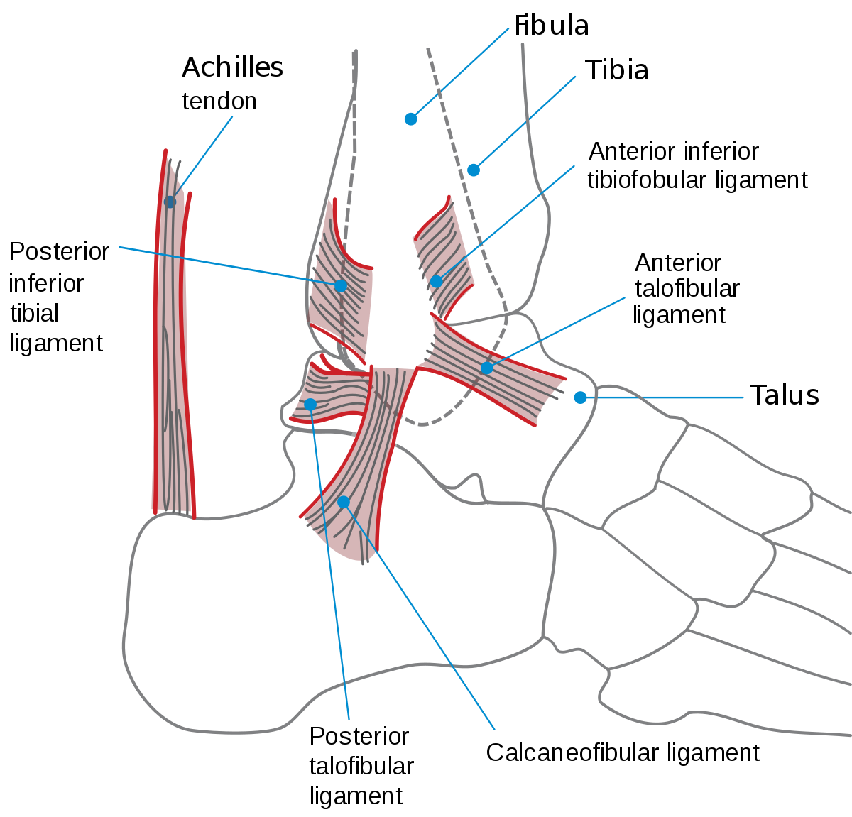
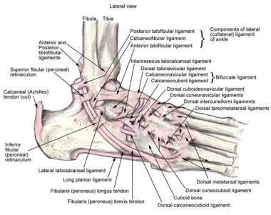
These all work together to bear weight allow movement and provide a stable base for us to stand and move on. Lateral side of the ankle joint capsule.
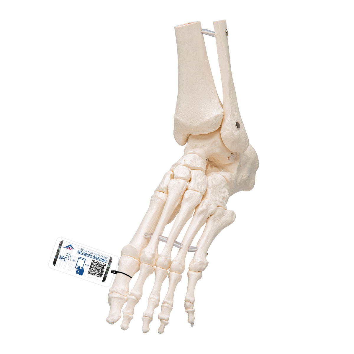 Foot Ankle Skeleton Elastic Mounted 3b Smart Anatomy
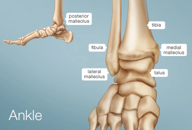 Ankle Human Anatomy Image Function Conditions More
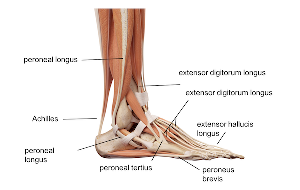 Ankle Foot Anatomy
Rheumatoid Arthritis Of The Foot And Ankle Orthoinfo Aaos
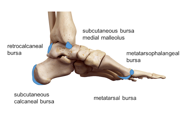
 Bursitis Ankle Bursa Care And Prevention

 Ankle Foot Anatomy
 Ankle Foot Atlas Of Anatomy

 Ankle Wikipedia
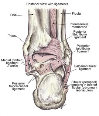
 Ligaments Of The Foot Muscles Tendons Ligaments Of The
 Ankle Foot Anatomy
 Ankle Foot Anatomy
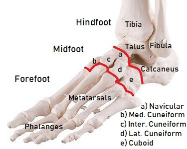 Foot Bones Anatomy Injuries Foot Pain Explored
 Ankle Joint Anatomy Overview Lateral Ligament Anatomy And
 Applied Anatomy Of Ankle And Foot
 Ankle Sprains
 Why Ankle Pain Treatments Chronic Ankle Pain Ankle Joint
![]() Ankle Joint Anatomy Bones Ligaments And Movements Kenhub
Ankle Joint Anatomy Bones Ligaments And Movements Kenhub
 The Ankle Joint Articulations Movements Teachmeanatomy
 Foot Bones Anatomy Conditions And More

Ankle Joint Anatomy Overview Lateral Ligament Anatomy And
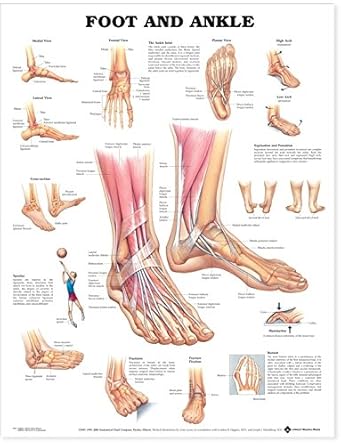 Foot And Ankle Anatomical Chart
 Ankle Foot Anatomy
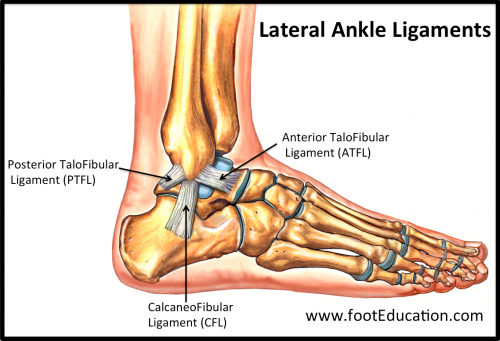 Ligaments Of The Foot And Ankle Overview Footeducation

 Crossfit Bones Of The Foot And Ankle
Belum ada Komentar untuk "Anatomy Of Ankle And Foot"
Posting Komentar