Anatomy Of A Dogs Eye
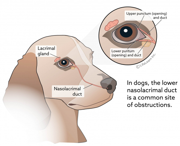
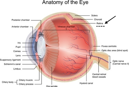
Anatomy of the eye. In other cases problems with the eyes themselves like poor tear production or abnormal anatomy can put dogs at risk for corneal damage.
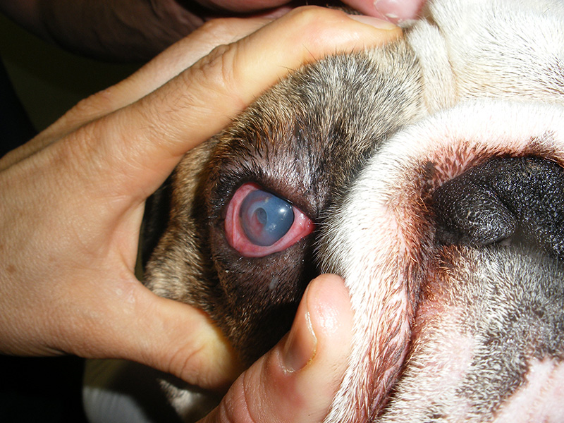
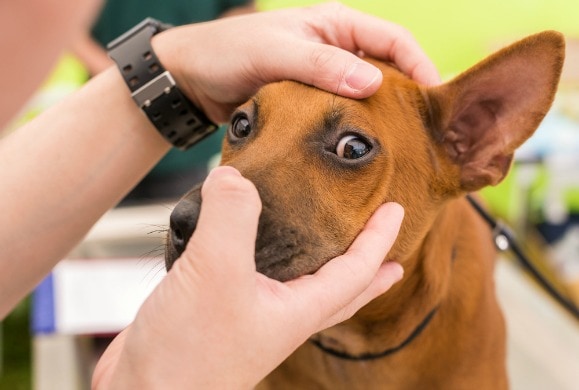
To examine the eyes the head is cupped between both hands with one thumb on the upper eyelid and the other thumb on the lower eyelid.

Anatomy of a dogs eye. Dog rabbit rat mouse primate. Rabbits are able to resist blinking for long intervals because they have a very stable tear film. To see the parts of the eye beneath the upper eyelid pull the upper eyelid up with your thumb which will open the eye widely.
Eyelashes are absent from the lower lid of carnivores. A dogs eyelids have a number of special features. 8 common eye problems in dogs.
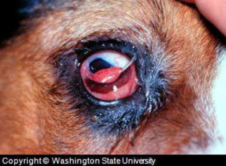
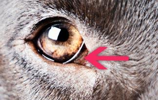
This is the circular. Dogs have a third eyelid on each eye known as the haw or. In the first detailed analysis comparing the anatomy and behavior of dogs and wolves researchers found that the facial musculature of both species was similar except above the eyes.
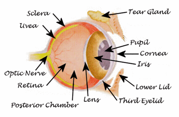
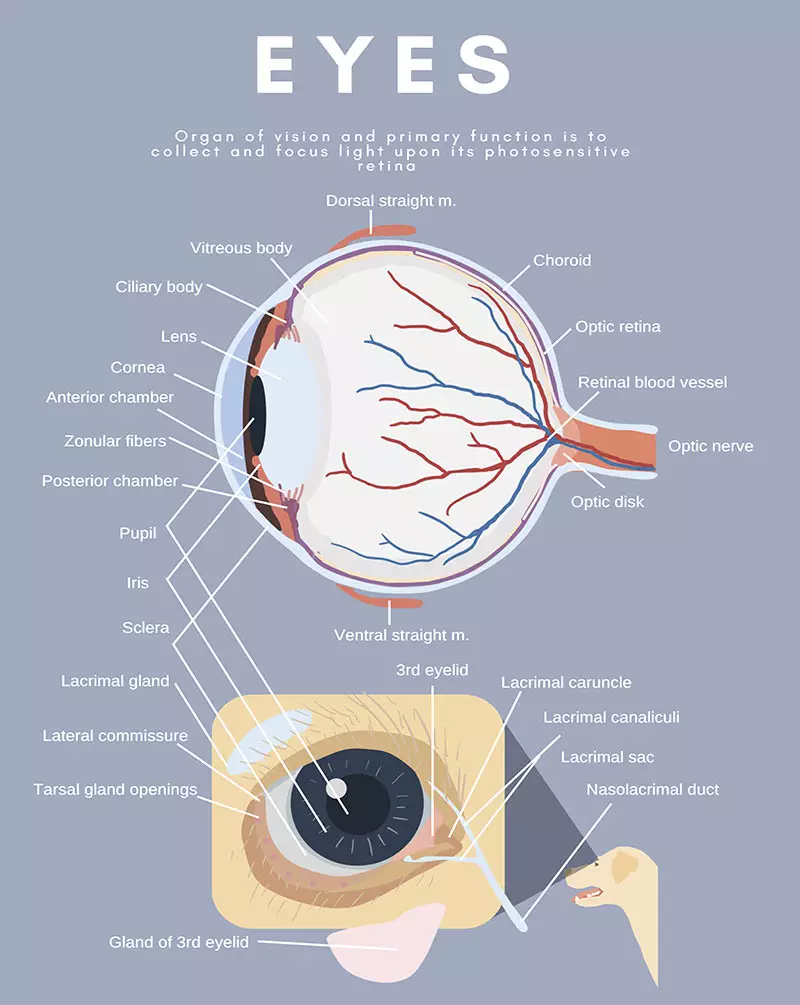
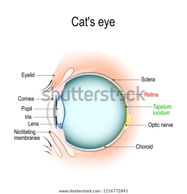
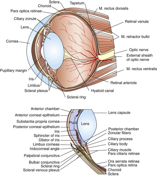
Anatomy of the normal dog eye. The eye the three coats of the eye. Many anatomical terms used to describe parts of a dog are similar to the ones used for horses.
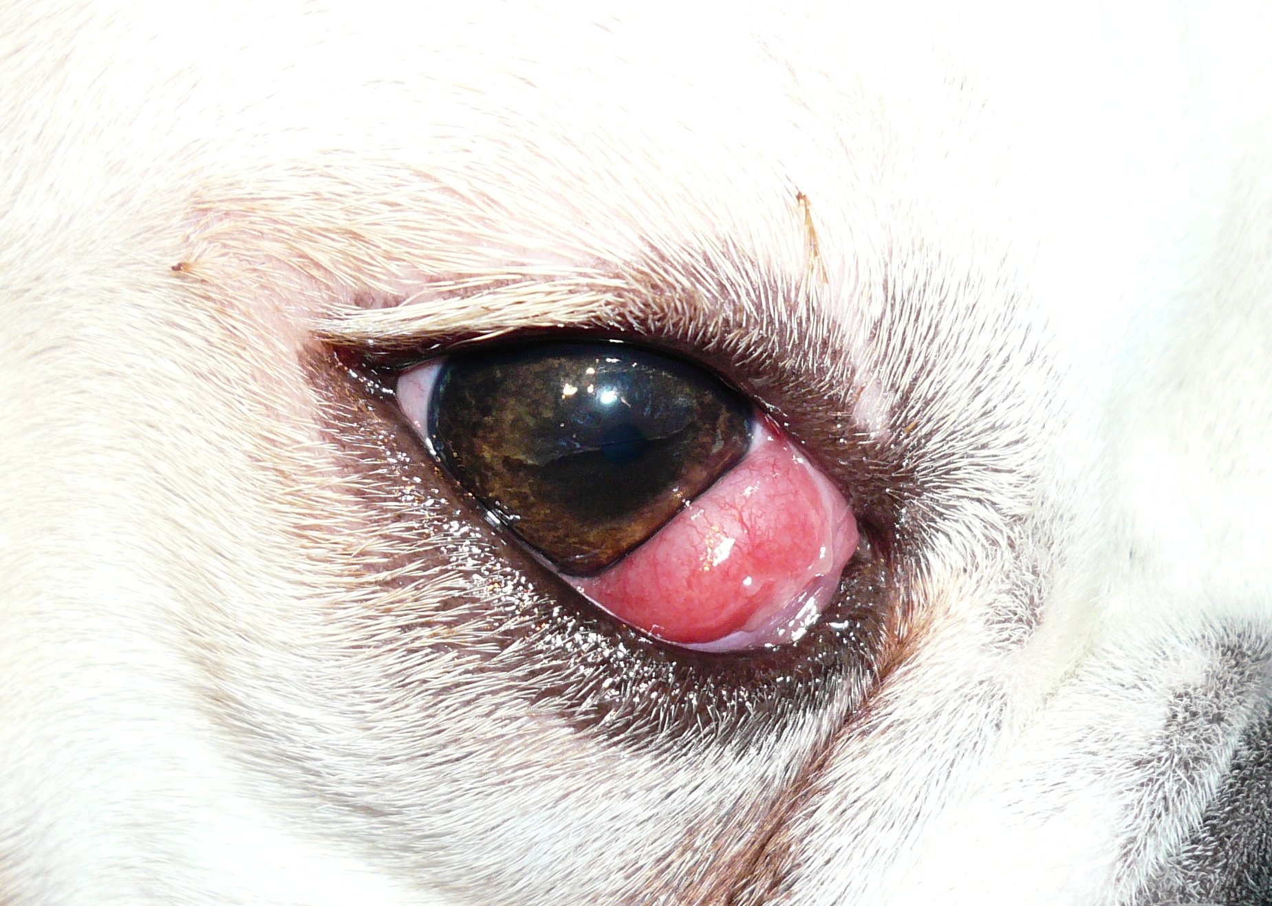
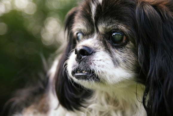
The eye may also be red and have excessive drainage. This is likely due to the contribution of a lipid contribution from the prominent hardarian gland. A dog with a corneal wound will often rub at the affected eye and squint because of pain.
Some canine anatomical names may be familiar to you dogs have elbows and ears and eyes but other names may be downright foreign. In a healthy dog eye the dog conjunctiva color should match the color of the dogs gums. These are the outer most protective layers of the dog eye anatomy.
Signs of dog eye problems include blood vessels that look engorged any bruises around the eye or a sclera which is yellow could be dog jaundice and discharge such as mucous. The dogs eye is made up of three layers. Dog anatomy from head to tail.
The eyes of a dog are protected not only by the same types of eyelids that people have but also by the nictitating membrane which is sometimes called the third eyelid. Tough outer protective layers of the eye the sclera is covered by a thin membrane. Blinking also helps spread tears over the surface of the eye keeping it moist and clearing away small particles.
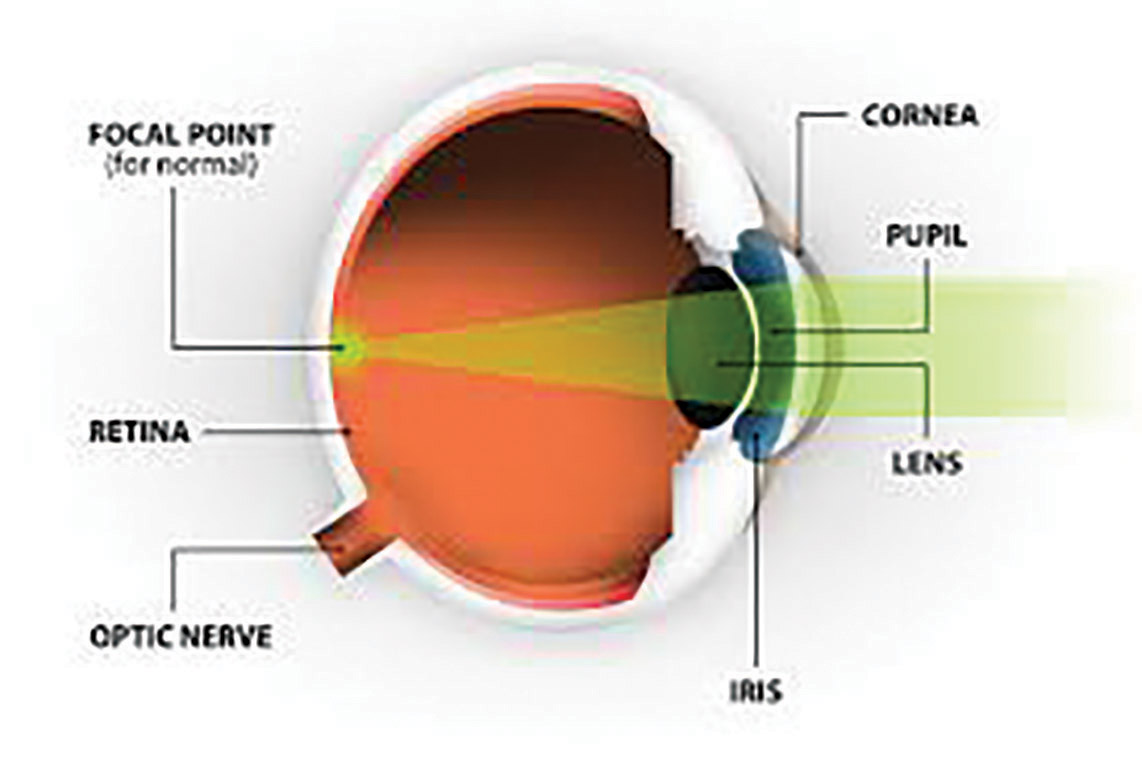
Dog grooming for dummies. The cornea is the central dome shaped area of the eye surface.
 Dog Eye Care Pets World
 Nictitating Membrane An Overview Sciencedirect Topics
 Petmd Mobile Petmd Slideshows
 Collie Eye Anomaly In Dogs Cats Symptoms Causes
 Brachycephalic Dogs Are Most Susceptible To Corneal
 Nictitating Membrane An Overview Sciencedirect Topics
 Canine Eye Notes Pt 1 By Tamberella On Deviantart Dog
 Eye Discharge Epiphora In Dogs Vca Animal Hospital
 Amazon Com Ambesonne Educational Pet Mat For Food And Water
 Dog Eye Pictures And Treatment Eye Problems And Diseases
 Canine Uveitis And The Veterinary Technician Today S
 Canines Evolved Puppy Dog Eyes To Woo Human Companions
Shih Tzu Keratoconjunctivitis Sicca Ufaw
 Dogs Evolved Sad Eyes To Manipulate Their Human Companions
 Cherry Eye Wikipedia
 Dog Anatomy Wikipedia
 Anatomy Cats Dogs Eye Vertical Section Stock Vector Royalty

 Progressive Retinal Atrophy In Dogs Pra
 Petmd Mobile Petmd Slideshows
 Petmd Mobile Petmd Slideshows
 Surgery Of The Eye Veterian Key
 A Guide To Cherry Eye In Dogs Is This A Dog Eye Infection
 Nictitating Membrane An Overview Sciencedirect Topics
Belum ada Komentar untuk "Anatomy Of A Dogs Eye"
Posting Komentar