Anatomy Of Rat Brain
The rat atlas is a three dimensional 3d computerized map of rat brain anatomy created with digital imaging techniques. Access online via elsevier 2006.
 Rat Brain Anatomy
Photographs of sufficient magnification are included to permit investigators to judge for themselves the veracity of the atlas delineations.

Anatomy of rat brain. The is based on high resolution isotropic ex vivo t2 weighted mri and dti data acquired at the duke center for in vivo microscopy at resolutions of 39 μm and 78 μm respectively. Three principal strains are now commonly used for scientific study. The waxholm rat atlas is an open access volumetric atlas of the sprague dawley rat brain.
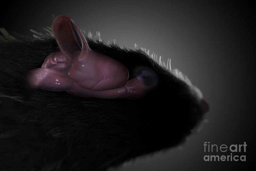
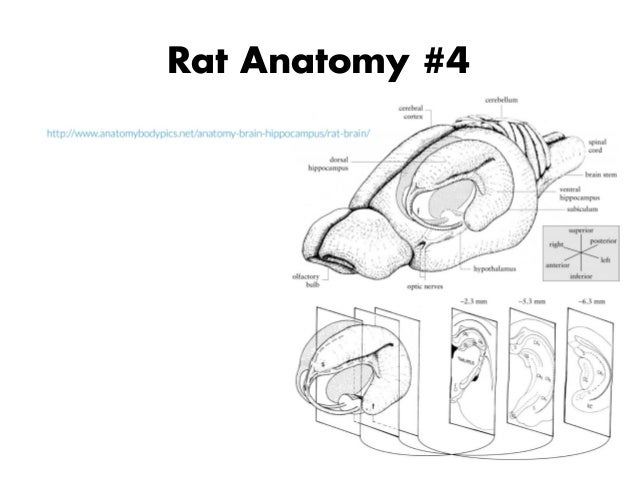
The laboratory rat was developed from the norwegian rat rattus norvegicus by an american physiologist henry donaldson who started a breeding colony in 1906 at the wistar institute in philadelphia. A tool by matt gaidicamatt gaidica. Rat brain pictures dorsal aspect of brain and rostral two ventral aspect of the brain and junction of segments of spinal cord.
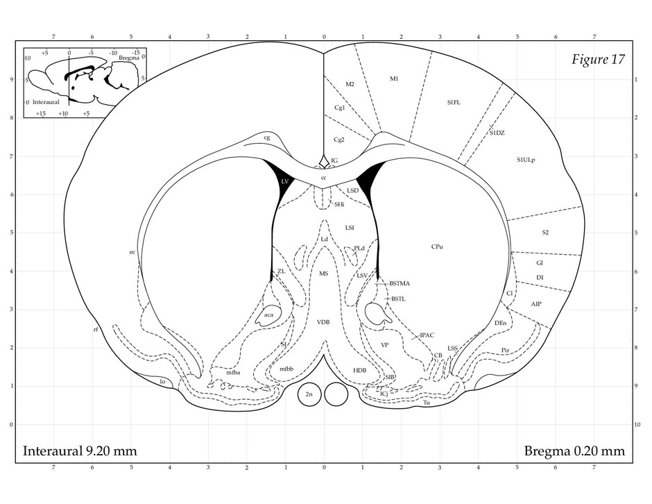
Thus after normalizing an individual image to the rat brain template the intracranial tissues could be conveniently extracted. Paxinos george and charles watsonthe rat brain in stereotaxic coordinates. Developed at the wistar institute.
The rat brain is analyzed through stereotaxic localization of discrete brain areas and the subdivisions of many areas of rat brain are mapped using plates and diagrams. Electronic sharing and interactive use are benefits afforded by a digital format but the foremost advantage of this 3d map is its whole brain integrated representation of rat in situ neuroanatomy. The specimen is an 80 day old male sprague dawley rat.
Medulla with spinal cord. In addition an intracranial rat brain mask was formed from this canonical brain by assigning 1 for intracranial voxels and 0 for others as shown in figure 1b.
 Figure 1 From Design And Evaluation Of Phased Array
 Depicted Adapted Diagram Of Da Pathways In Basal Ganglia In
 Schematic Representation Of Gross Anatomical Brain Areas Of
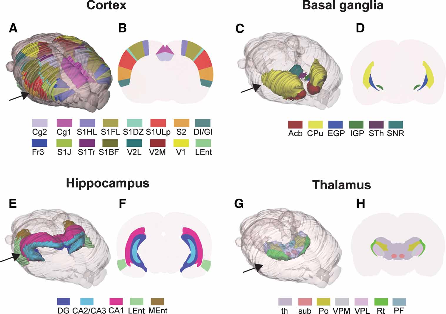 Frontiers Three Dimensional Atlas System For Mouse And Rat
 Frontiers Three Dimensional Atlas System For Mouse And Rat
 The Diencephalon Of The Albino Rat Studies On The Brain Of
 Science Source Rat Brain Anatomy
 Rat Brain Images Stock Photos Vectors Shutterstock
 Rat Brain Enamel Pin Science Pin Lapel Pin Anatomy Physiology Medicine Doctor Pin Doctor Gift Scientist Gift Rat Lover Dissection
Rat Brain Pictures
 Pdf Mitotic Activity In Adult Rat Brain Induced By
 Quantitative Rodent Brain Receptor Imaging Springerlink
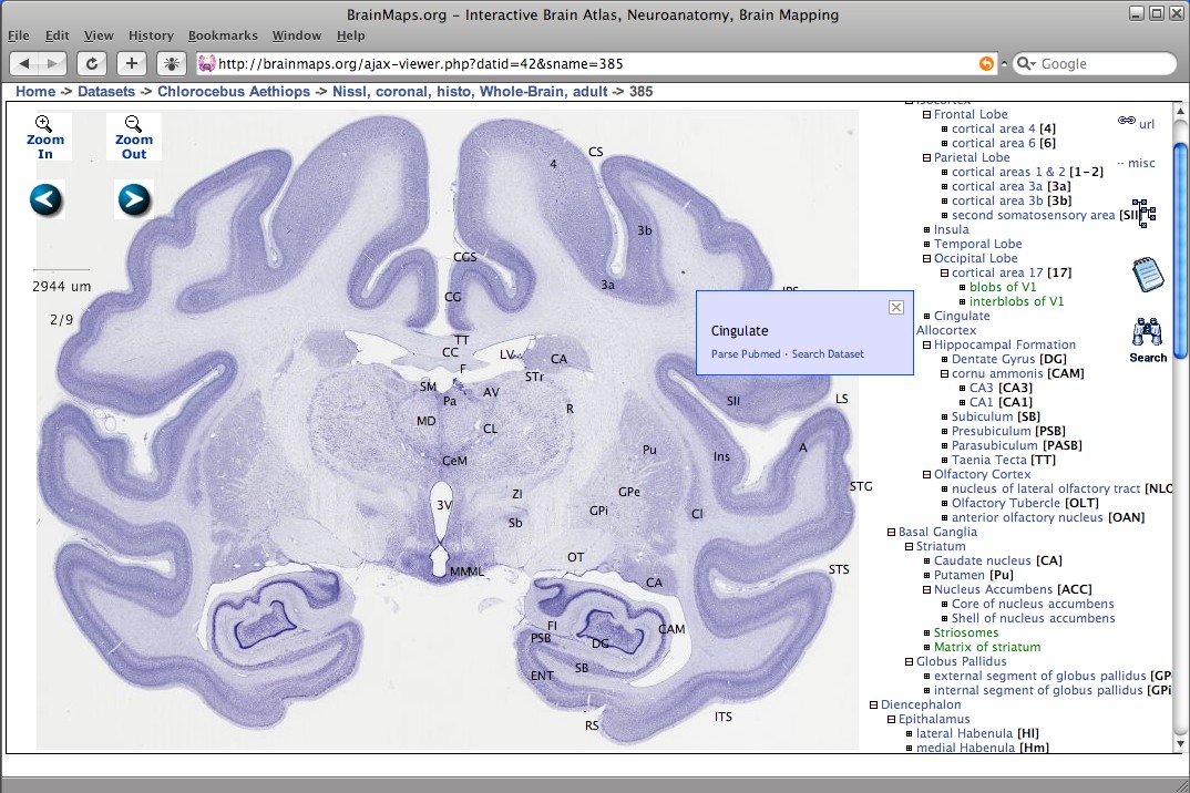 Brainmaps Wikipedia
 Enlarge
 Nervous System Sciencedirect
 Sucrose Intensity Coding And Decision Making In Rat

 Rat Brain Anatomy
 Brain Maps 4 0 Structure Of The Rat Brain An Open Access

Figure 4 9 The Distribution Of Opioid Receptors In The Rat Brain

 Focused Ultrasound Neuromodulation
 A Vascular Brain Anatomy Of The Rat Reproduced From 10
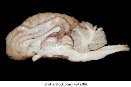
 Sagittal A And Coronal B And C Drawings Of The Rat Brain
80331-7.fp.png) Diazepam Receptor Specific Binding Of 3h Diazepam And 3h
Diazepam Receptor Specific Binding Of 3h Diazepam And 3h
 Voltage Gated K Channel B Subunits Expression And

 Rat Brain Comparative Anatomy
 Anatomy Of Corpus Callosum In Prenatally Malnourished Rats
Anatomy Of The Hippocampal Formation A Overview Of
Rat Brain Pictures
Belum ada Komentar untuk "Anatomy Of Rat Brain"
Posting Komentar