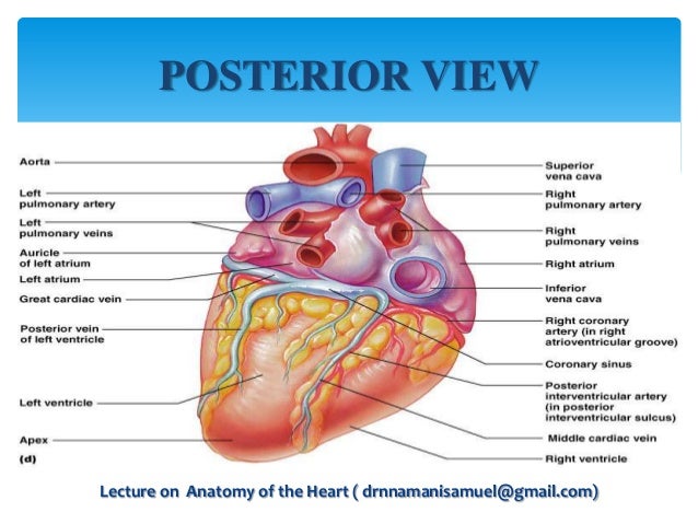
Heart Anatomy Posterior
It is divided by a partition or septum into two halves and the halves are in turn divided into four chambers. The heart has five surfaces.
 Heart External Anatomy Anterior And Posterior Diagram
This amazing muscle produces electrical impulses that cause the heart to contract.

Heart anatomy posterior. The structures initially seen from this perspective include the superior vena cava right atrium right ventricle pulmonary artery and aorta. Anterior ventral refers to the front and posterior dorsal refers to the back. From the right ventricle the blood is pumped through the pulmonary semilunar valve into the pulmonary trunk.
Posterior view of heart anatomy in this image you will find superior vena cava right pulmonary artery right pulmonary veins right atrium inferior vena cava coronary sinus right coronary artery coronary sulcus posterior interventricular artery in it. On the posterior surface of the heart the right coronary artery gives rise to the posterior interventricular artery also known as the posterior descending artery. Just pick an audience or yourself and itll end up in their incoming play queue.
Intraoperatively the anatomy of the heart is viewed from the right side of the supine patient via a median sternotomy incision. The anatomy of the heart. Heart valves separate atria from ventricles and ventricles from great vessels.
Blood flow through the heart. The heart is situated within the chest cavity and surrounded by a fluid filled sac called the pericardium. 0 0000 a shoutout is a way of letting people know of a game you want them to play.
Putting this in context the heart is posterior to the sternum because it lies behind it. Putting this in context the heart is posterior to the sternum because it lies behind it. Blood flow through the heart.
Araneae normally have eight eyes in four pairs. The blood in the lungs returns to the heart through the pulmonary veins. The blood flow through the heart is quite logical.
Anatomy of the human heart posterior view. The pulmonary trunk carries blood to the lungs where it releases carbon dioxide and absorbs oxygen. Base posterior diaphragmatic inferior.
It runs along the posterior portion of the interventricular sulcus toward the apex of the heart giving rise to branches that supply the interventricular septum and portions of both ventricles. Because of the unusual nature and positions of the eyes of the araneae spiders and their importance in taxonomy evolution and anatomy special terminology with associated abbreviations has become established in arachnology. Retrolateral refers to the surface of a leg that is closest to the posterior end of an arachnids body.
 Cardiovascular Anatomy Anatomy Of The Heart Flashcards
 Heart Anatomy Flashcards Memorang
 Gross Anatomy Glossary Heart Coronary Arteries Draw It
 Solved A Heart Posterior View La Aorta H Apex Of Heart
 Cardiac Skeleton An Overview Sciencedirect Topics
 Overview Of Sinoatrial And Atrioventricular Heart Nodes
 Heart Anatomy Structure Valves Coronary Vessels Kenhub
 Posterior View Of Human Heart Anatomy With Annotations Stock
 Procedure B Dissection Of A Sheep Heart Human Anatomy
 Heart Anatomy Structure Valves Coronary Vessels Kenhub
 Heart Anatomy Posterior Stock Photos And Images Agefotostock
 Coronary Arteries And Cardiac Veins Anatomy And Branches
 Solved 2 Label The Major Arteries And Veins On The Poste
 Cardiac System 1 Anatomy And Physiology Nursing Times
 Coronary System Tutorial What Is The Coronary System
 Cardiac Anatomy And Physiology Anesthesia Key
 Solved Which Of The Following Statements Are False Concer
 Posterior Interventricular Sulcus Lab 3 Heart And Vessels
 General Heart Anatomy Posterior View Diagram Quizlet
 Cardiovascular Media Library Watch Learn Live
 Science Source Heart Illustration Posterior View
 Coronary Arteries And Cardiac Veins Anatomy And Branches
 Special Circulations Of The Cardiovascular System Online
 Posterior View Of Heart Stock Photos Posterior View Of


Belum ada Komentar untuk "Heart Anatomy Posterior"
Posting Komentar