Ultrasound Anatomy
Otero and diego a. There are two main types of fetal ultrasound exams.
 Abdominal Ultrasound Registry Review
Ultrasound evaluation of normal fetal anatomy.
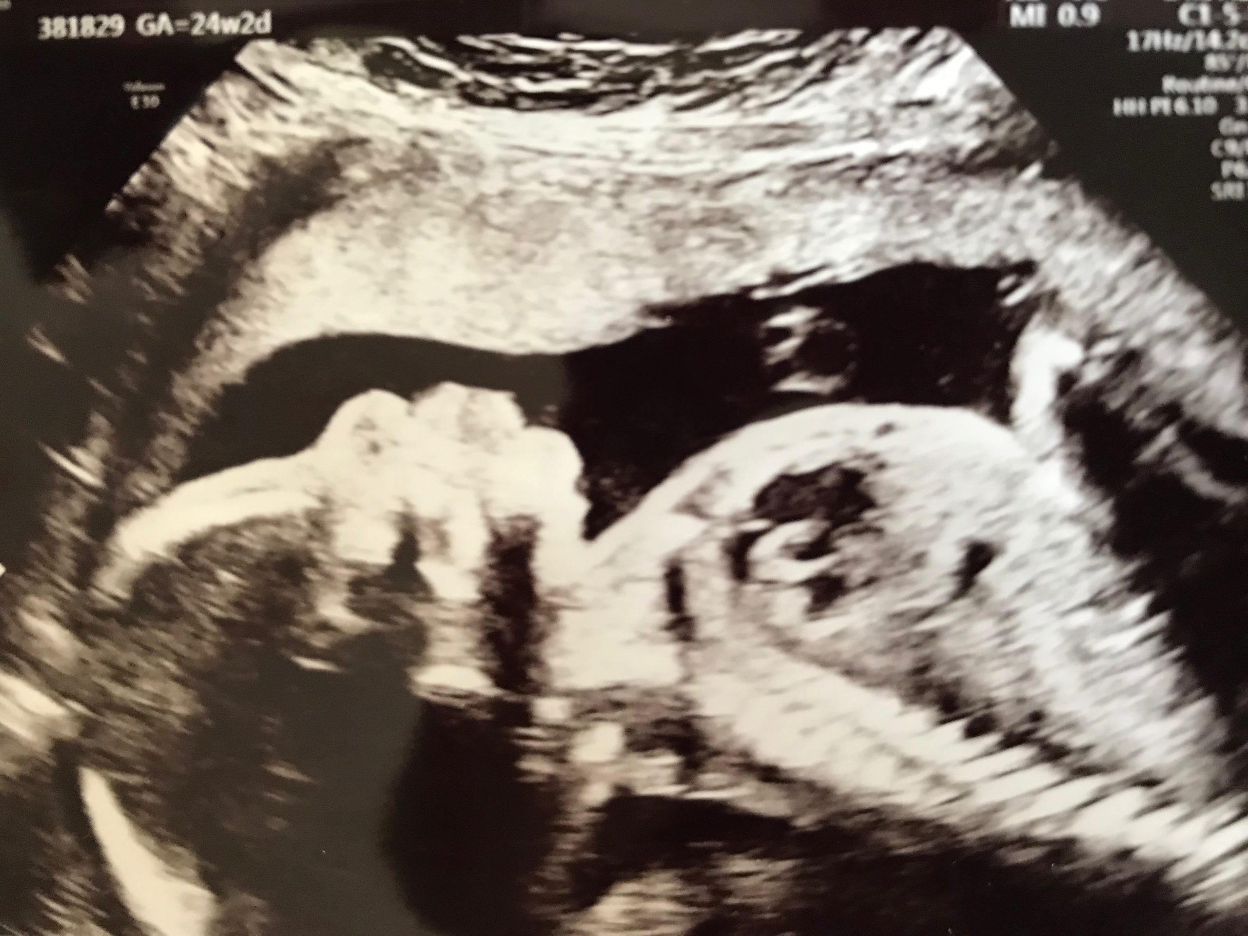
Ultrasound anatomy. The second trimester scan is a routine ultrasound examination in many countries that is primarily used to assess fetal anatomy and detect the presence of any fetal anomalies. Heart rate rhythm 4 chamber views. During the scan we will examine each part of the fetal body in detail.
The anatomy scan is a level 2 ultrasound which is typically performed on pregnant women between 18 and 22 weeks. Skull shape integrity bpd and hc measurements. The second trimester extends from 13 weeks and 0 days to 27 weeks and 6 days of gestation although the majority of these studies are performed between 18 and 23 weeks.
Manual of small animal regional anesthesia. Our understanding of normal fetal anatomy as seen on sonograms continues to be an area of considerable growth. A transabdominal fetal ultrasound is done by moving a transducer.
The purpose of the 20 to 22 week ultrasound is to look at all of the fetuss anatomy and to determine if all looks normal. The gender of your babybabies can usually be determined at this ultrasound. Most anatomy scans are performed in the second trimester of pregnancy typically at 20 weeks but they can be done anytime between 18 weeks and 22 weeks.
The following fetal parts are checked during the anatomy ultrasound. Description examining your growing babybabies. Brain ventricles choroid plexus mid brain posterior fossa cerebellum cisterna magna.
If you have a condition that needs to be monitored such as carrying multiples you may have more than one detailed ultrasound. Illustrated anatomy for nerve stimulation and ultrasound guided nerve blocks by pablo e. Things that cant be seen in earlier scans such as spinal cord.
Liver ultrasound showing education liver segments normal liver anatomy portal vein hepatic veins the biliary tree and ultrasound scanning protocol worksheets. When a level 2 ultrasound is done. Portela jul 1 2019 hardcover.
Neck nuchal fold thickness. Those who want to can find out the sex of the baby if desired. In special cases a detailed examination of the fetal heart and.
Instrumentation has improved steadily yielding both improved and more consistent image quality. With this type of fetal ultrasound a wandlike device called.
 The Anatomy Ultrasound Everything You Should Know
 Aahs Defining The Anatomy Of The Dorsal Scapholunate
 Normal Anatomy Of Fetal Spine
 Free Chapter Normal Cns Ultrasound Brain Anatomy Ob Images
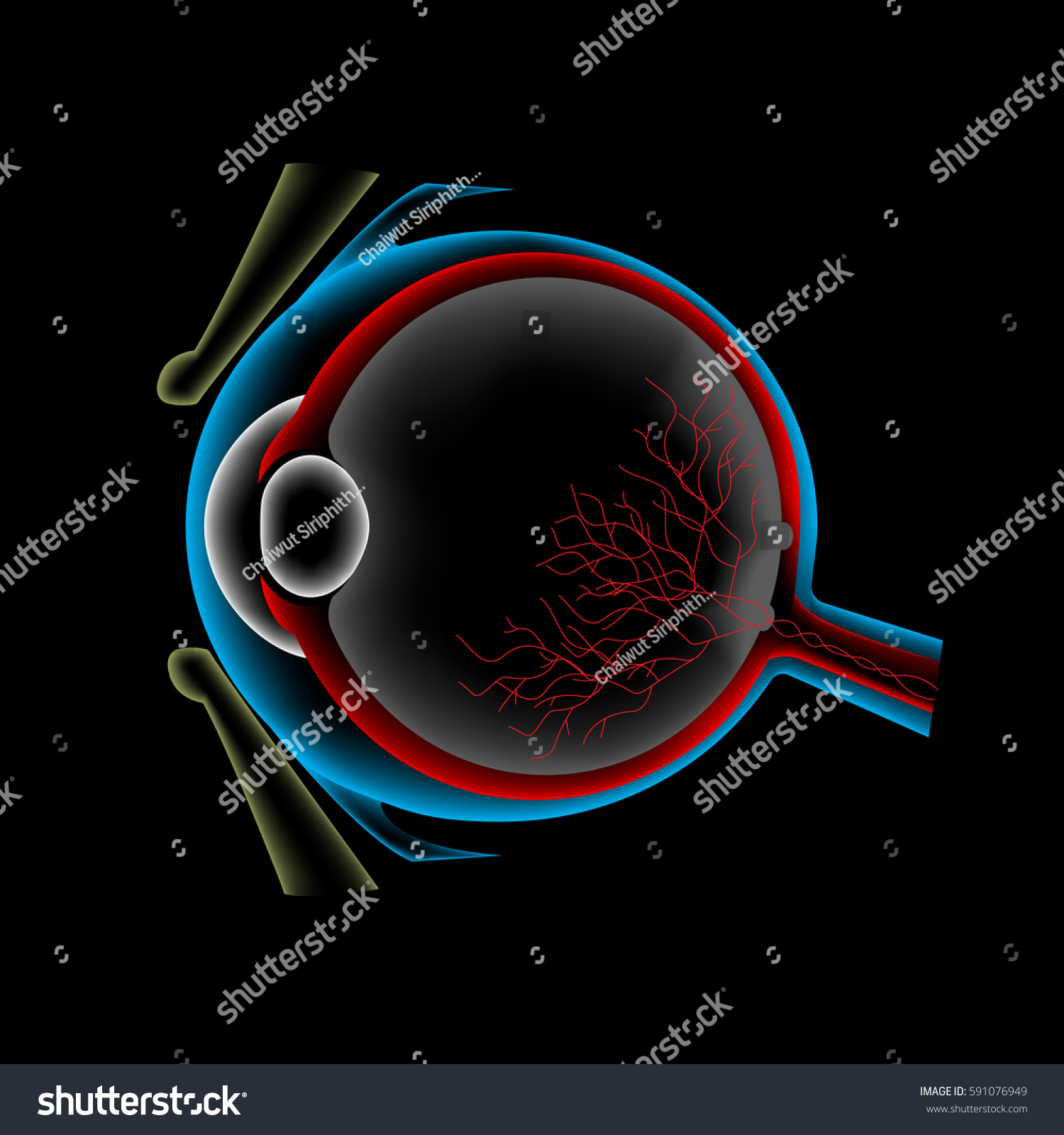 Ocular Ultrasound Eye Anatomy Scan Stock Vector Royalty

 What To Expect At Your Anatomy Scan Ultrasound Lifes
 Musculoskeletal Nuem Blog
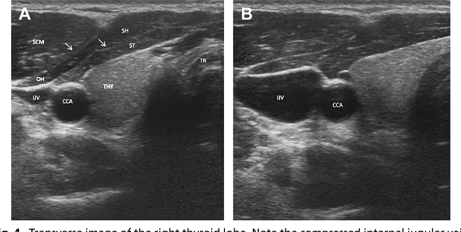 Figure 7 From Head And Neck Anatomy And Ultrasound
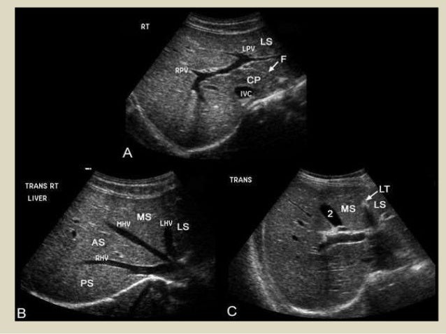
 Liver Anatomy And Segments By Ultrasound In Arabic
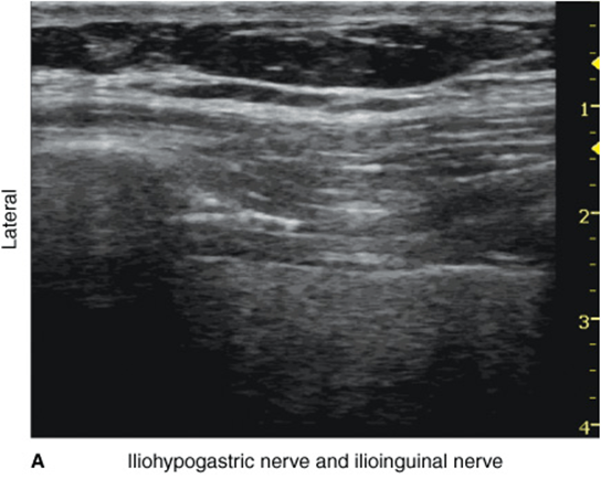 Ultrasound Guided Iliohypogastric And Ilioinguinal Nerve
 Anatomy Scan And 4d Ultrasound In Japan Tiny Tot In Tokyo
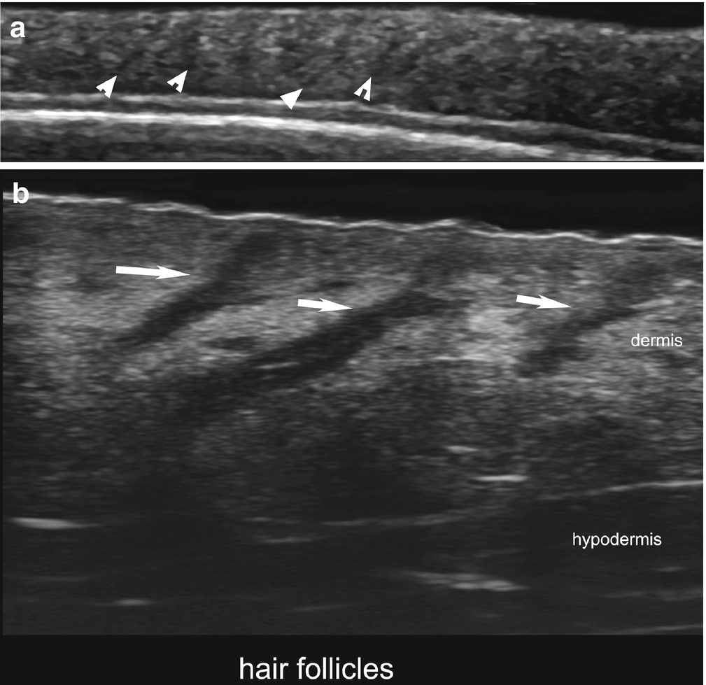
Normal Ultrasound Anatomy Of The Skin Nail And Hair

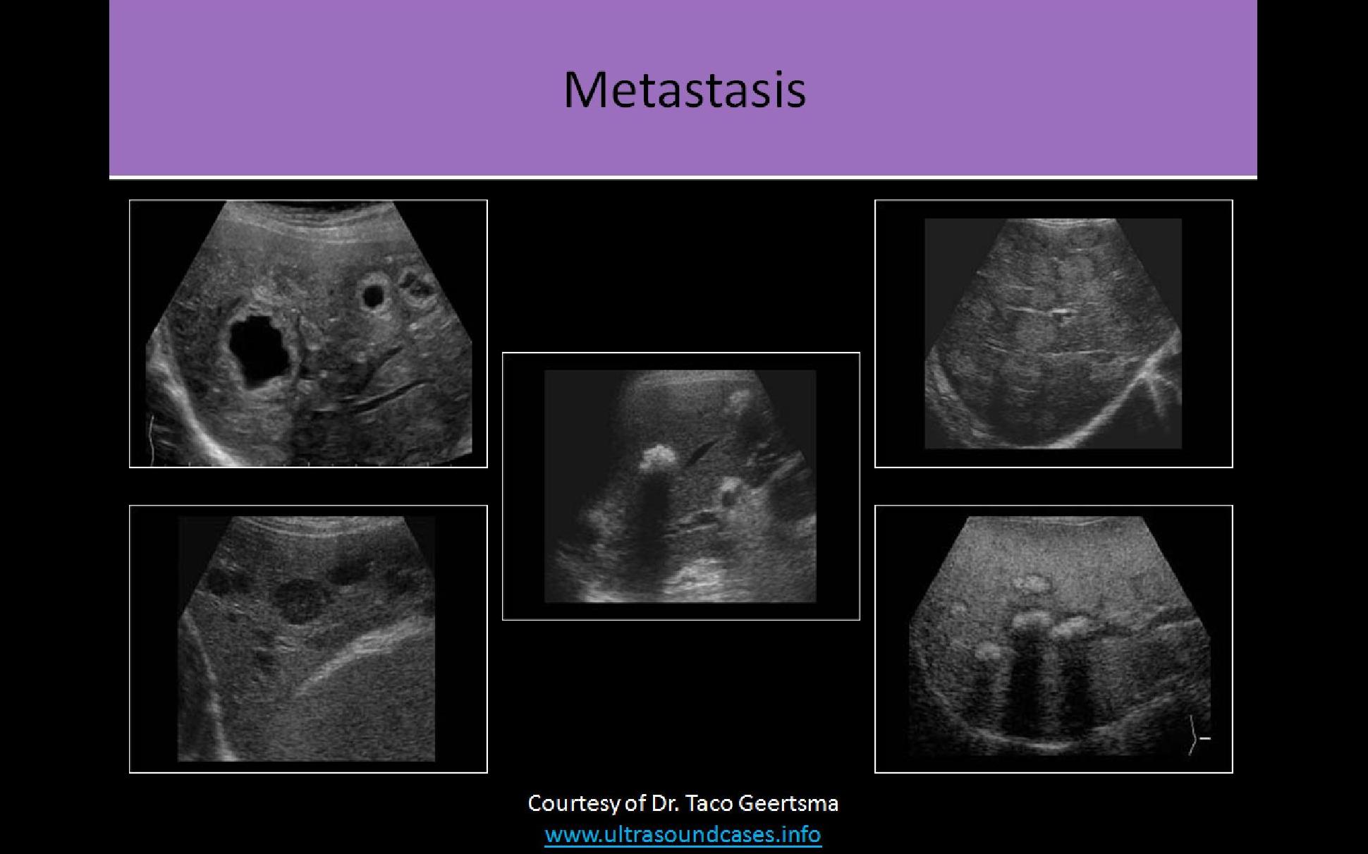
 Presentation1 Abdominal Ultrasound Anatomy
 1 Normal Ultrasound Anatomy Seen Above The Asis Download

 19 Weeks Our Anatomy Ultrasound The Love Notes Blog

 20 Week Anatomy Ultrasound Youtube
 Normal Ultrasound Anatomy Of The Eye In The Correct Plane


 Pocket Atlas Of Normal Ultrasound Anatomy Radiology Pocket
Belum ada Komentar untuk "Ultrasound Anatomy"
Posting Komentar