Anatomy Of Bony Pelvis
The pelvis is formed by four bones which include a pair of hip bones otherwise known as innominate bones. The bony pelvis is formed by the sacrum and coccyx and a pair of hip bones os coxae or innominate bones comprising the ischium pubis and ilium and are part of the appendicular skeleton.
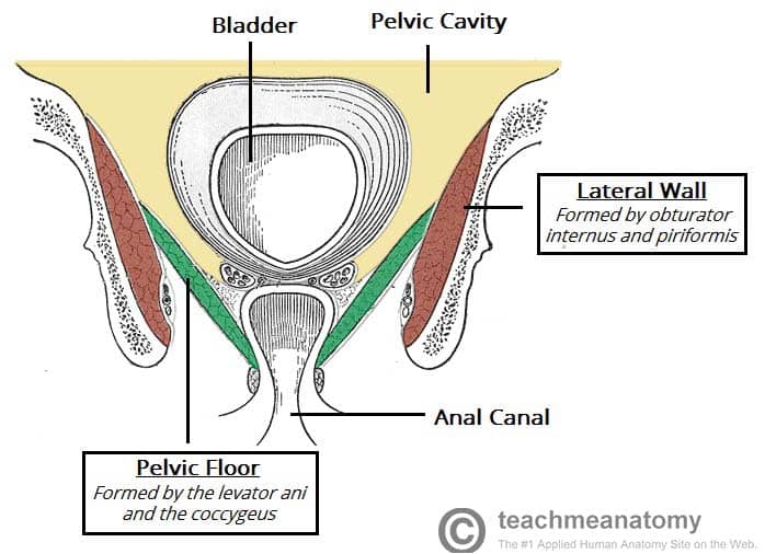 The Pelvic Floor Structure Function Muscles
The hip bone or coxal bone forms the pelvic girdle portion of the pelvis.
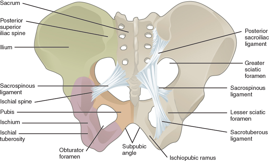
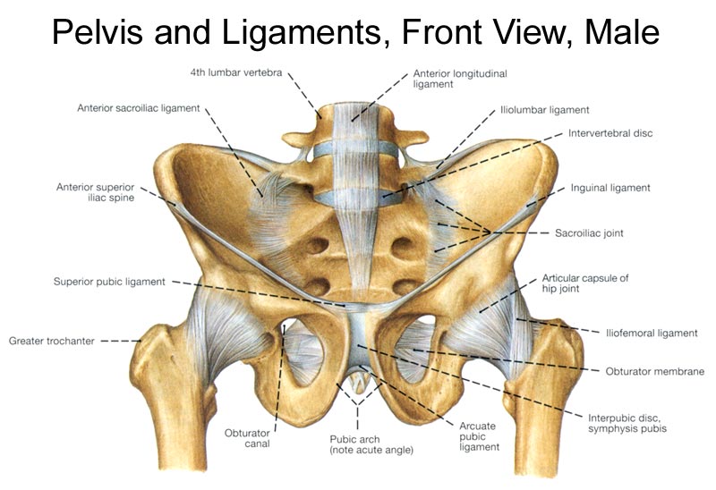
Anatomy of bony pelvis. The two main ligaments of the pelvis are the sacrotuberous and sacrospinous ligaments. These form the lateral and anterior walls of the bony pelvis. There are four articulations within the pelvis.
Its primary function is the transmission of forces from the axial skeleton to the lower limbs as well as supporting the pelvic viscera. The three bones and three joints composing the pelvic ring have no inherent stability without vital ligamentous structures. It is a small bone that forms the lower part of posterior wall.
The right and left hip bones plus the sacrum and the coccyx together form the pelvis. This area provides support for the intestines and also contains the bladder and reproductive organs. At birth the ilium ishium and pubis are all separate bones.
The pelvis is the lower part of the torso. The pelvic girdle is formed by a single hip bone. This is what can make the anatomy of the pelvis a little confusing.
The hip bone attaches the lower limb to the axial skeleton through its articulation with the sacrum. The sacrum and two innominate bones. These foramina are created by the positioning of bony.
The pelvic floor or pelvic diaphragm below the pelvic cavity. Its located between the abdomen and the legs. The hip bone is made up of the ilium ishium and pubis.
Structure the pelvic cavity typically defined as a small part of the space enclosed by the bony pelvis. Sacroiliac joints x2 between the ilium of the hip bones and the sacrum. As we mature they fuse together and become the hip bone also known as the os coxae.
It forms most of the posterior wall. The perineum below the pelvic floor. Anatomy of bony pelvis the pelvis is a ring structure made up of three bones.
The bony pelvis consists of the two hip bones also known as innominate or pelvic bones the sacrum and the coccyx.
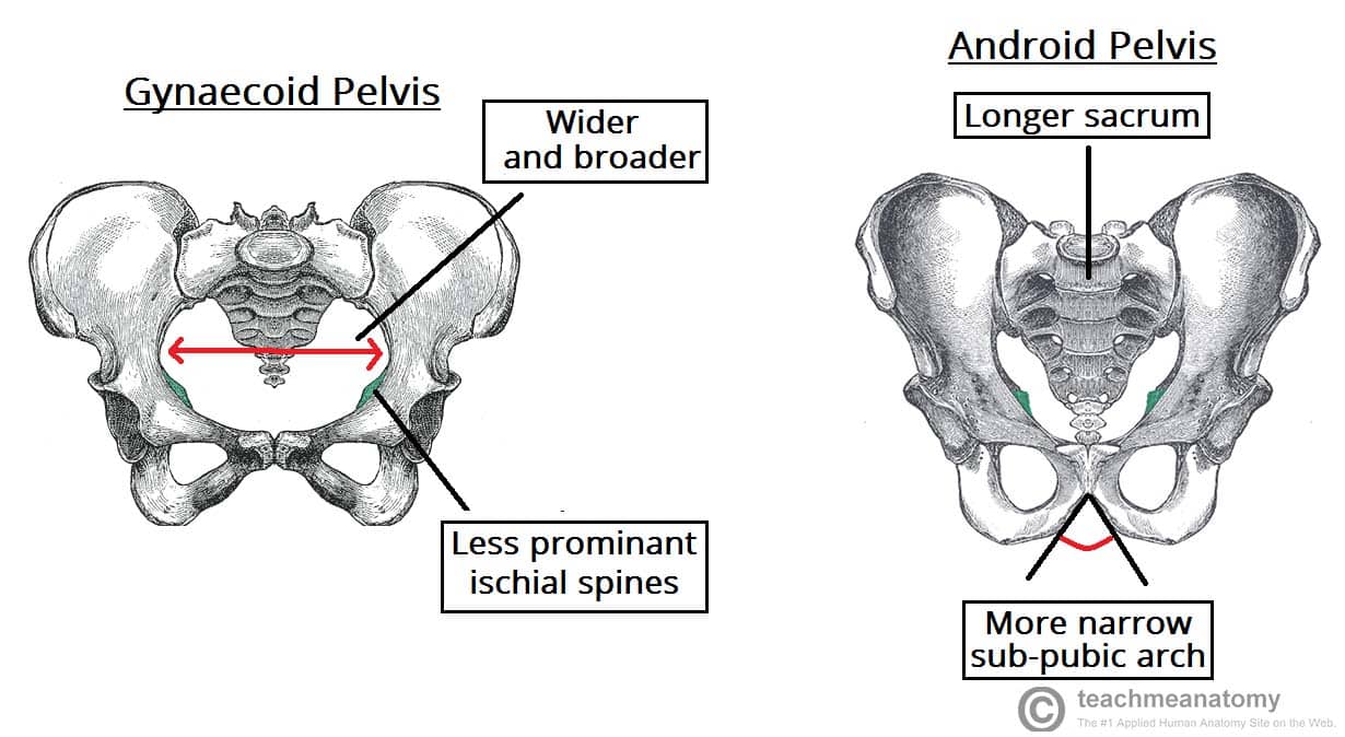
 Anatomy Of The Female Bony Pelvis Fetal Skull Ppt Video
 Pelvis Wikipedia
 8 3 The Pelvic Girdle And Pelvis Anatomy And Physiology
 Bony Pelvis
 Bony Pelvis Pelvic Cavity Part 1
 Pelvic Bone Anatomy Illustrated Bony Pelvis Stock
 Antenatal Care Module 6 Anatomy Of The Female Pelvis And
 Antenatal Care Module 6 Anatomy Of The Female Pelvis And
 Hip Joint
 Bony Pelvis Anatomy Website
 Bony Pelvis Anatomy Bone And Spine
 Image 1 Diagram Of Pelvis And Sacrum With Bony Landmarks
 Fig 7 27 Bones Of The Bony Pelvis Susuliban Flickr
 Anatomy Of Bony Pelvis
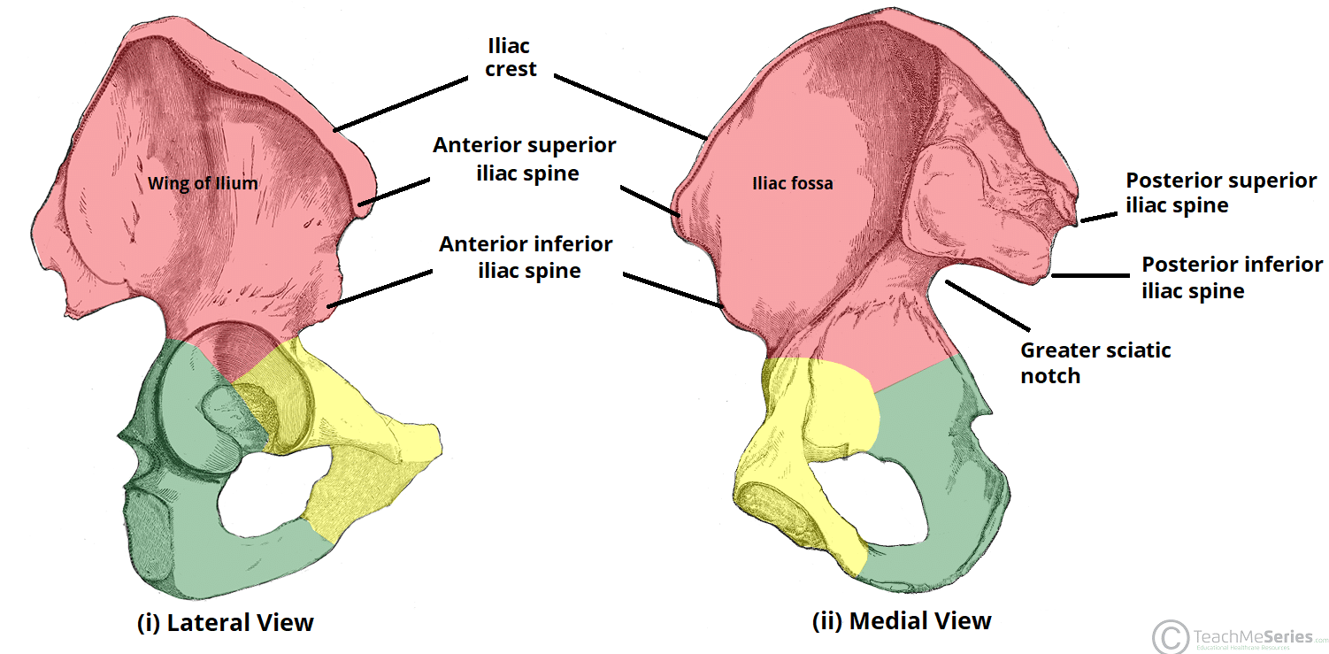 The Hip Bone Ilium Ischium Pubis Teachmeanatomy
 The Pelvic Girdle And Pelvis Anatomy And Physiology I
 Female Bony Pelvis And Fetal Skull For Undergraduate
 Pelvis Wikipedia
Female Pelvic Anatomy
 Bony Pelvis Radiology Reference Article Radiopaedia Org
 A Anatomy 1 Bony Pelvis Anatomy Pelvic Fracture And
The Pelvic Girdle Structure Function Assessment
 Bony Pelvis
 Bony Pelvis Anatomy Bone And Spine

 Pubis Bone Wikipedia
 Antenatal Care Module 6 Anatomy Of The Female Pelvis And
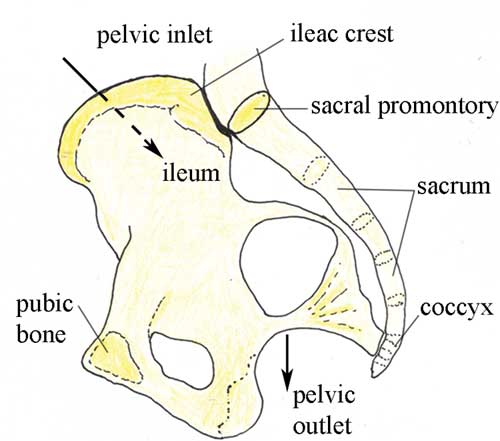
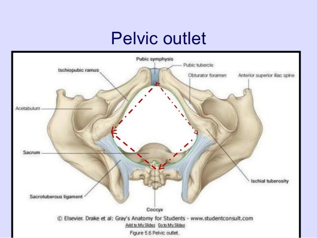
 Pelvic Inlet An Overview Sciencedirect Topics
 A Anatomy 1 Bony Pelvis Anatomy Pelvic Fracture And
 Bony Pelvis
 8 3 The Pelvic Girdle And Pelvis Anatomy And Physiology
 Bony Pelvis
Belum ada Komentar untuk "Anatomy Of Bony Pelvis"
Posting Komentar