Right Atrium Anatomy
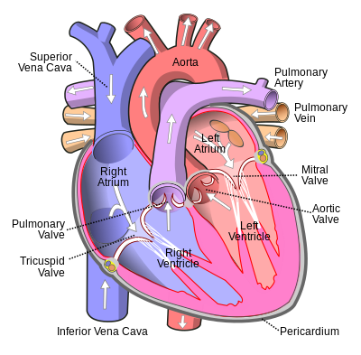
The right atrium contains the sinoatrial nodesa node which helps the heart in regulating its rhythm. Deoxygenated blood enters the right atrium through the inferior and superior vena cava.
 Cardiac Anatomy Radiology Key
The sa node is connected to the brain via the autonomic nerves which control the heart rate in maintaining blood pressure oxygen and carbon dioxide homoeostasis.
Right atrium anatomy. The atrium is the upper chamber through which blood enters the ventricles of the heart. Within the right atrium youve essentially got two spaces. All animals with a closed circulatory system have at least one atrium.
The atrium was formerly called the auricle. There are two atria in the human heart the left atrium receives blood from the pulmonary circulation and the right atrium receives blood from the venae cavae. The right atrium receives oxygen poor blood from three veins.
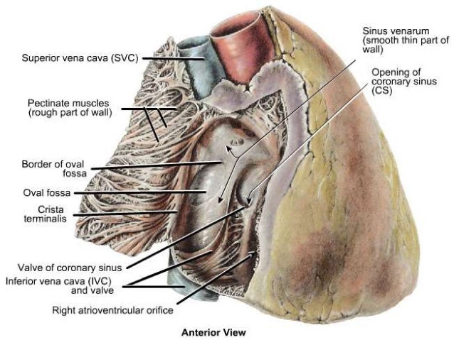
Internally this corresponds to the crista terminalis. Humans have two atria. The heart is comprised of two atria and two ventricles.
That term is still. The atrium is lined by pectinate muscles to the left of this crest and these extend into the right atrial appendage. It acts as a pacemaker and contracts the cardiac muscles.
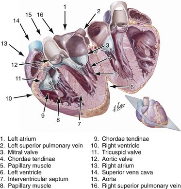
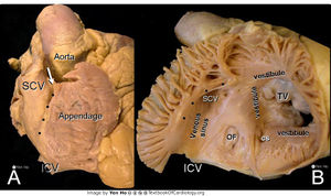
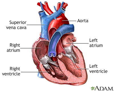
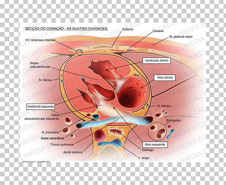
The superior vena cava inferior vena cava and coronary sinus figures 1 and 2. The right atrium is the receiving chamber for oxygen poor blood deoxygenated returning from the systemic circuit. The right atrium forms the entire right border of the human heart.
The inferior aspect of the right atrium is taken up by the inferior vena cava. Contains the sinoatrial node. Blood enters the heart through the two atria and exits through the two ventricles.
The right side of the heart then pumps this deoxygenated blood into the pulmonary arteries around the lungs. The upper part is made p by the right auricle which overlies the upper part of the atrioventricular groove. The atria receive blood while relaxed then contract to move blood to the ventricles.
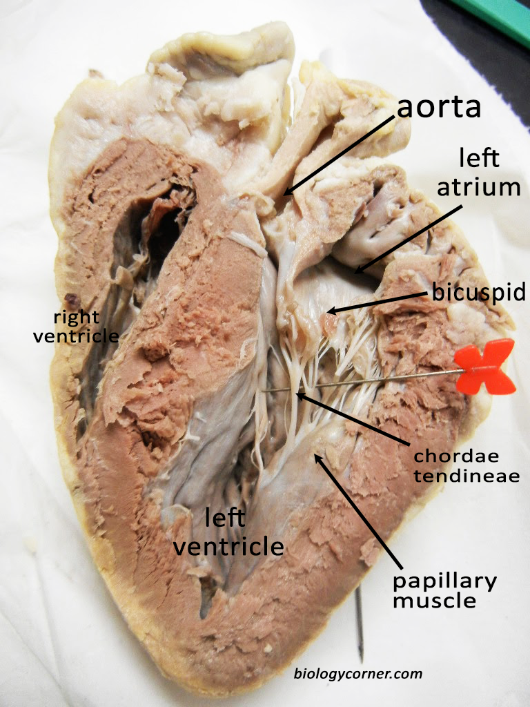
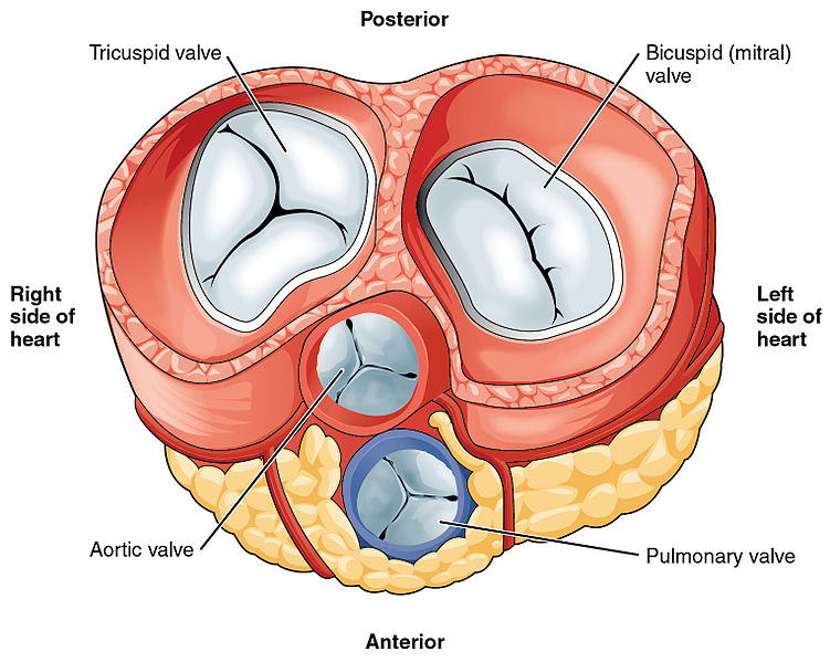
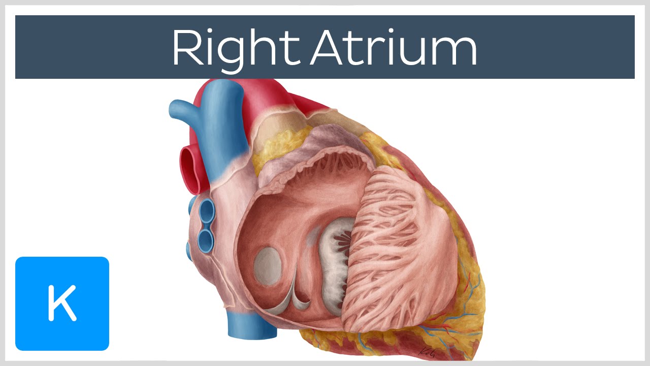
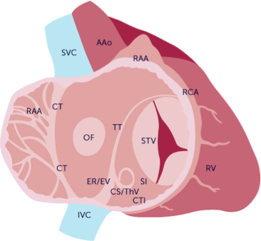
Deoxygenated blood entering the heart through veins from the tissues of the body first enters the heart through the right atrium before being pumped into the right ventricle. One of the main anatomic landmarks of the right atrium the crista terminalis is a muscular ridge on the anterior aspect of the chamber. The right atrium leads into the right ventricle through the tricuspid valve.
A groove is sometimes formed between the termination of the svc and the auricle the sulcus terminalis. The right atrium is located in the upper portion of right side of heart consisting of the sinus venosus and the right atrial appendage. The right atrium is one of the two atria of the heart which function as receiving chambers for blood entering the heart.
Theyre divided by something called the sulcus terminalis on the external surface of the heart. This structure separates the smooth posterior wall from the ridged muscular anterior wall. Right atrium atlas of human cardiac anatomy.
 Right Atrium Human Heart
:max_bytes(150000):strip_icc()/heart_inner_section-577d5c673df78cb62c939314.jpg) Atria Of The Heart Function
Atria Of The Heart Function
 Heart Anatomy Left Atrium And Ventricle
 Heart Detail Picture Image On Medicinenet Com
 Anatomy Of The Heart Textbook Of Cardiology
 Right Atrium And Right Ventricle
 Right Atrium The Anatomy Of The Heart Visual Atlas Page
 Cardiac Anatomy The Right Atrium Daily Med Fact
![]() Heart Right And Left Atrium Anatomy And Function Kenhub
Heart Right And Left Atrium Anatomy And Function Kenhub

 Cv Physiology Cardiac Anatomy
 Pulmonary Vein Wikipedia
 Heart Chambers Medlineplus Medical Encyclopedia Image
 Queensland Cardiovascular Group Anatomy Of The Heart
Interior Of Right Atrium
![]() Heart Right And Left Atrium Anatomy And Function Kenhub
Heart Right And Left Atrium Anatomy And Function Kenhub
 Right Atrium Location Anatomy Function Human Anatomy Kenhub
 Pediagenosis
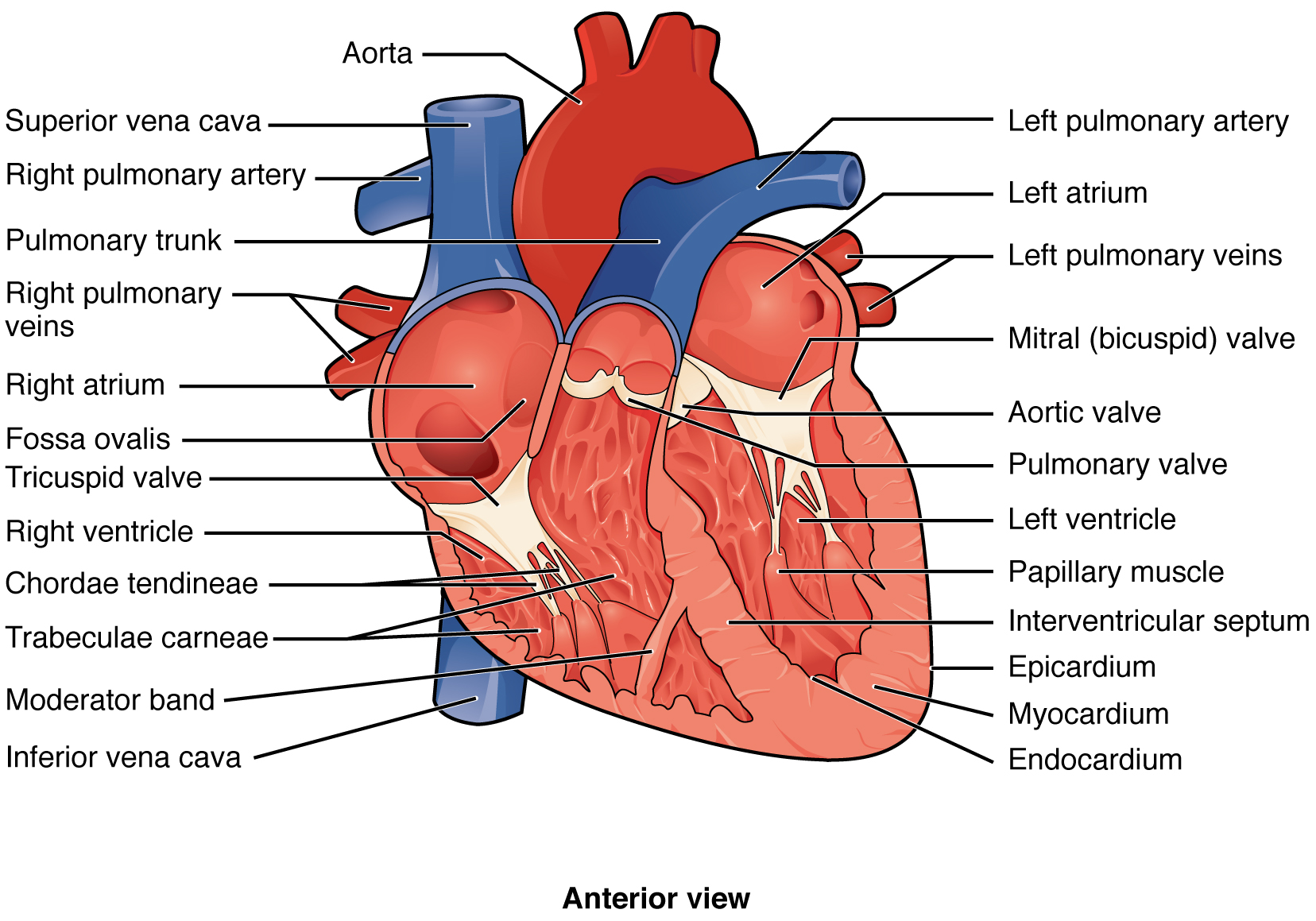
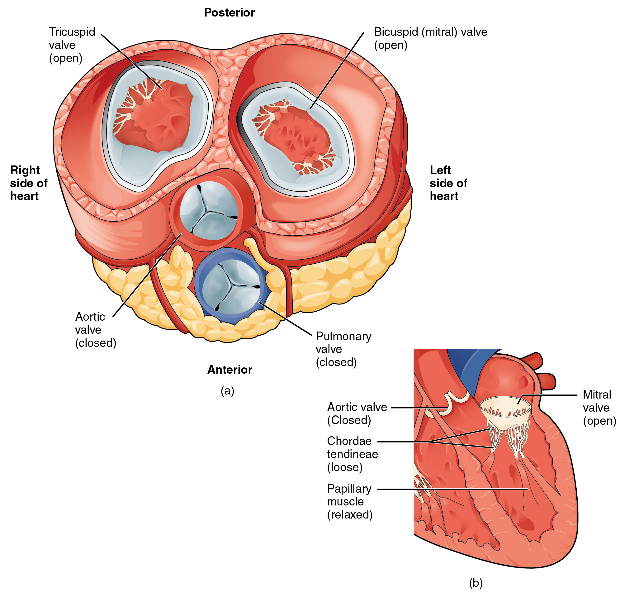 19 1 Heart Anatomy Anatomy And Physiology
 Heart Internal Features Anatomy Qa
![]() Heart Right And Left Atrium Anatomy And Function Kenhub
Heart Right And Left Atrium Anatomy And Function Kenhub
![]() Heart Ventricles Anatomy Function And Clinical Aspects
Heart Ventricles Anatomy Function And Clinical Aspects
 The Heart Human Anatomy
 Heart Anatomy Chambers Valves And Vessels Anatomy

 1 Heart Anatomy From The Anterior View Left And Interior
 Heart Anatomy Healthlink Bc
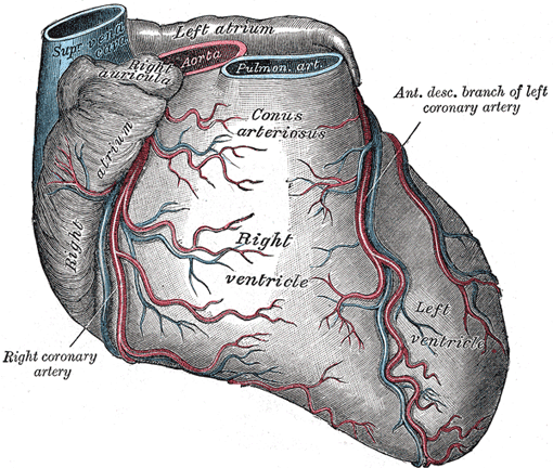
 1 Anatomy Of The Heart Showing Right Atrium Ra Right
 3 5 The Right Atrium 123sonography
 Heart Valve Right Atrium Aorta Anatomy Png Clipart Anatomy
 Trabeculae Carneae Wikipedia
 Normal Heart
Belum ada Komentar untuk "Right Atrium Anatomy"
Posting Komentar