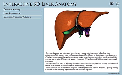
Ultrasound Anatomy Of Liver
This lecture is a part of basic radiologic anatomy series. Scan plane left lobe of liver.
Ultrasound Of The Liver
Left lobe of liver.

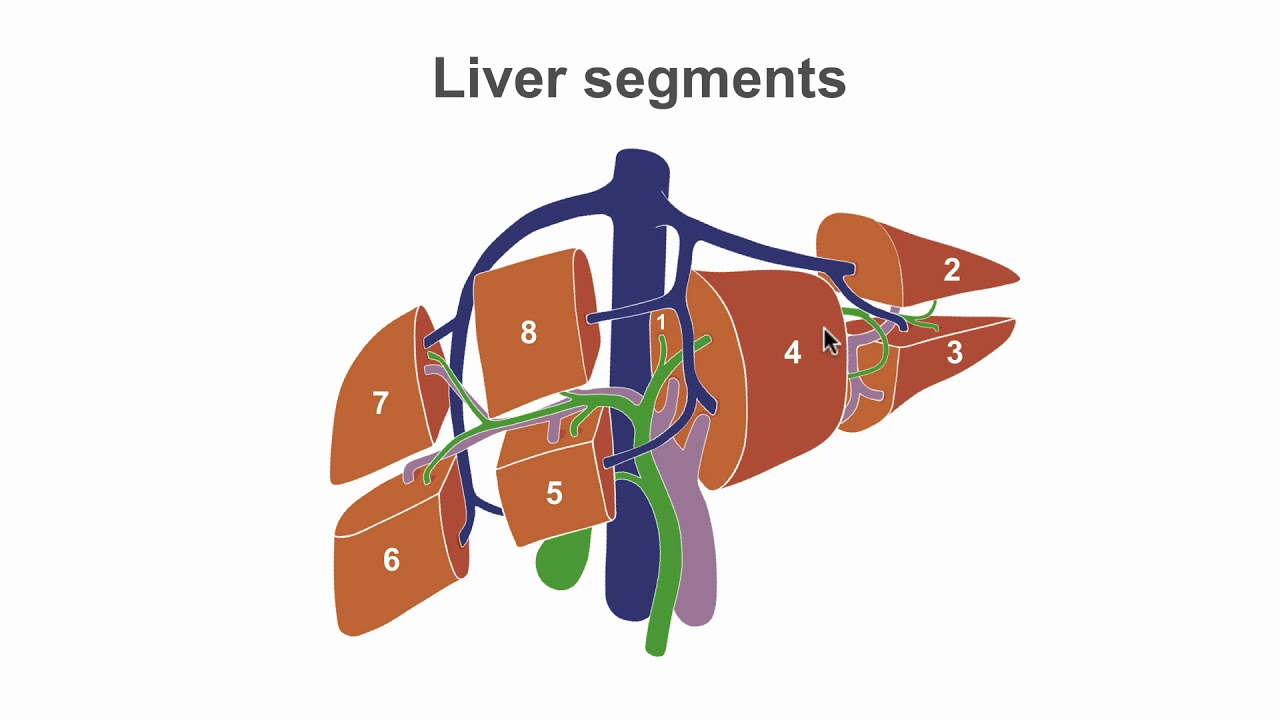
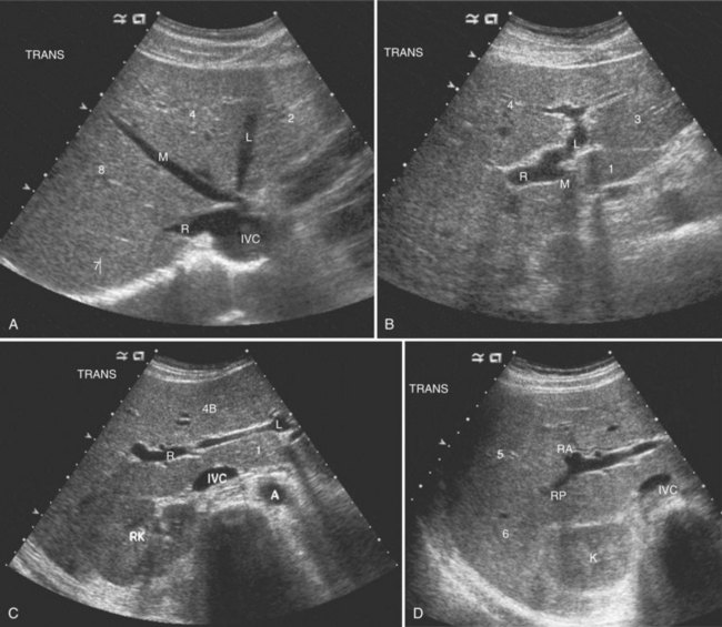
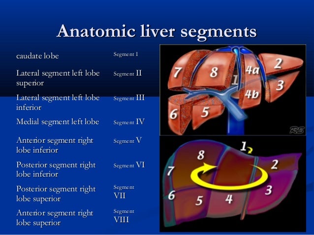
Ultrasound anatomy of liver. The couinaud classification pronounced kwee no is currently the most widely used system to describe functional liver anatomy. Local anaesthetic is injected on the mid axillary line where on percussion there is dullness. It is the preferred anatomy classification system as it divides the liver into eight independent functional units ter.
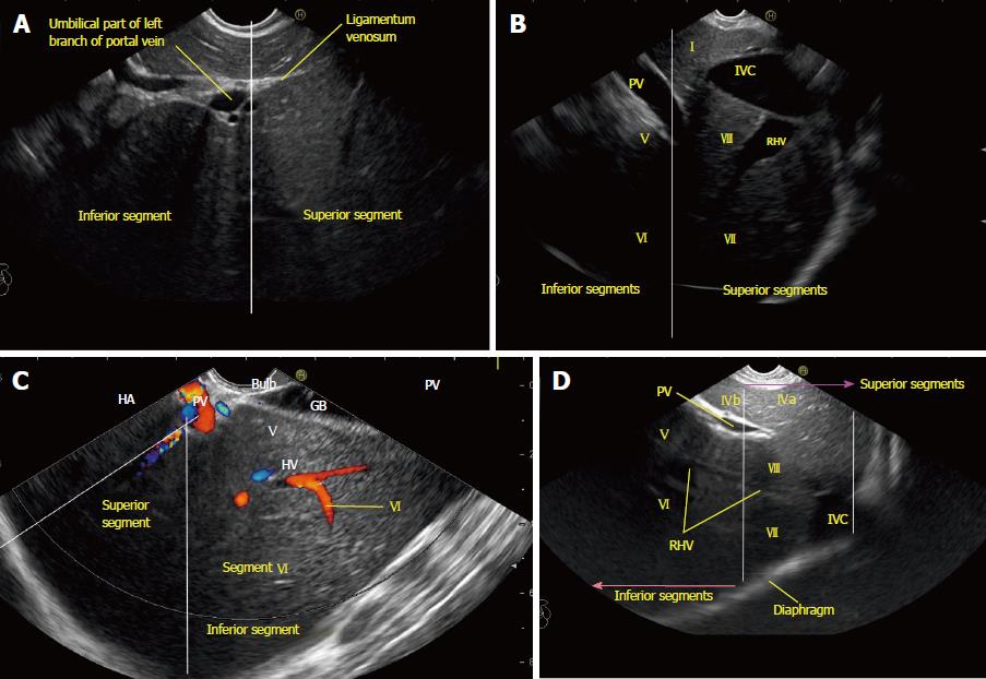
During the procedure the liver is located via ultrasound. Transverse view shows right rtleft ltand caudate cl lobes of the liver. An abdominal ultrasound can test for many liver conditions including cancer cirrhosis or problems from gallstones.
The lecture discussing the basic sonographic anatomy of the hepatobiliary system including normal anatomical variants and measurements. Figure 2 2 lobes of the liver. Liver ultrasound showing education liver segments normal liver anatomy portal vein hepatic veins the biliary tree and ultrasound scanning protocol worksheets googhywoiu9839t543j0s7543uw1.
Each segment has its own vascular inflow outflow and biliary drainage. The probe is in the epigastric region just below the sternum. The patient is asked to deeply expire avoiding damage to the lungs and the needle biopsy is taken during held expiration.
The couinaud classification of liver anatomy divides the liver into eight functionally indepedent segments. It is angled cephalad to view the left lobe in its entirety. Anechoic structures white arrows represent normal vesselsthe diaphragm black arrow is seen superiorly.
The echo reflection pattern of the liver is similar to or slightly higher than that of the renal cortex. The liver allows for effective ultrasound imaging. The probe may need to be angled towards the left side to see the most medial edge of the left lobe.
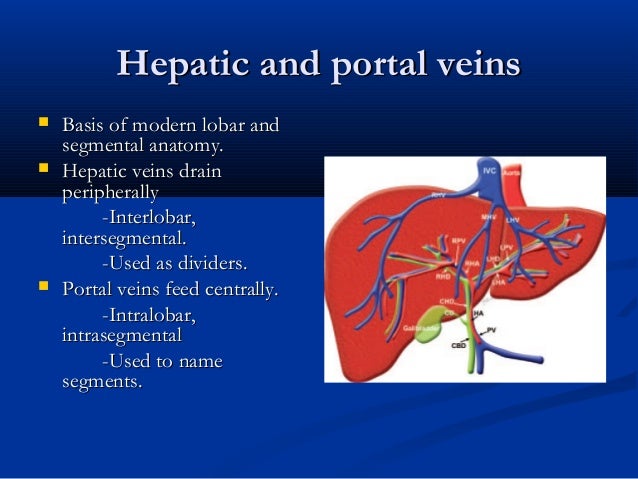
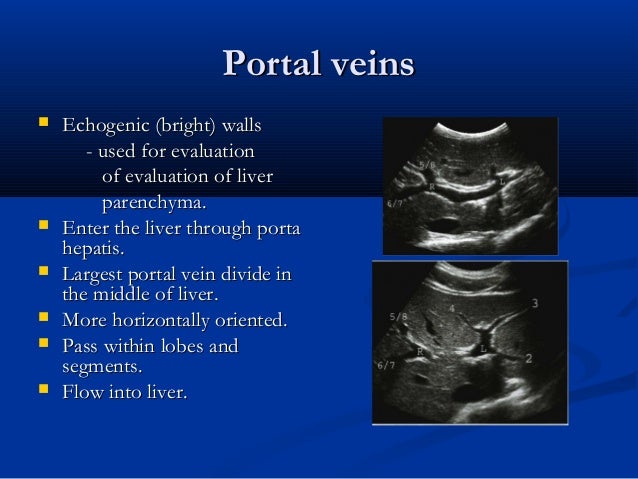
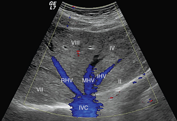
Normal anatomy seen in the transverse view of the left lobe. A longitudinal sonogram demonstrates a homogeneous liver with midlevel echoes. In the centre of each segment there is a branch of the portal vein hepatic artery and bile duct.
A healthy liver has a homogeneous echo reflection pattern and smooth contours. Despite their limitations and the difficulties that can arise serial ultrasound studies of the liver are an inevitable part of current therapies liver transplant tips radiofrequency ablation procedures etc especially in critically ill patients.
 Couinaud Classification Of Hepatic Segments Radiology
 Mastering Liver Anatomy Before The Ultrasound
![]() Us Study Of The Liver Longitudinal And Transverse Scan
Us Study Of The Liver Longitudinal And Transverse Scan
 Long Liver Ultrasound Sonography Ultrasound Technician
 Liver Ultrasound

 Stepwise Evaluation Of Liver Sectors And Liver Segments By
 Ultrasound Of Liver Segments Anatomy Ultrasound Vascular
 Boundaries Between Subsegments Iva And Ivb In The Human
 Liver Ultrasound
 Chapter 7 Hepatobiliary Ultrasound Surgical And
Ultrasound Of The Liver
 Hepatic Vein An Overview Sciencedirect Topics
Ultrasound Of Liver Segments Anatomy
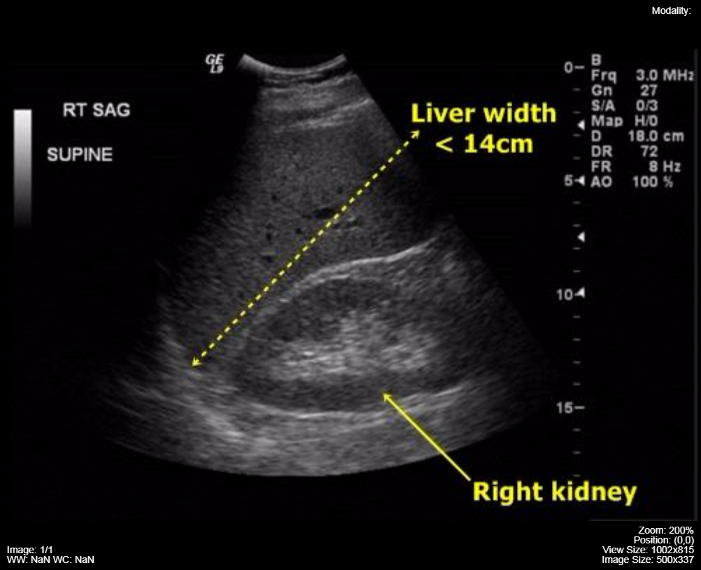
 Liver Span Wikipedia
 Startradiology
 3 8 Ultrasound Us Medicine Libretexts
 Ultrasound Of Liver Segments Anatomy
 Startradiology

 Ultrasound Of The Liver Biliary Tract And Pancreas
 Visual Guide To Liver Cancer
 Liver Anatomy And Segments By Ultrasound In Arabic
 Liver Ultrasound
 Hepatobiliary Ultrasound Radiology Key

 Ultrasound Of Liver Segments Anatomy

Belum ada Komentar untuk "Ultrasound Anatomy Of Liver"
Posting Komentar