Bony Anatomy Of Foot
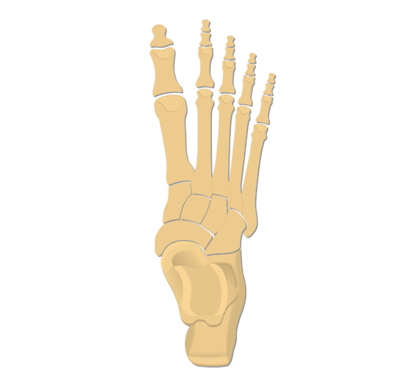
The bones of the foot are organized into the tarsal bones metatarsal bones and phalanges. The talus bone is the second largest bone in the entire foot and unlike the rest bones there is no attachment of muscles.
Human Being Anatomy Skeleton Foot Image Visual
Learn this topic now at kenhub.
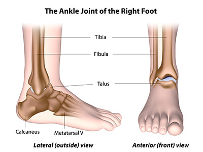
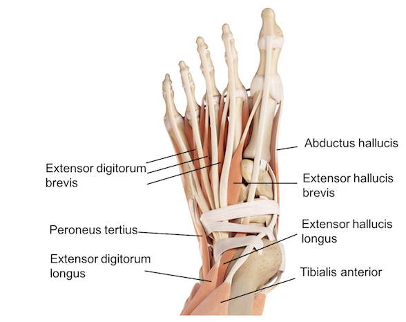
Bony anatomy of foot. Muscles tendons and ligaments run along the surfaces of the feet allowing the complex movements needed for motion and balance. This is an article covering the articular surfaces ligaments and muscles that produce movement at the joints of the feet. These make up the toes and broad section of the feet.
The foot is responsible for balancing the bodys weight on two legs a feat which modern roboticists are still trying to replicate. The calcaneus heel bone is the largest bone in the foot. The bones of the foot are organized into rows named tarsal bones metatarsal bones and phalanges.
Bones and main ligaments of the foot. The foot begins at the lower end of the tibia and fibula the two bones of the lower leg. The tarsal bones are 7 in number.
The end of the leg on which a person normally stands and walks. This may sound like overkill for a flat structure that supports your weight but you may not realize how much work your foot does. Tarsals five irregularly shaped bones of the midfoot that form the foots arch.
The calcaneus is the largest of the tarsal bones located in the heel of the foot and bears the weight of the body as the heel hits the ground. Tarsal bones gross anatomy. Talus the bone on top of the foot that forms a joint with the two bones of the lower leg the tibia and fibula.
At the base of. It forms the lower part of the ankle formed collectively by the tibia fibular and talus bones. The foot is an extremely complex anatomic structure made up of 26 bones and 33 joints that must work together with 19 muscles and 107 ligaments to execute highly precise movements.
The bones of the feet are. The foot contains 26 bones 33 joints and over 100 tendons muscles and ligaments. The other bones of the foot that create the.
Calcaneus the largest bone of the foot which lies beneath the talus to form the heel bone. They are named the calcaneus talus cuboid navicular and the medial middle and lateral cuneiforms. This is an article covering the muscle attachments blood supply innervation and ossification of the phalanges of the foot.
:background_color(FFFFFF):format(jpeg)/images/library/11040/537_m_lumbricales_fkt.png) Ankle And Foot Anatomy Bones Joints Muscles Kenhub
Ankle And Foot Anatomy Bones Joints Muscles Kenhub
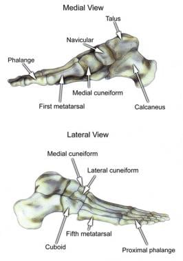 Foot Bone Anatomy Overview Tarsal Bones Gross Anatomy
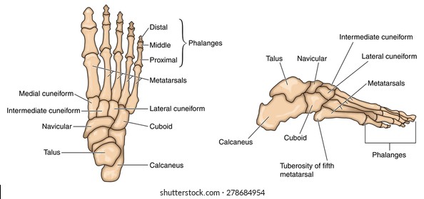 Foot Bones Photos 40 827 Foot Stock Image Results
 Foot Anatomy Detail Picture Image On Medicinenet Com
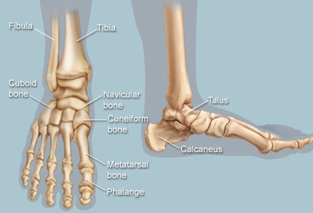 Feet Human Anatomy Bones Tendons Ligaments And More
Anatomy Of The Foot And Ankle Orthopaedia
 Bones Of The Foot Illustrations Foot Anatomy Illustrations
:watermark(/images/watermark_5000_10percent.png,0,0,0):watermark(/images/logo_url.png,-10,-10,0):format(jpeg)/images/atlas_overview_image/807/FbJOmFqYRQYurlWMoIubQ_foot-bones-and-ligaments_english.jpg) Diagram Pictures Bones Of The Foot Anatomy Kenhub
Diagram Pictures Bones Of The Foot Anatomy Kenhub
 The Foot Advanced Anatomy 2nd Ed
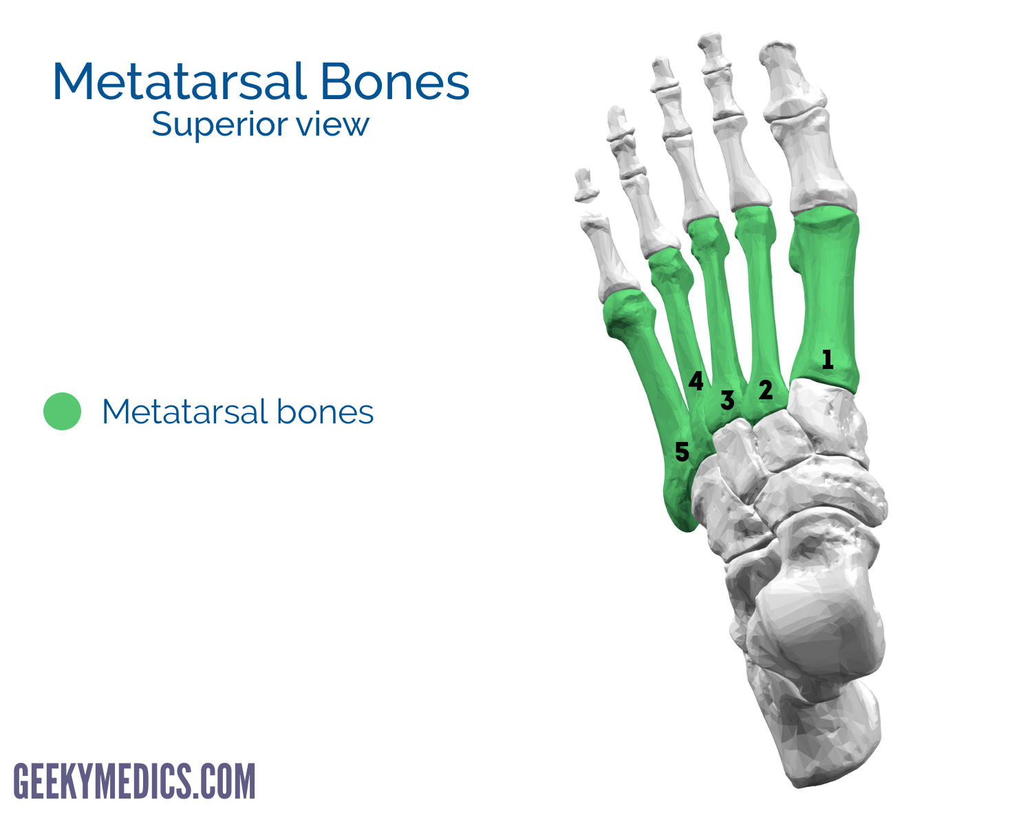 Bones Of The Foot Tarsal Bones Metatarsal Bone Geeky
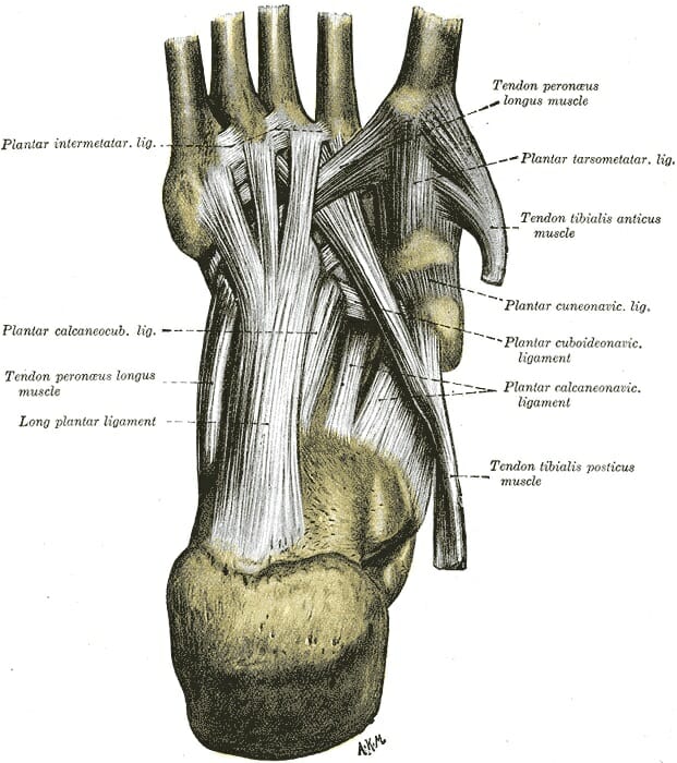 Foot Anatomy Bones Ligaments Muscles Tendons Arches
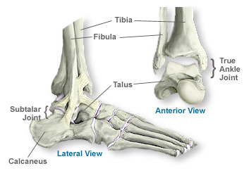 Anatomy Of The Ankle Southern California Orthopedic Institute
 Bones Of Foot Human Anatomy The Diagram Shows The Placement
 Foot Bones Anatomy And Mnemonic Tarsals Metatarsals
 Bones Of The Foot Illustrations Foot Anatomy Illustrations
 Ankle Cartilage Preservation Towson Orthopaedic Associates
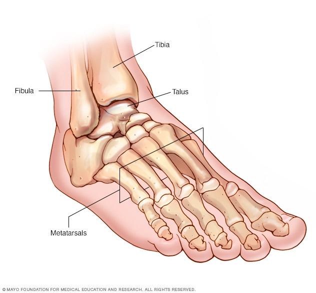 Foot And Ankle Bones Mayo Clinic
Human Being Anatomy Skeleton Foot Image Visual
 Tarsal Metatarsal And Phalanges Of The Foot
 Ball Of Foot Pain Do The Bottoms Of Your Feet Toes Hurt



Belum ada Komentar untuk "Bony Anatomy Of Foot"
Posting Komentar