Internal Jugular Anatomy
The sigmoid sinus arises in the posterior cranial fossa and exits the cranium through the jugular foramen located at the base of the skull. Internal jugular vein anatomy and function inferior petrosal and sigmoid dural venous sinuses which are venous channels located between the layers of the dura mater the outermost membrane that envelops the brain and the spinal cord unite to form the internal jugular veins.
 Common Carotid Artery L Common Carotid Artery L Internal
The internal jugular vein is a continuation of this system downward through the neck.
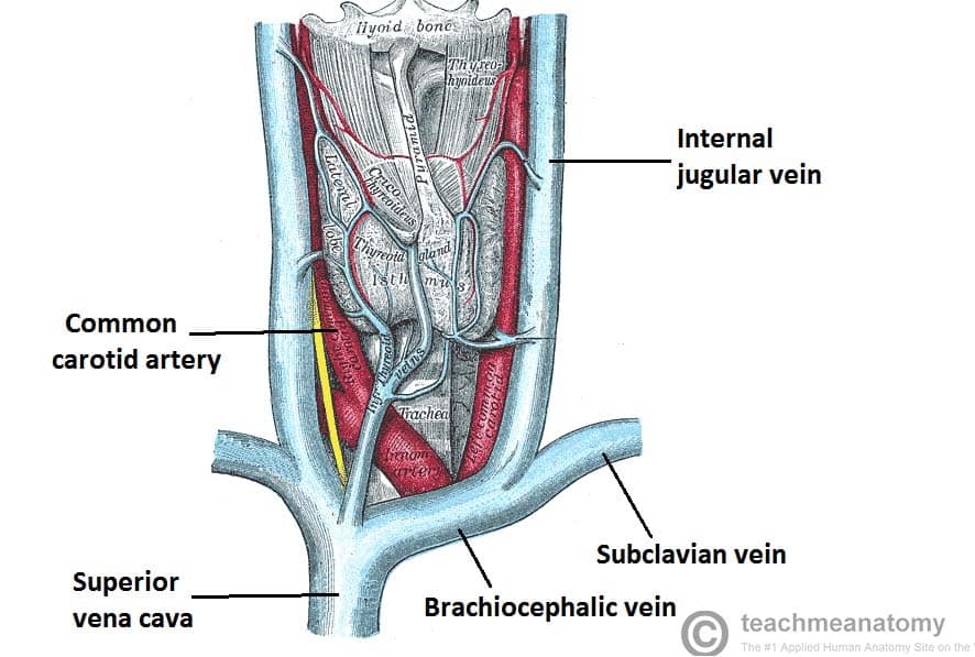
Internal jugular anatomy. The vein runs in the carotid sheath with the common carotid artery and vagus nerve. The internal jugular vein is a paired jugular vein that collects blood from the brain and the superficial parts of the face and neck. It receives blood from parts of the face neck and brain.
The internal jugular vein ijv is the major venous return from the brain upper face and neck. The internal jugular vein functions as a guide for surgeons during removal of deep cervical lymph nodes. Gross anatomy origin and course.
Khatri md sam wagner sevy rvt rdcs manuel h. They each rest beside the thyroid gland at the center of the neck just above the collarbone and near the trachea or windpipe. The facial or common facial vein is the most essential tributary of the internal jugular vein for it acts as a useful landmark in the removal of the jugulodigastric tonsillar and upper anterior group of deep cervical lymph nodes.
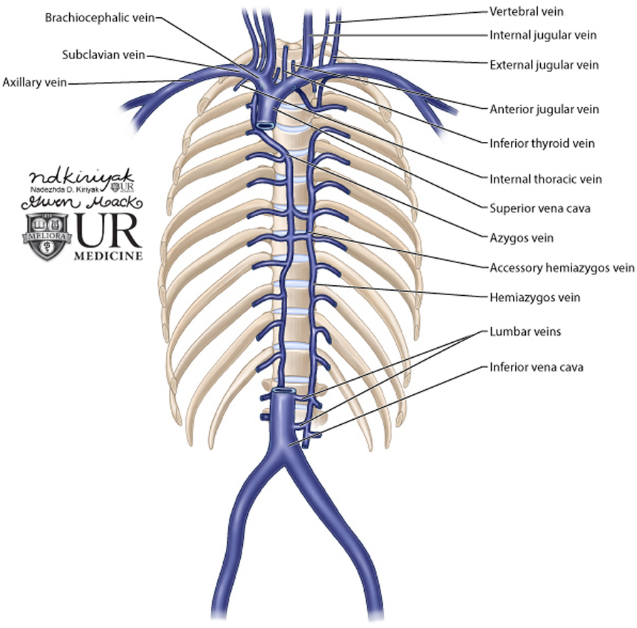
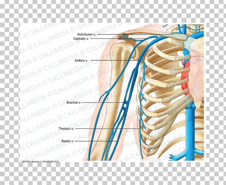
At approximately the level of the collarbone each unites with the subclavian vein of that side to form the innominate veins. The internal jugular vein is a run off of the sigmoid sinus. It descends in the carotid sheath with the internal carotid artery.
The internal jugular vein is a major blood vessel that drains blood from important body organs and parts such as the brain face and neck. It is formed by the union of inferior petrosal and sigmoid dural venous sinuses in or just distal to the jugular foramen forming the jugular bulb. A prospective study vijay p.
The internal jugular vein maintains its regional anatomy and patency after carotid endarterectomy. Anatomically there are two of these veins that lie along each side of the neck. Espinosa md and jay b.
As the internal jugular vein runs down the lateral neck it drains the branches of the facial retromandibular and the lingual veins. The internal jugular veins lie deeper and are larger than the external jugular veins which lie closer to the surface.
 Left Internal Jugular Vein The Anatomy Of The Veins Visu
 External And Internal Jugular Vein
 Blood Finds A Way Pictorial Review Of Thoracic Collateral
Useful Notes On The Internal Jugular Vein Of Human Neck

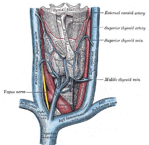 Figure Veins And Arteries Of The Statpearls Ncbi
 Central Venous Access Radiology Key
 Subclavian Vein Internal Jugular Vein External Jugular Vein
 Are The Jugular Vein And Carotid Artery Present On Both
 Internal Jugular Vein Wikipedia
 Venous Drainage Of The Head And Neck Dural Sinuses
 Figure 1 From Ultrasound Guidance For Internal Jugular Vein
Internal Jugular Vein Stepwards
 Misplaced Central Venous Catheters Applied Anatomy And
 The Neck Anatomy An Essential Textbook 1st Ed

Cerebral Oxygenation Monitoring And Imaging Part 1
 Internal Jugular Vein Canvas Prints Page 14 Of 16 Fine
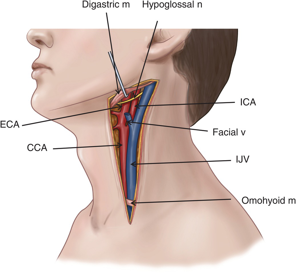 Carotid Artery And Internal Jugular Vein Injuries Chapter 8
 Internal Jugular Vein Diagram And Functions
 Solved 4 Label The Specific Veins Bringing Blood Back Fr
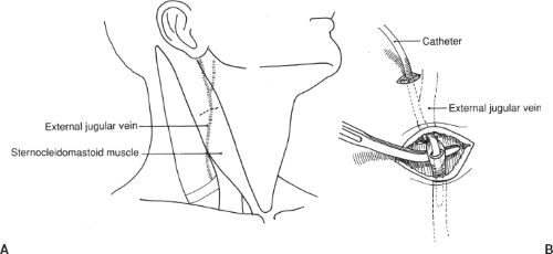 Venous Access External And Internal Jugular Veins
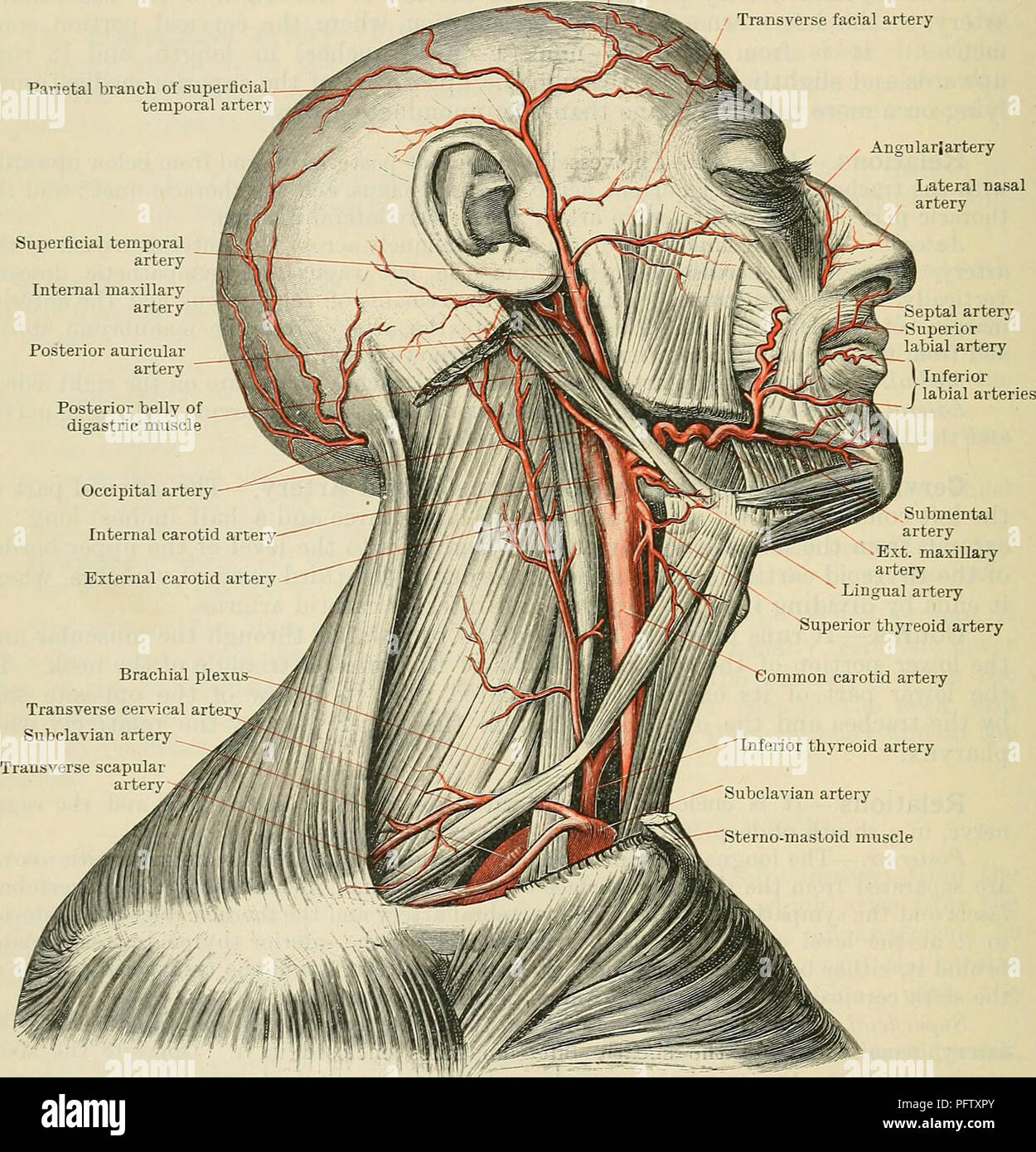 Cunningham S Text Book Of Anatomy Anatomy 890 The Vasculae
Internal Jugular Vein Cardiovascular Anatomyzone


Belum ada Komentar untuk "Internal Jugular Anatomy"
Posting Komentar