Fetal Anatomy Ultrasound
In some cases the baby may have their legs crossed or be facing away from the abdomen and thus the sexual organs will not be visible during the anatomic ultrasound. Those who want to can find out the sex of the baby if desired.

Sectional fetal anatomy in ultrasound the basis for a sufficient ultrasound examination in obstetric diagnosis is a knowledge of the normal systematic and sectional topographic anatomy of the fetus.
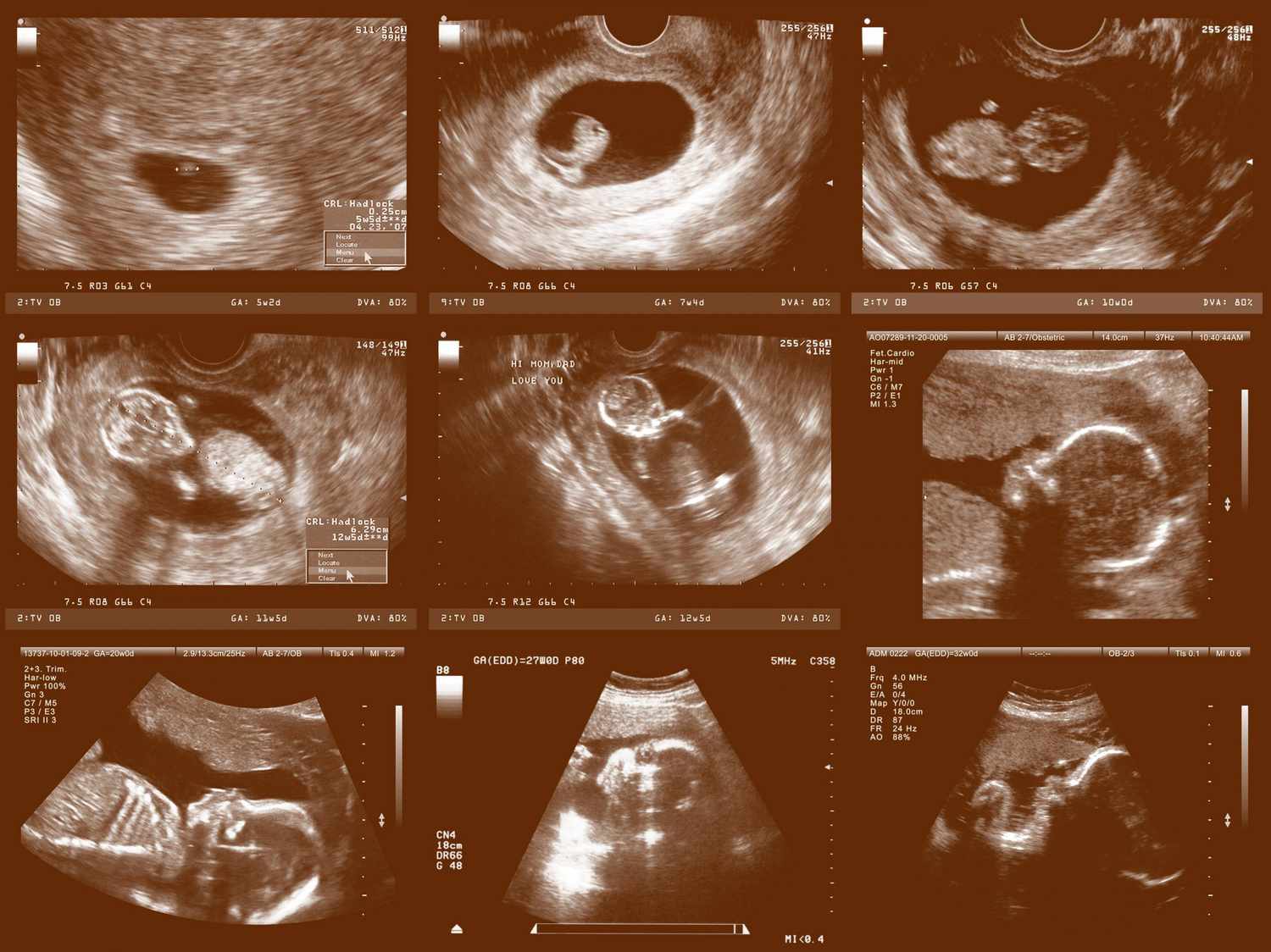
Fetal anatomy ultrasound. By the 20th week of pregnancy the baby can weigh up to 11 ounces and measure 10 inches outstretched. 9 1 and abdominal fig. The second trimester scan is a routine ultrasound examination in many countries that is primarily used to assess fetal anatomy and detect the presence of any fetal anomalies.
A fetal ultrasound sonogram is an imaging technique that uses sound waves to produce images of a fetus in the uterus. Fetal ultrasound images can help your health care provider evaluate your babys growth and development and monitor your pregnancy. This sonogram is used to determine fetal anomalies the babys size and weight and also to measure growth to ensure that the fetus is developing properly.
Not the least of these has been the surge in ultrasonic imaging of premature neonates12 these tiny neonates are the equivalent of second trimester fetuses as early at times as 24 weeks. When the pregnancy hits the 20th week of gestation an anatomy ultrasound is often ordered. Continued advancement of ultrasound technology including increase in frequency and choice of focal position have improved visualization of fetal anatomy and therefore have also increased the required anatomic knowledge of these structures for those performing and interpreting fetal sonograms.
In some cases fetal ultrasound is used to evaluate possible problems or help confirm a diagnosis. The anatomy scan is a level 2 ultrasound which is typically performed on pregnant women between 18 and 22 weeks. The second trimester extends from 13 weeks and 0 days to 27 weeks and 6 days of gestation although the majority of these studies are performed between 18 and 23 weeks.
Determining fetal sex the gender of your babybabies can usually be determined at this ultrasound. Medical books sectional fetal anatomy in ultrasound. 9 ultrasound evaluation of normal fetal anatomy.
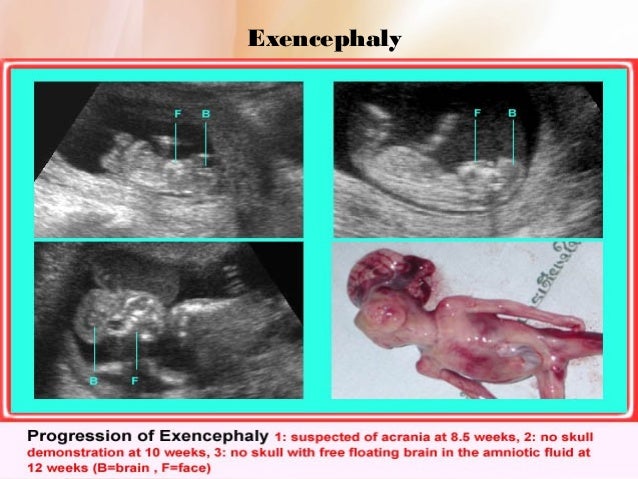
Visualization of their brain 12 fig.
 Fetal Lateral Ventricle Measurements How To Measure Posterior Ventricle For Ventriculomegaly
 Anomaly Scan Wikipedia
 A Gallery Of High Resolution Ultrasound Color Doppler 3d
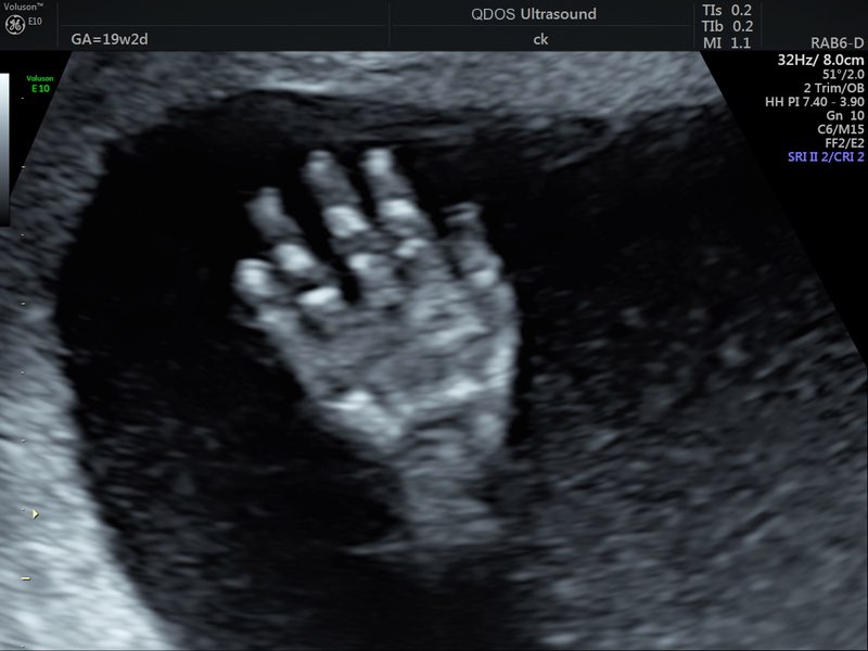 19 Week Scan Perth Mid Trimester Ultrasound Pregnancy
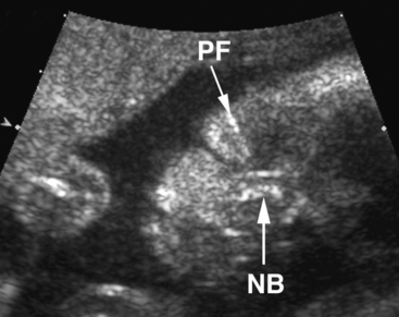 Ultrasound Evaluation Of Normal Fetal Anatomy Radiology Key
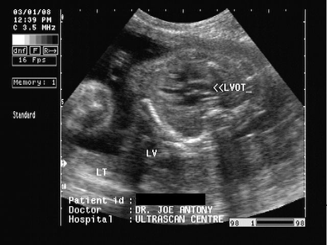
 Image Result For Fetal Heart Outflow Tracts Ultrasound
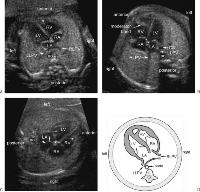 Ultrasound Evaluation Of The Fetal Heart Radiology Key

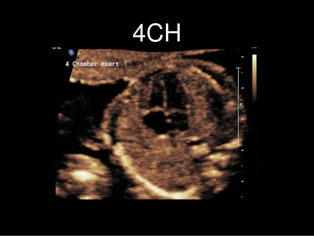
 The Basic Fetal Heart Scan Youtube
 Normal Fetal Anatomy Fetal Aorta Ultrasound Services In
 Routine Fetal Anatomy Scan At 18 23 Weeks
 All About The 20 Week Ultrasound Parents
 Labelled Fetal Heart Ultrasound
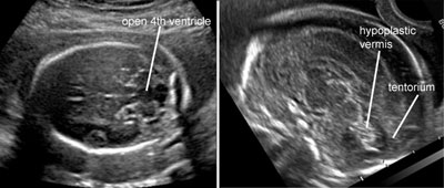
 Ultrasound Of The Fetus At 11 To 18 Weeks Fleischer S
 Ultrasound Atlas Glowm
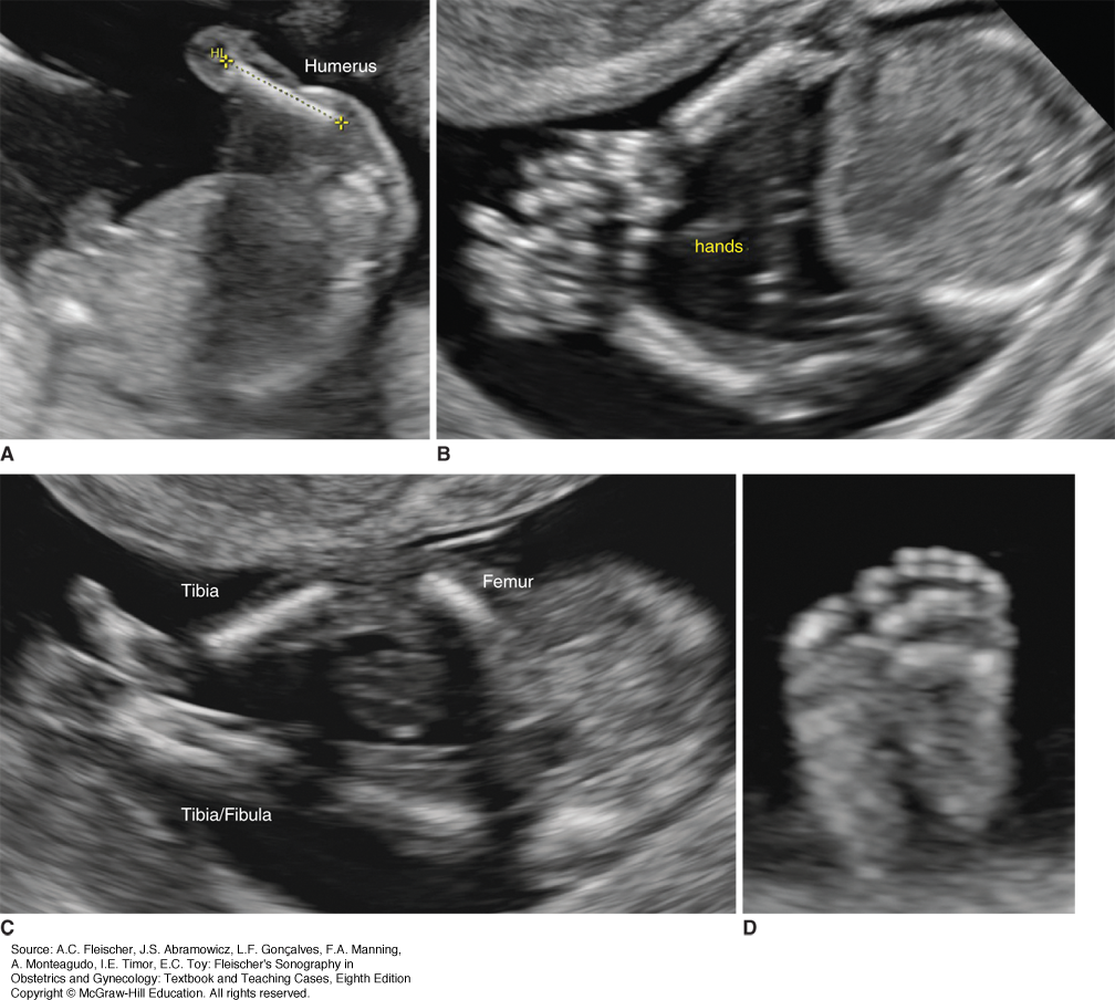 Ultrasound Of The Fetus At 11 To 18 Weeks Fleischer S
 Second Trimester Ultrasound Scan Radiology Reference
 Why More Women Should Decline Their First Trimester

 What Is An Anatomy Ultrasound During Pregnancy Babymed Com
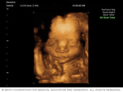 Fetal Ultrasound Mayo Clinic
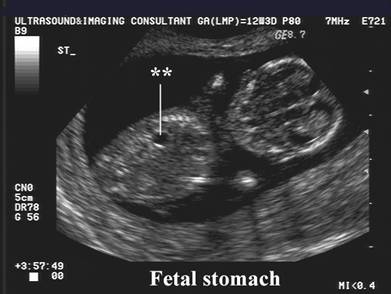
 Ultrasound Of The Fetus At 11 To 18 Weeks Fleischer S
 Fetal Heart Ultrasound How To

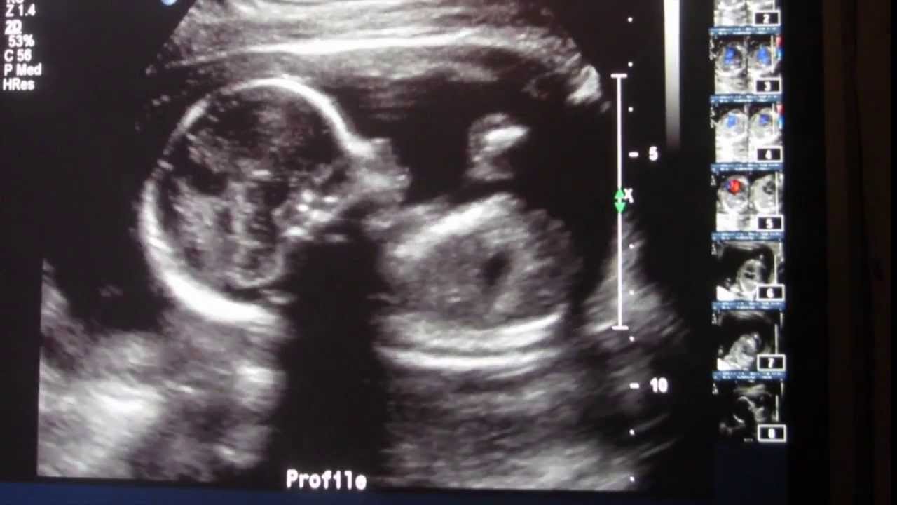
Cute Clear Ultrasound Of Baby Lucky 19 Weeks Ultrasound Anatomy Scan
 Fetal Heart Ultrasound How To
Belum ada Komentar untuk "Fetal Anatomy Ultrasound"
Posting Komentar