Pelvic X Ray Anatomy
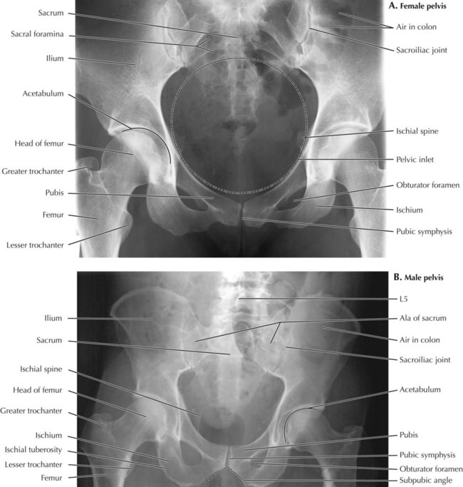
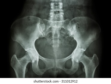
An x ray is a common imaging test that has been used for decades to help doctors view the inside of the body without having to open it up using surgery. Ap view of pelvis showing important anatomical lines the five bones that comprise the pelvis are the ilium ischium pubis sacrum and coccyx.
 X Ray Of The Pelvis Purpose Procedure And Risks
Anatomy the bony pelvis comprises the two hemi pelvis bones which are bound anteriorly at the pubic symphysis and posteriorly at the sacroiliac joints.

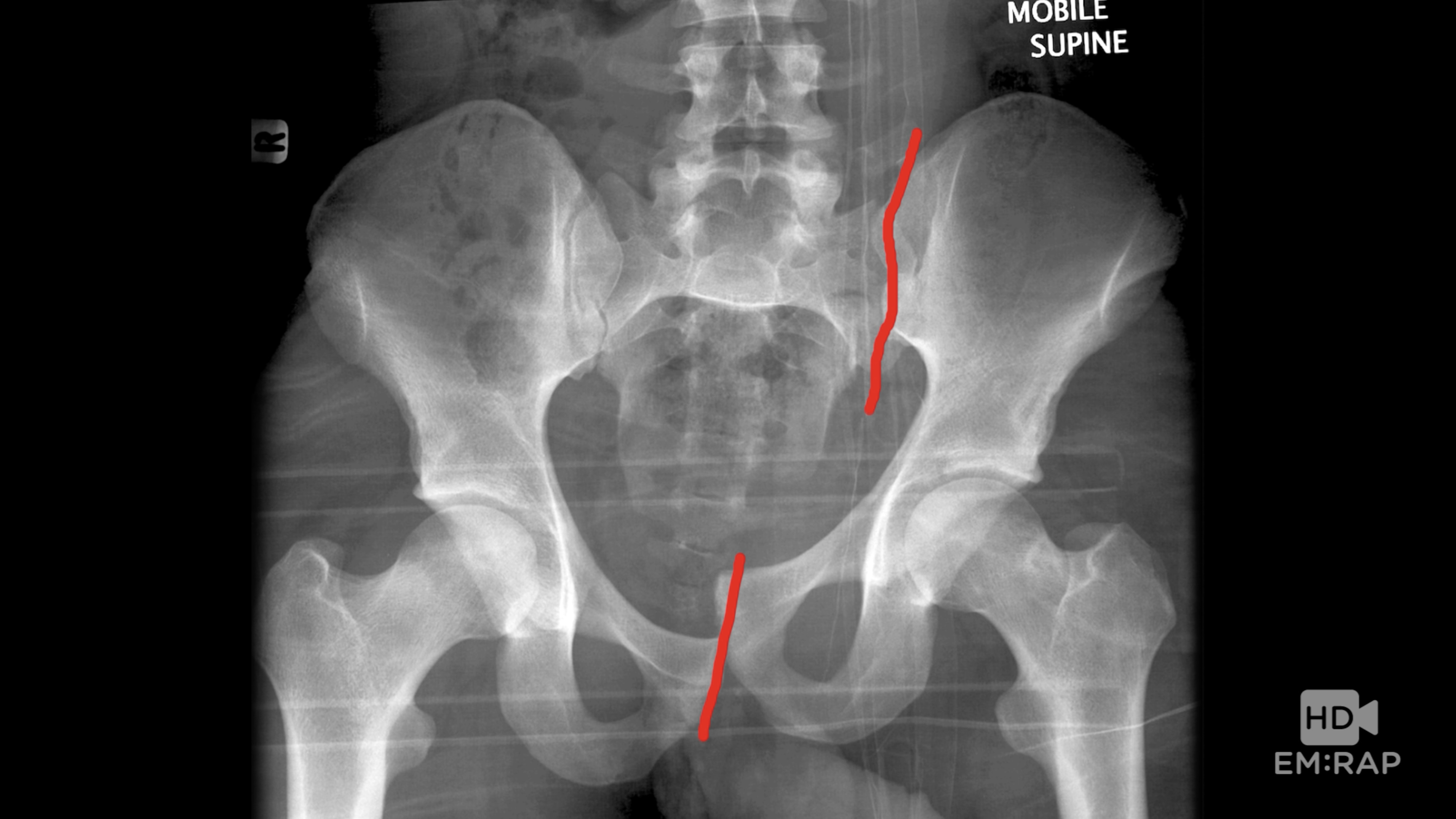
Pelvic x ray anatomy. The bony pelvis is a ring structure in which isolated fractures are rare. When reviewing a pelvic x ray detection of a single break in the ring mandates search for a second injury. Interpretation of the pelvic x ray.
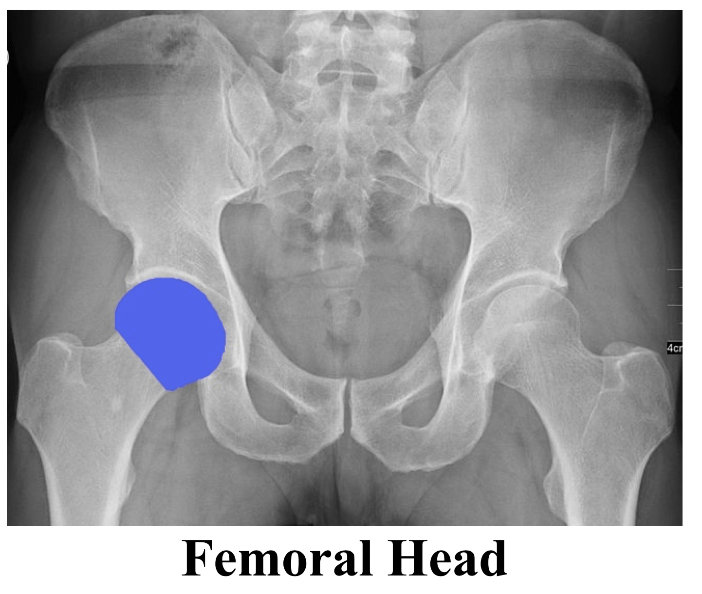
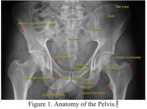
Pelvis x ray anatomy in this image you will find the sacroiliac joint acetabular obturator foramina greater trochanter pubic symphysis femoral heads lesser trochanters in it. What is an x ray of the pelvis. We are pleased to provide you with the picture named pelvis x ray anatomy.
As with other anatomical bone rings if a fracture is seen in one place a careful check should be made for a second fracture or for disruption of the pubic symphysis or sacroiliac joints. Mands thorough break down of this commonly used ed diagnostic the pelvic xr. Pelvic xrays are a key component of trauma fractures and dislocations seen every day in the ed but when is the last time you went back over the anatomy and radiographic tips and tricks of the pelvic radiograph.
Most trauma to the pelvis and hips can be evaluated with an ap projection of the pelvis and hips.
 Pelvis And Perineum Radiology Key

 Back To Basics Pelvic Xrays Taming The Sru
 Abdomen X Ray Annotated Radiology Case Radiopaedia Org
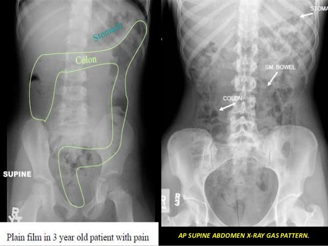 Presentation1 Pptx Radiological Anatomy Of The Abdomen And
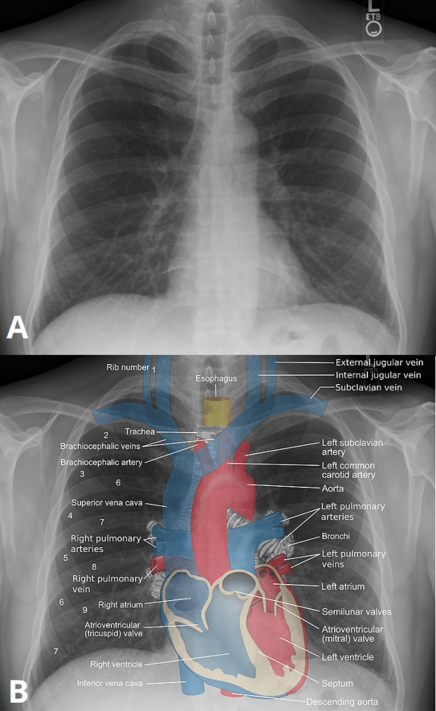 Plain Film X Ray Principles Interpretation Teachmeanatomy
 X Ray Pelvis Images Stock Photos Vectors Shutterstock
 Ao Surgery Reference
 Radiographs Of The Dog
 Pelvic Ring Fractures Trauma Orthobullets
 Film Critique Of The Lower Extremity Part 1
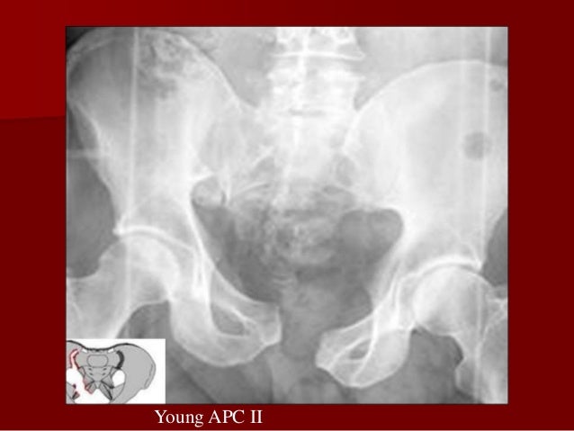
 Hd Pelvic Fractures Em Rap
 Film Critique Of The Lower Extremity Part 1
 Pelvis Acetabulum Anatomy Imaging Classification
 Ao Surgery Reference
 Labeled Radiographic Anatomy Of The Male Bottom Image And
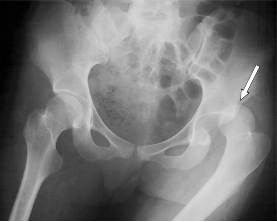 Dislocated Hip Symptoms Diagnosis And Treatments Hss
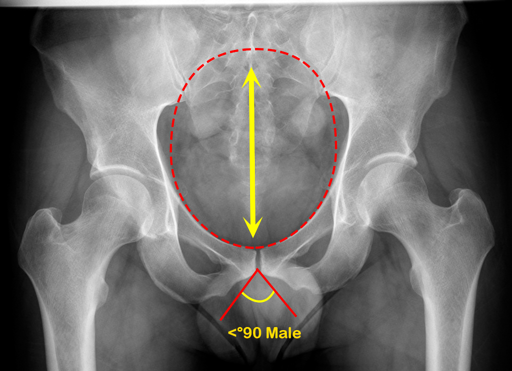 Normal Pelvis Gender Differences Radiology Case
Startradiology
 Radiographs Of The Dog
 X Ray Pelvis Labeling Questions Radiology Case
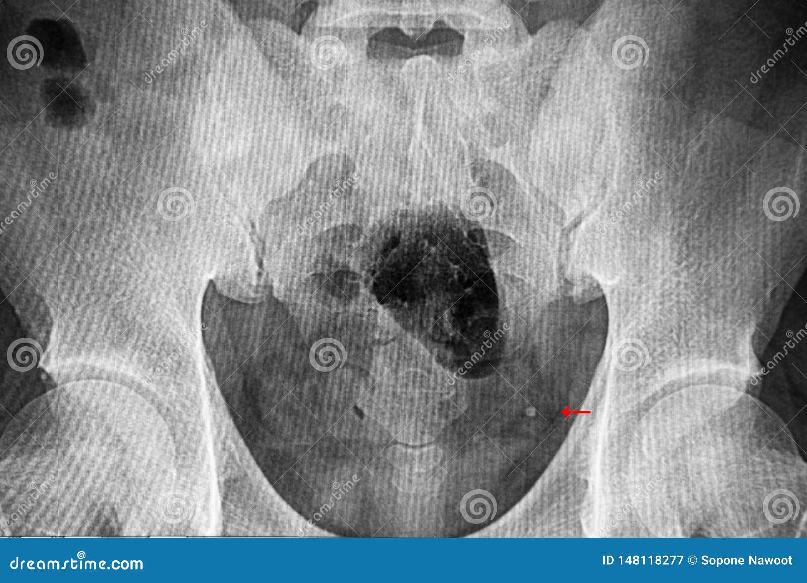 Pelvic Xray Film Showing Distal Ureterc Stone Stock Image
 Ao Surgery Reference
 Pelvis X Ray Ap View Showing Left Sided Dysplastic Hip
 Chapter 8 Plain Film Of The Abdomen Basic Radiology 2e
 Additional Radiographic Views Of The Pelvis And Pelvic Limb
Belum ada Komentar untuk "Pelvic X Ray Anatomy"
Posting Komentar