Venous Arm Anatomy
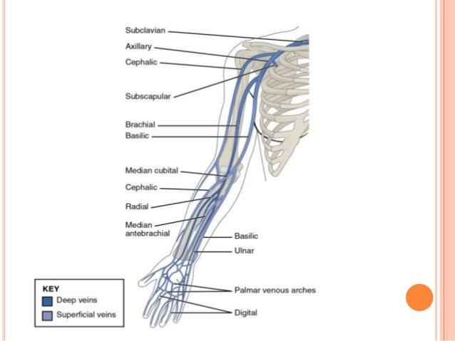
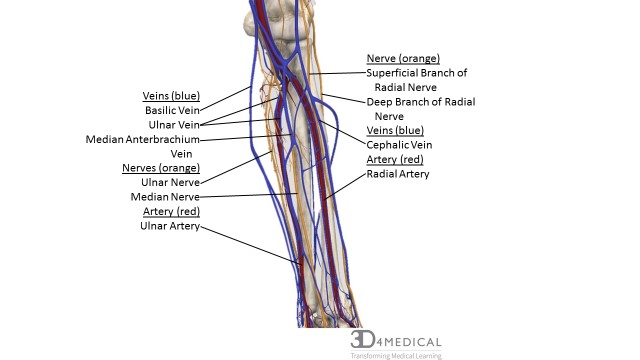
The vessels of the arms are part of the circulatory system which provides nutrients to the tissues. Within the venaecomitantes are both radian and ulnar veins with the ulnar veins typically existing as larger in size while the radial veins interact with the dorsal metacarpal veins.
 Basilic Vein Wikipedia
Anatomy physiology module provides a broad spectrum of adult male and female normal anatomy cases with varying body morphologies to maximize training efficacy.

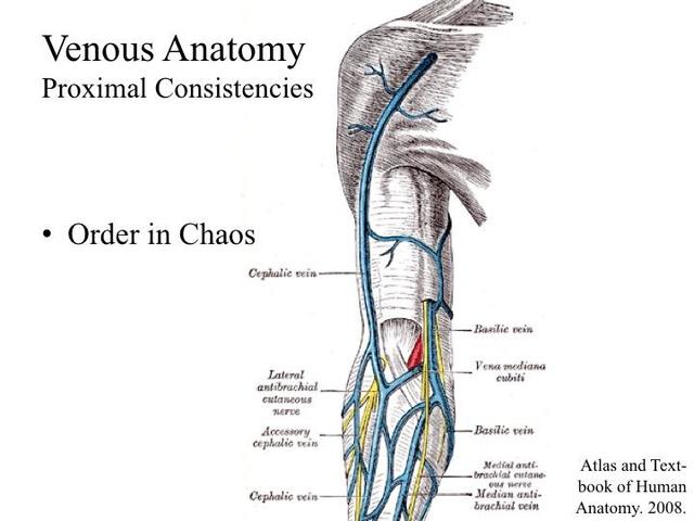
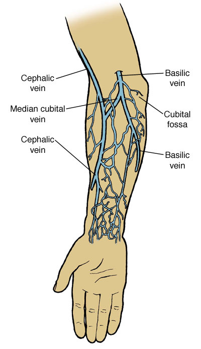
Venous arm anatomy. The basilic vein is typically larger than the. The veins of the arm may be divided into two groups. As you reach the proximal arm the axillary vein will divide into the basilic and brachial veins.
In the upper arm the basilic and cephalic veins are the major routes for superficial venous drainage with ultimate runoff into the deep system figs. Upper arm veins brachial basilic the basilic vein is the larger and is more superficial. Each individual hands on training case is accompanied by image window specific expert instruction and probe positioning guidance.
On the other hand the ulnar veins interact more with tributaries that deal with deep volar venous arches causing them to have more to do with the wrist area of a human being. The venae comitantes of the brachial artery ie the deep veins of the arm or brachial veins are joined by the basilic vein above the lower border of the posterior wall of the axilla to form the axillary vein. Chapter 77 venous anatomy of the extremities.
The venous system of the upper limb drains deoxygenated blood from the arm forearm and hand. The primary venous return from the arm is through the axillary vein which continues centrally as the subclavian and brachiocephalic innominate veins before emptying into the superior vena cava. The arteries deliver freshly oxygenated blood to muscles and bone.
And the other into the cephalic median cephalic vein. It can anatomically be divided into the superficial veins and the deep veins. Deep veins and superficial veins.
There are three parts of the axillary vein the first distal part into which the cephalic vein enters at a point just superior to the pectoralis minor muscle and the second and third parts which give off branches corresponding to the tributaries off the axillary artery.
Upper Limb Anatomy
 Handcare Org Anatomy Vessels
 Vector Isolated Illustration Of Human Arterial And Venous Circulatory
 Dentistry And Medicine Blood Supply Venous Drainage
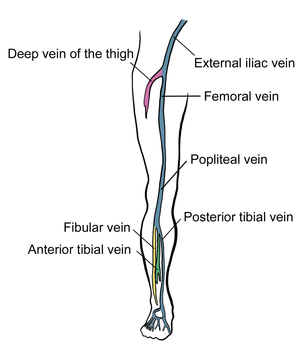
 Pdf Lower Extremity Venous Anatomy Semantic Scholar
 Us Iv Technique Proceduralist Org
 Basilic Vein An Overview Sciencedirect Topics
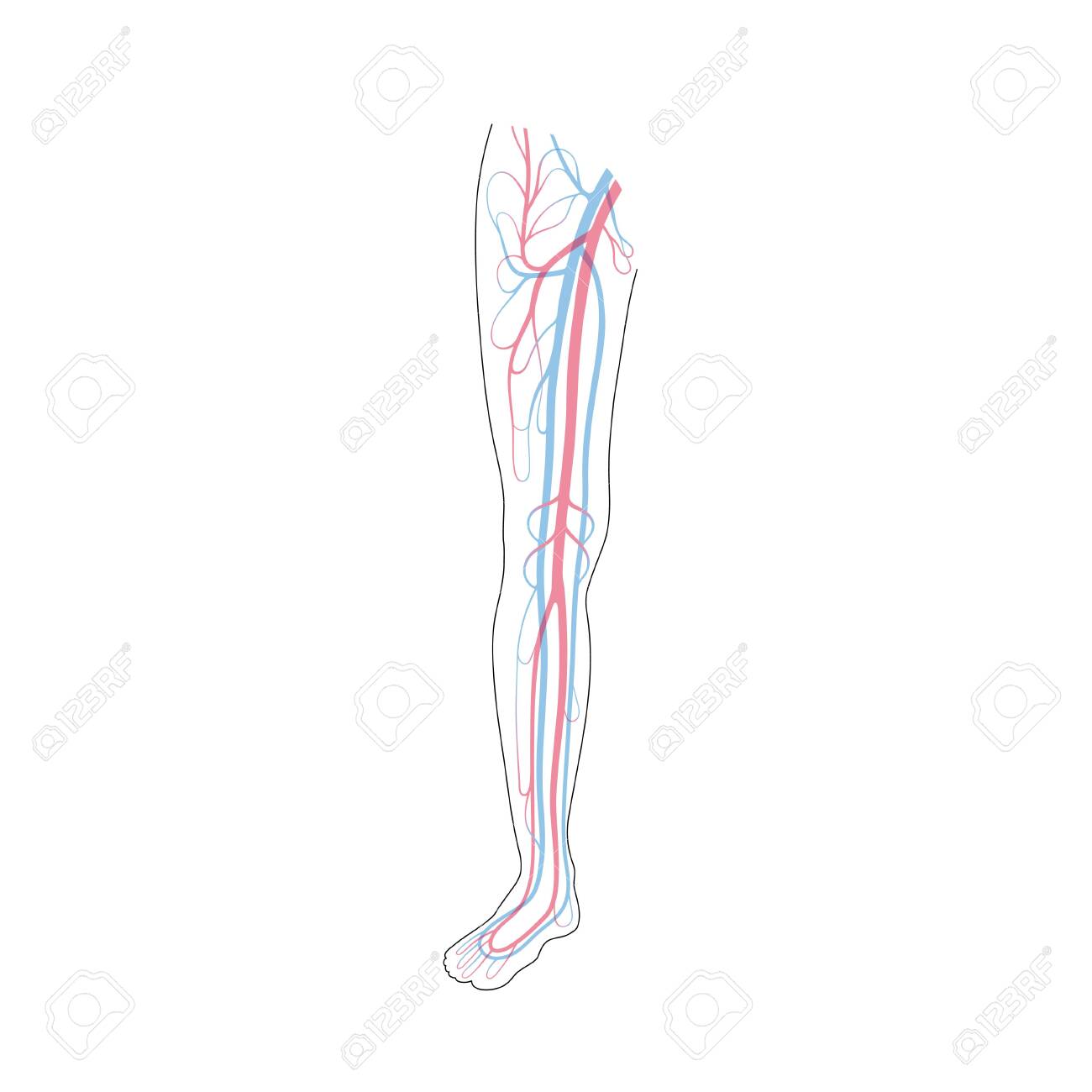
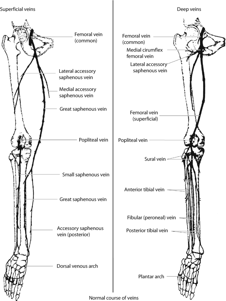
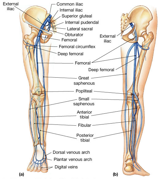 Venous Drainage Of The Lower Extremity Anatomy
 Anatomy Of Gsv And Ssv With Common Variants Of Ssv Gsv

 Extremity Veins Springerlink
 Venous Lymphatic Drainage Of Upper Limb
 Online Cme Upper Extremity Venous Evaluation
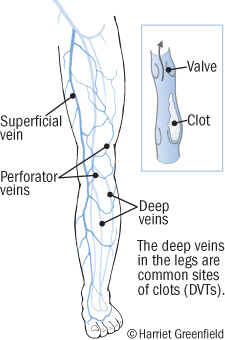
 Deep Vein Thrombosis Rcemlearning
 Instant Anatomy Diagram
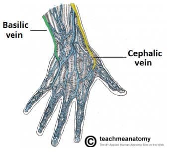
![]() Veins Of The Upper Limb Anatomy Kenhub
Veins Of The Upper Limb Anatomy Kenhub
 Upper Extremity Venous Anatomy Vascular Ultrasound
 Venous Drainage Of The Upper Limb Basilic Cephalic
 Emergency Ultrasound
 F0 Pngfuel Com Png 705 706 Circulatory System Vein
 Chapter 54 Peripheral Venous Cutdown Emergency Medicine

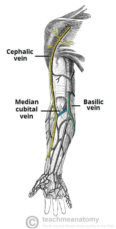 Venous Drainage Of The Upper Limb Basilic Cephalic
 Arm Dvt Normal Ultrasoundpaedia
Venous Hemodynamics What Happens When Flow Is Wrong
![]() Veins Of The Upper Limb Anatomy Kenhub
Veins Of The Upper Limb Anatomy Kenhub
 Clinical Education Intravenous Therapy Skills
 Cardiovascular System Human Veins Arteries Heart
 Anatomy Atlases Anatomy Of First Aid A Case Study Approach
 Nerves Blood Vessels And Lymph Advanced Anatomy 2nd Ed
 Superficial Venous Thrombosis Heart And Blood Vessel
 Picc Line Vein Anatomy Upper Limb Anatomy Anatomy Images
 Deep Vein Thrombosis Blood Clots In Your Veins Harvard Health
Belum ada Komentar untuk "Venous Arm Anatomy"
Posting Komentar