Outer Eye Anatomy
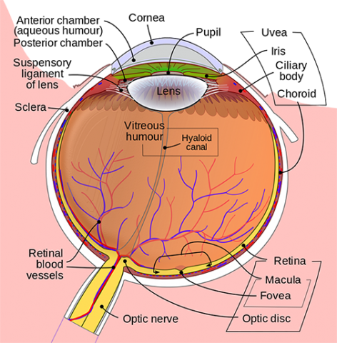
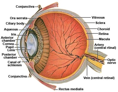
Behind the eye your optic nerve carries. Human eye is spherical about 25 cm in diameter.
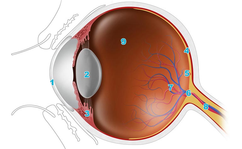
 Human Eye Anatomy And Main Barriers To Ocular Drug Delivery
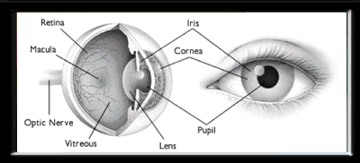
The inside lining of the eye is covered by special light sensing cells that are collectively called the retina.
Outer eye anatomy. In particular it is essential to identify the anterior and posterior lamellae. These muscles move the eye up and down and side to side and rotate the eye. Here is a tour of the eye starting from the outside going in through the front and working to the back.
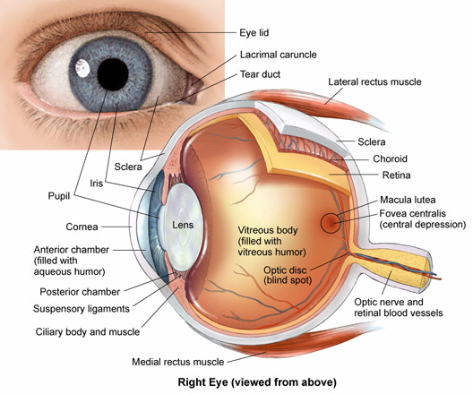
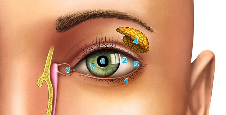
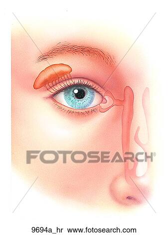
The outer layer of the eye consists of 8 eye parts. The eye sits in a protective bony socket called the orbit. Tear drains from the eyes in to the nose through the tear duct.
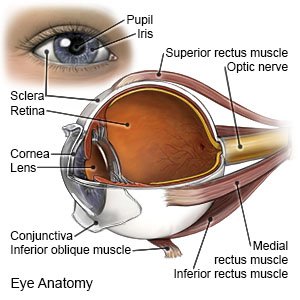
Extraocular muscles help move the eye in different directions. The iris the colored part of the eye controls how much light the pupil lets in. The cornea is like a window it helps to focus light onto the retina.
Behind the septum are a number of different other structures a knowledge of which is essential if surgery is to be performed. The eye is surrounded by the orbital bones and is cushioned by pads of fat within the orbital socket. Anatomy of the eye the ottawa hospital.
The orbital septum differentiates the orbital tissue from the lid. It is situated on an orbit of skull and is supplied by optic nerve. The cornea is the outer covering of the eye.
This is why a teary eye is usually accompanied by a runny nose. The lens works together with the cornea to focus light correctly on the retina. Eye part 1 cornea.
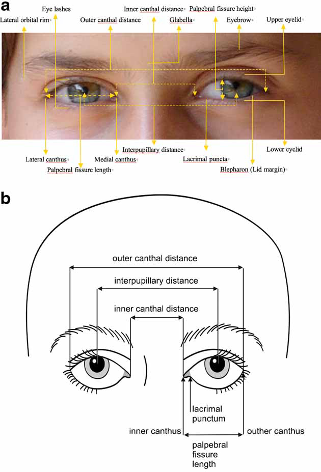
The eyelids the nose is the inner canthus and the other is the outer canthus. To understand the diseases and conditions that can affect the eye it helps to understand basic eye anatomy. This is a small tube that runs from the eye to the nasal cavity.
Next light passes through the lens a clear inner part of the eye. Anatomy of the eye. This dome shaped layer protects your eye from elements that could cause damage to the inner parts of the eye.
Nerve signals that contain visual information are transmitted through the optic nerve to the brain. In the diagram above anatomy of the eye the artery is shown in red while the vein is shown in blue. The eye is the photo receptor organ.
Anatomy of the eyelid. 1 the skin containing glands that open onto the surface of the lid margin and the eyelashes. There are 6 sets of muscles attached to outer surface of eye ball which helps to rotate it in different direction.
When light passes through the eye the cornea refracts the light rays in a way so that it can land directly on the retina. The lid may be divided into four layers. 2 a muscular layer containing principally the orbicularis oculi muscle responsible for.
It converts light into electrical impulses. Six extraocular muscles in the orbit are attached to the eye. Anatomy parts and structure.
Some of this light enters the eye through an opening called the pupil pyoo pul. The transparent dome like structure that is covering the iris and the pupil. The cornea is shaped like a dome and bends light to help the eye focus.
 How The Human Eye Works Live Science
 How The Eye Works Fighting Blindness
 Anatomy And Structure Of The Eye Brightfocus Foundation
An Easy Guide To Your Eye S Anatomy Lenstore Co Uk
 Eye Anatomy Detail Picture Image On Medicinenet Com
 Eye Anatomy And How The Eye Works
 Elements Of Morphology Human Malformation Terminology
 Corneal Flash Burns What You Need To Know
 Anatomy Of The Eye Vision Direct Uk
 Eye Anatomy And Function
Parts Of The Eye American Academy Of Ophthalmology
 Cornea Definition And Detailed Illustration
 Know Your Eye Ahalia
 Anatomy Of The Eye Kellogg Eye Center Michigan Medicine
 Anatomy Of The Eye The Ottawa Hospital
 Eye Anatomy Mydr Com Au
 Vision And The Eye S Anatomy Healthengine Blog
 Iris Outer Border Of Iris Pupil Folds Of Iris In 2019
 What Are The 3 Layers That Make Up The Wall Of The Eyeball
 Eye Anatomy Central Florida Retina
 An Easy Guide To Your Eye S Anatomy Lenstore Co Uk
 Iris Anatomy Wikipedia
 An Easy Guide To Your Eye S Anatomy Lenstore Co Uk
 Right Eye Outer Anatomy Unlabeled Stock Illustration
 Eye Anatomy Ocular Anatomy Vision Conditions Problems


Belum ada Komentar untuk "Outer Eye Anatomy"
Posting Komentar