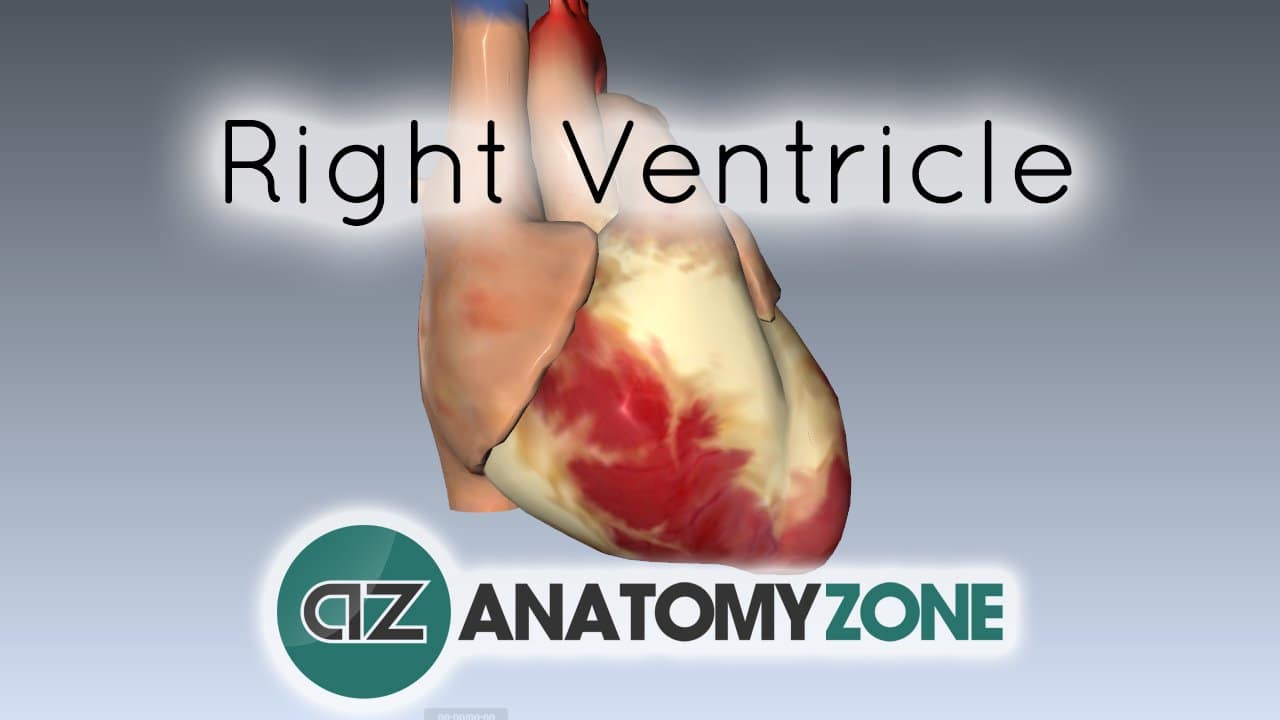
Right Ventricular Anatomy
Posterior originates from the inferior wall of the right ventricle. The mesh surface is the left ventricle.
 Left Vs Right Ventricle Difference Between
The right ventricle is one of the hearts four chambers.

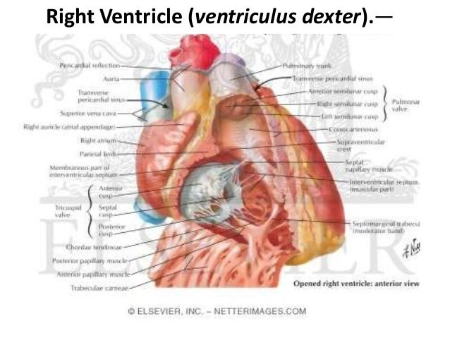
Right ventricular anatomy. The right ventricle extends from the right atrium to the apex of the heart. Forms the major portion of the anterior surface of the heart. Anterior is the largest of the three muscles.
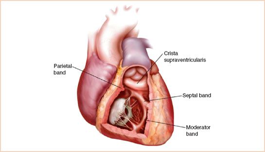
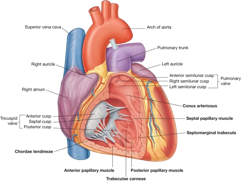
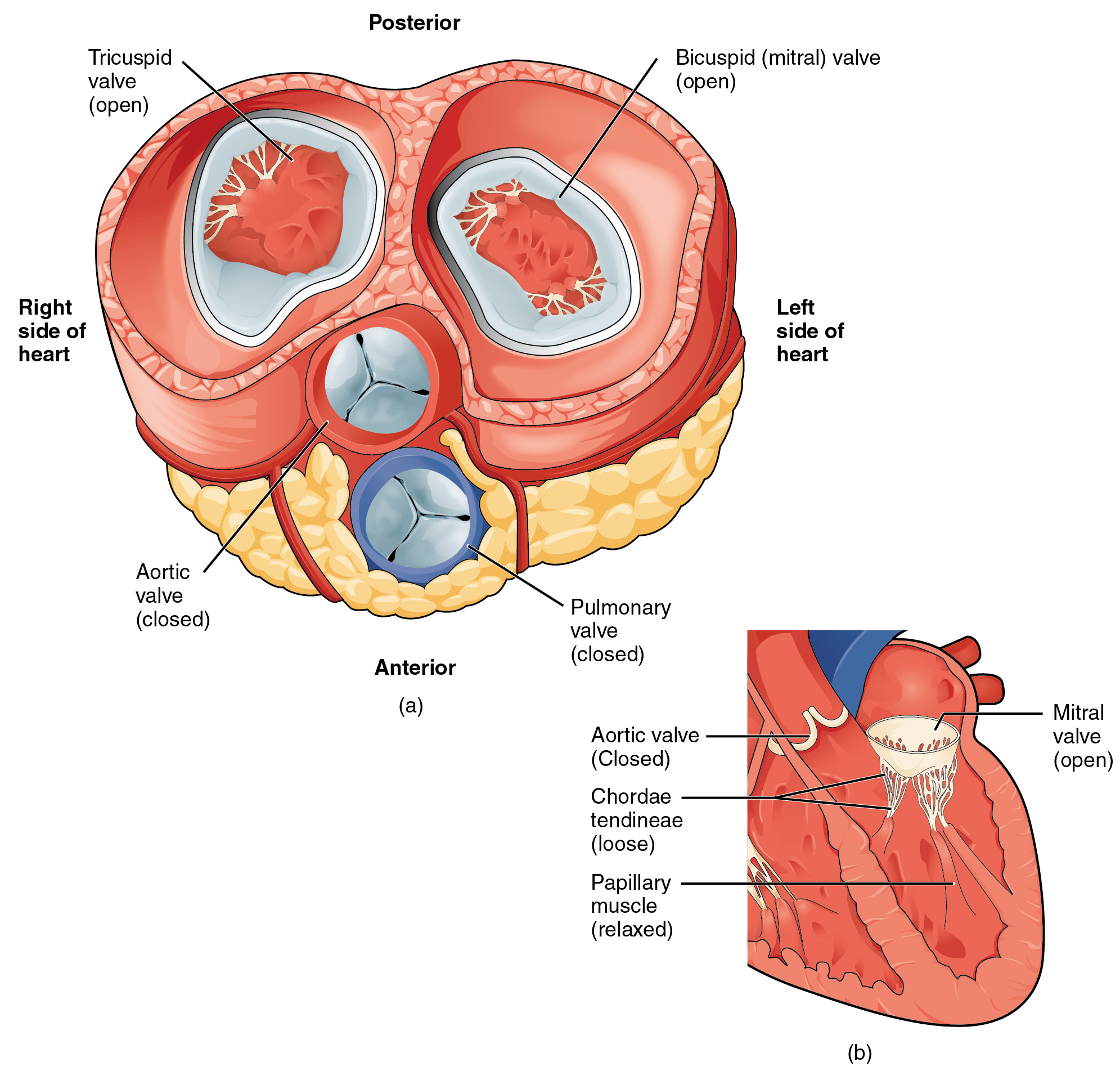
The moderator band is located in the right ventricular apex that connects the interventricular septum to the anterior papillary muscle. The point of the chordae tendineae and the papillary muscles is to prevent these cusps to prevent the tricuspid valve from being averted back into the right atrium when the ventricle contracts and the pressure increases and the back flow of blood forces against the tricuspid valve. The normal rv is anatomically and functionally different from the left ventricle which precludes direct extrapolation of our knowledge of left sided physiopathology to the right heart.
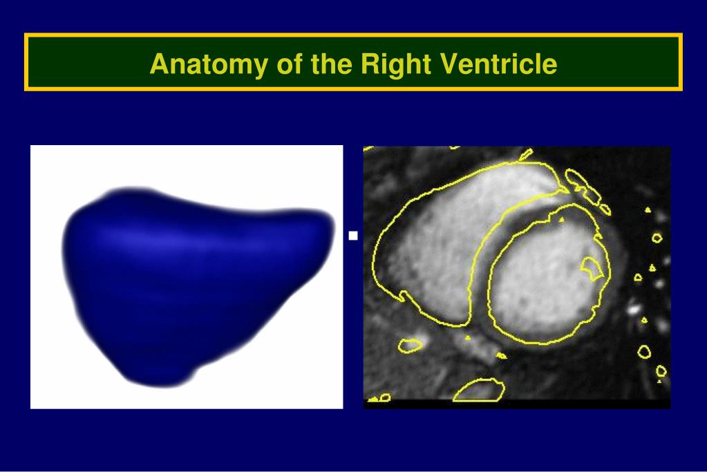
It is located in the lower right portion of the heart below the right atrium and opposite the left ventricle. The right ventricle projects to the left of the right atrium and when viewed in the cardiac short axis plane is semilunar in shape wrapping around the anterolateral aspect of the left ventricle lv. It also marks the inferior border of the cardiac silhouette.
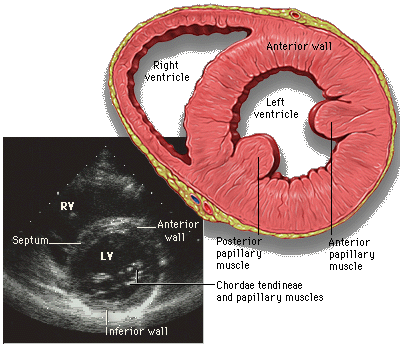
The other important internal features of the right ventricle are the papillary muscles of which there are three. It has thinner walls than the left ventricle due to lower right sided pressures compared to the left ventricle. Rv remodelling in diseased hearts can result in profound shape change as in this patient b with dilated rv due to severe pulmonary regurgitation following repair of tetralogy of fallot.
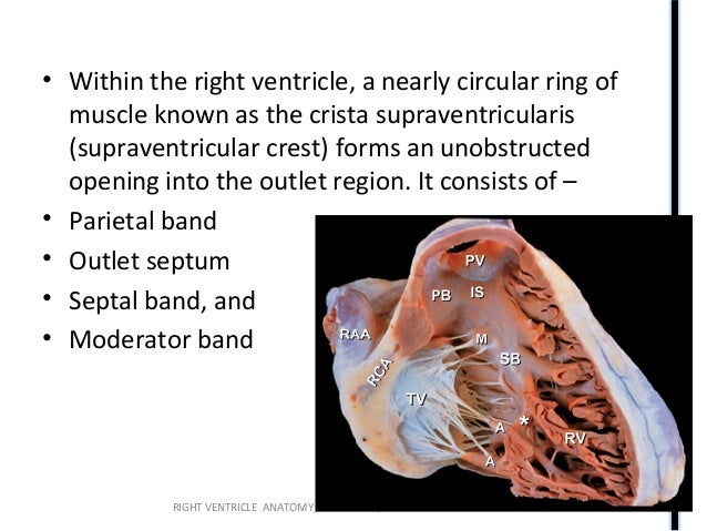
Septal has attachments to the interventricular septum and. The right ventricle in the normal heart is the most anteriorly situated cardiac chamber since it is located immediately behind the sternum. There is increasing recognition of the crucial role of the right ventricle rv in determining functional status and prognosis in multiple conditions.
As deoxygenated blood flows into the right atrium it passes through the tricuspid valve and into the right ventricle which pumps the blood up through. When looking within the heart it appears as any other trabecula would except that the moderator band does not.
 Single Ventricle Defects And The Fontan Acha
 Trabeculae Carneae Wikipedia
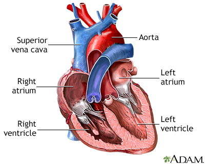 Heart Chambers Medlineplus Medical Encyclopedia Image
 Difference Between Left And Right Ventricle Definition
 Anatomical Specimens Of The Right Ventricle Rv A Section
 Right Ventricle Right Atrium Tricuspid And Pulmonic Valves
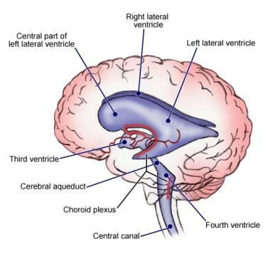 Ventricles Of The Brain Overview Gross Anatomy
 Illustration Picture Of Anatomical Structures Heart
 Internal Anatomy Of The Heart Fruits And Vegetables
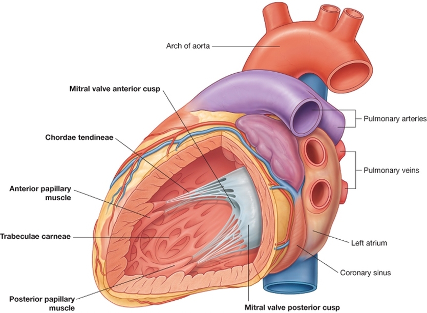
 Left Ventricle An Overview Sciencedirect Topics
 Assessment Of The Right Ventricle By Echocardiography Ppt
 Anatomy Of Heart

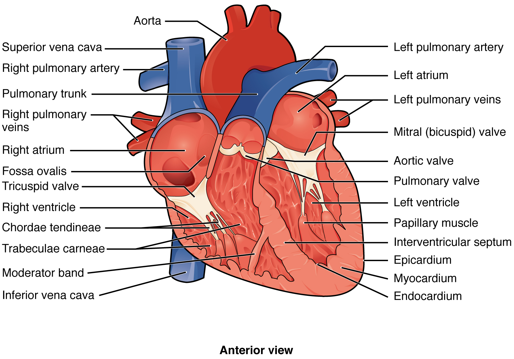
 19 1 Heart Anatomy Anatomy And Physiology
 Ppt Assessment Of The Right Ventricle By Echocardiography
Assessment Of The Right Ventricle By Echocardiography A
 Right Ventricle Rv Anatomy And Functions
 Anatomy Of The Heart Flashcards Quizlet
 Focus On Right Ventricular Outflow Tract Septal Pacing
 Right Ventricular Pulmonary Arterial Coupling Predicts

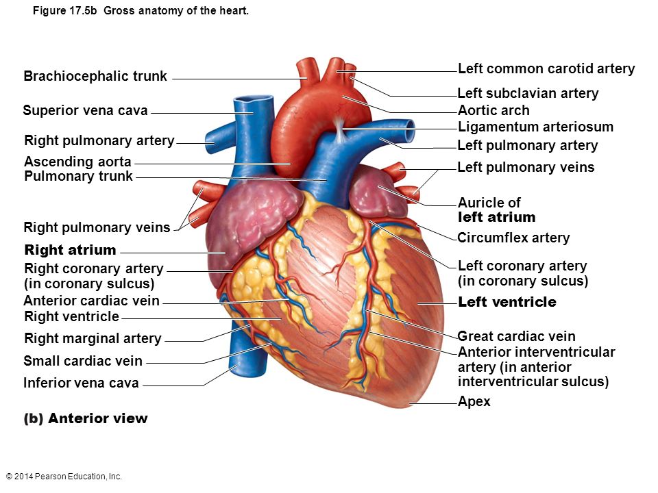
Figure 1 From Right Ventricular Septal Pacing A Paradigm
 Illustration Of The Heart Structures Left And Right Atria
![]() Heart Ventricles Anatomy Function And Clinical Aspects
Heart Ventricles Anatomy Function And Clinical Aspects
Belum ada Komentar untuk "Right Ventricular Anatomy"
Posting Komentar