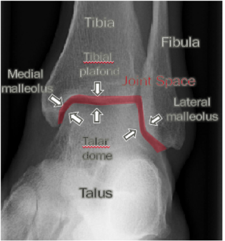
Ankle Xray Anatomy
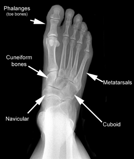
The rear foot the mid foot and the forefoot. The rearfoot is is composed of the talus and the calcaneus heel bone.
 Ankle X Rays
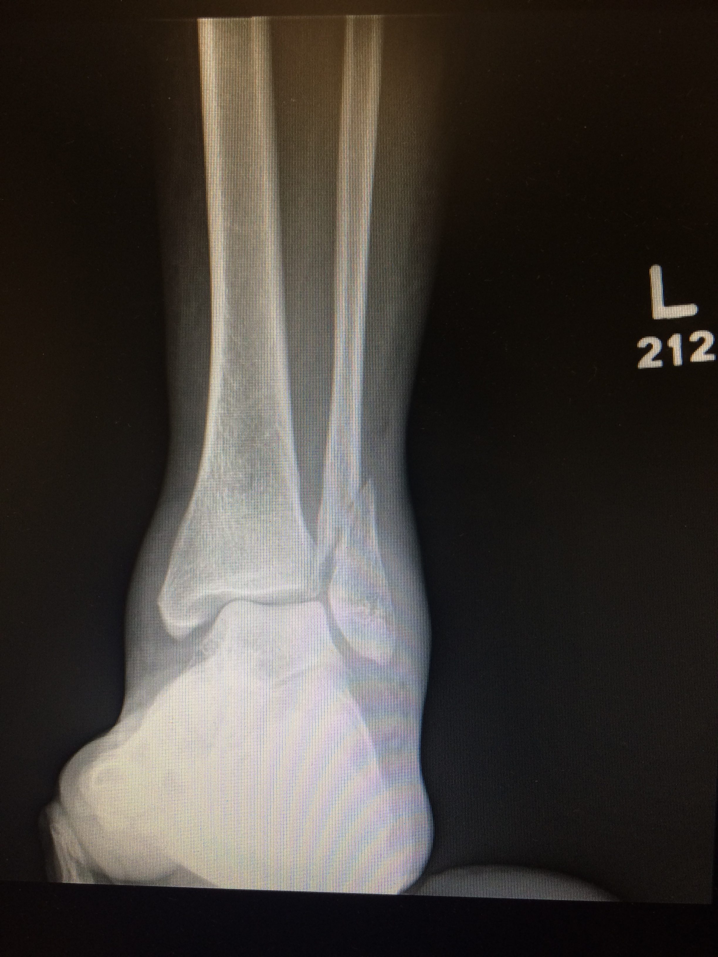
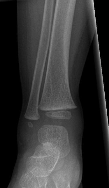
The tibia the fibula the talus and the calcaneus.

Ankle xray anatomy. A doctor puts pressure on an injured ankle and takes an x ray. The tibia and fibula make up just above the ankle. Click on a link to get sagittal view t1 axial view t2fatsat coronal view t2fatsat sagittal view t2fatsat.
It is the most complete reference of human anatomy available on web ipad iphone and android devices. The ankle is comprised of the talus the tibia and the fibula. An x ray film of the ankle is most commonly used to determine a fracture arthritis or other problems.
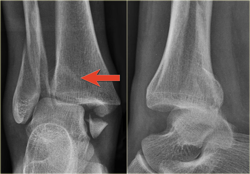
The ankle also consist of two joints the ankle joint where the tibia fibula and talus meet and the syndesmosis joint the joint between the tibia and fibula which is help together by ligaments. Anatomy the ankle is a synovial joint composed of the distal tibia and fibula as they articulate with the talus. The foot and ankle can be subdivided into 4 different parts.
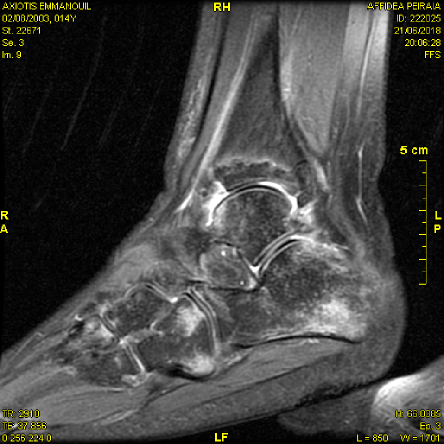
When calcaneal pathology is suspected an additional image can be made in axial direction. This webpage presents the anatomical structures found on ankle mri. The ankle consists of three bones the tibia the fibula and the talus.
Ankle ligament anatomy ankle injuries may involve bones or ligaments in isolation or a combination of bones and ligaments. The distal tibia and fibula articulate with each other at the distal tibiofibular joint which is more commonly referred to as the tibiofibular syndesmosis or simply the syndesmosis. E anatomy is an award winning interactive atlas of human anatomy.
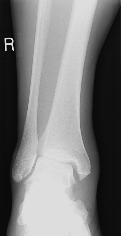
The ankle x ray is used primarily to demonstrateexclude a fracture. X rays directly visualise bone injury but understanding of the anatomical position of ligaments is required to appreciate the presence of ligament injuries which are not directly visualised. The ankle joint also known as talocrural joint is an example of a synovial joint and is formed by the bones tendons and ligaments found in the leg and the foot 1 2.
Ct mri radiographs anatomic diagrams and nuclear images. Explore over 5400 anatomic structures and more than 375 000 translated medical labels. There are several bones that make up the ankle.
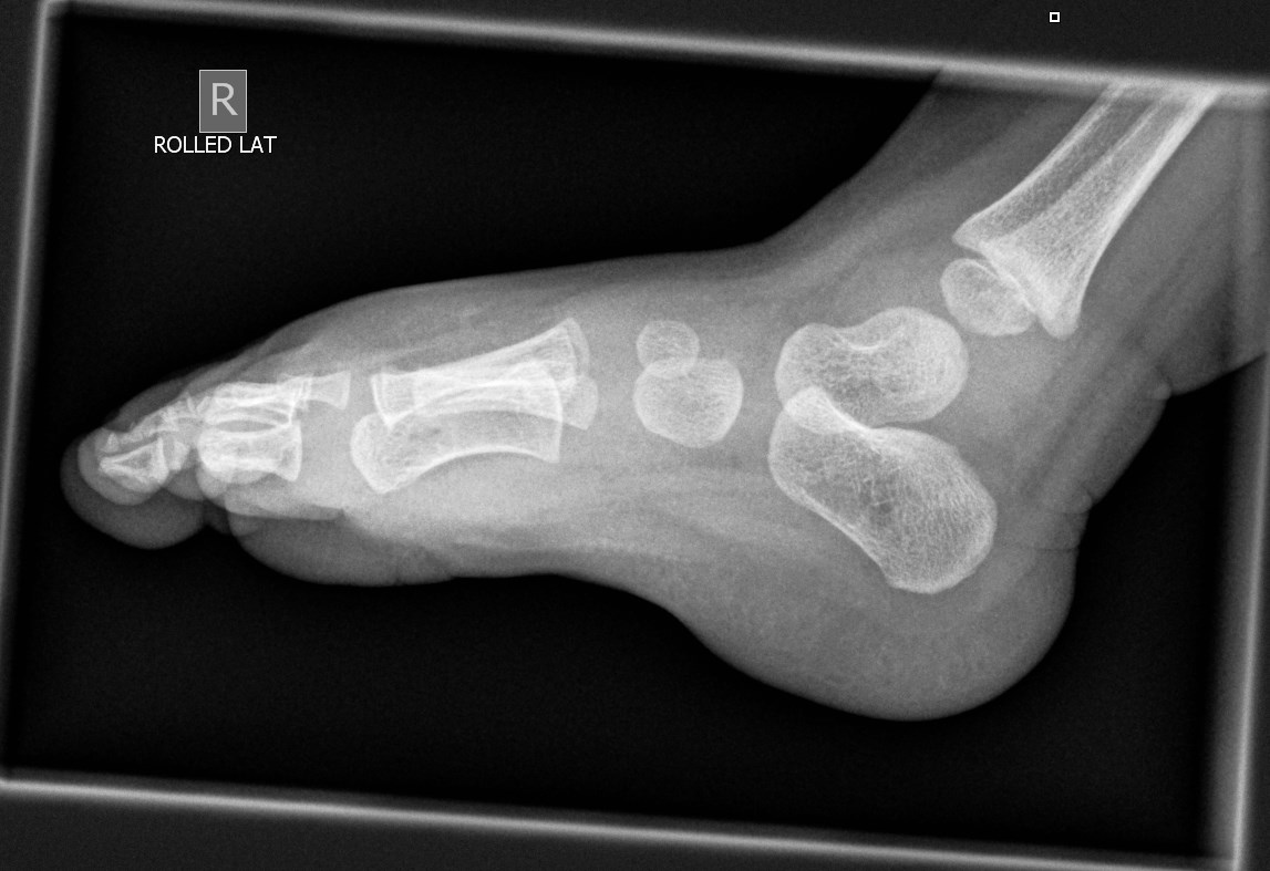
Normal foot and ankle x ray anatomy. Stanford bone tumor bayesian network issssr msk lectures for residents ocad msk cases from around the world stanford msk mri atlas has served almost 800000 pages to users in over 100 countries. A standard series includes an anteroposterior ap image a mortise image and a lateral image.
Mri of the ankle. Depending on the request various images can be made.
 The Radiology Assistant Ankle Fracture Mechanism And
 Ankle X Rays
 X Ray Of Human Ankle Metal Print
 Imaging In Ankle Fractures Overview Radiography Computed
 Update On Diagnosis And Management Of Cuboid Fractures
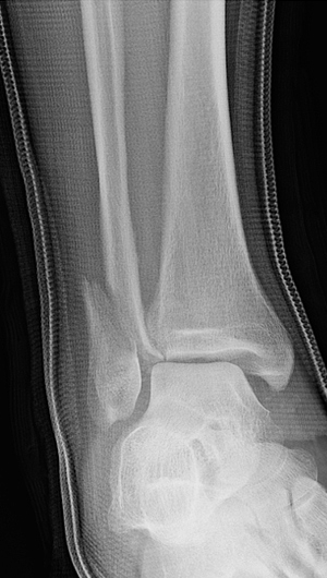
 X Enkel Startradiology
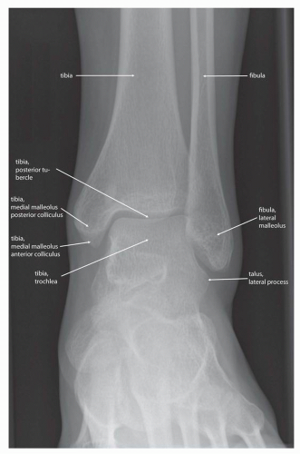
 Radiographic Anatomy Ankle Ap Medical Anatomy
 The Ankle
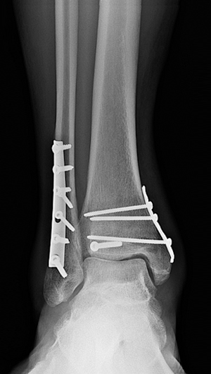 Broken Ankle Types Of Fractures Diagnosis Treatments

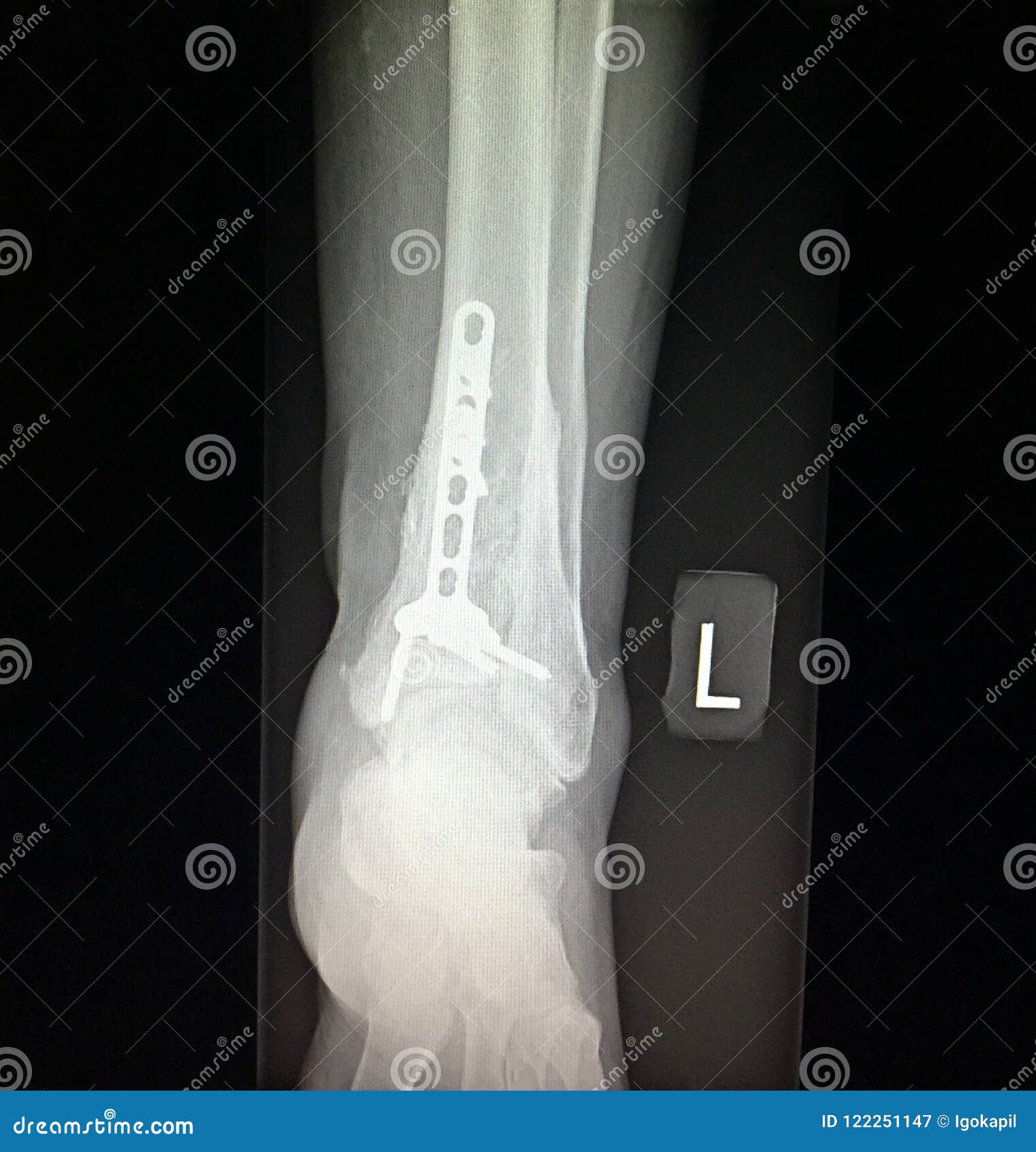 Ankle Left Tibia Distal Fracture Treatment Xray Stock Image
 X Enkel Startradiology
 Ankle X Rays
 The Radiology Assistant Ankle Fracture Mechanism And
 Broken Ankle Types Of Fractures Diagnosis Treatments
 X Ray Anatomy Lateral Ankle Diagram Quizlet
 The Radiology Assistant Ankle Fracture Mechanism And
 Diagnostic Imaging Techniques Of The Foot And Ankle
 A Normal Pa Chest X Ray Demonstrating The Normal Anatomy

 X Enkel Startradiology
 The Radiology Assistant Ankle Special Fracture Cases
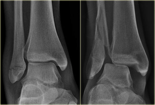
 Normal Ankle X Rays Radiology Case Radiopaedia Org
 Medial Malleolar Fracture An Overview Sciencedirect Topics
Ankle Fractures Broken Ankle Orthoinfo Aaos
 Ankle Stress Views Why When What Core Em
Belum ada Komentar untuk "Ankle Xray Anatomy"
Posting Komentar