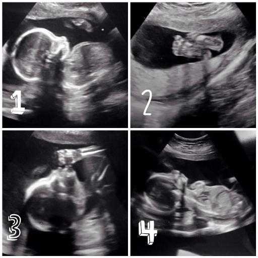
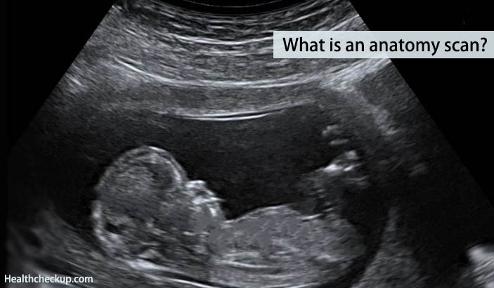
Fetal Anatomy Scan
We look at the brain the face the heart lungs diaphragm stomach intestines kidneys and the bladder. After all that we turn to the anatomy starting from the head to the toes.
 Second Trimester Ultrasound Scan Radiology Reference
When the pregnancy hits the 20th week of gestation an anatomy ultrasound is often ordered.
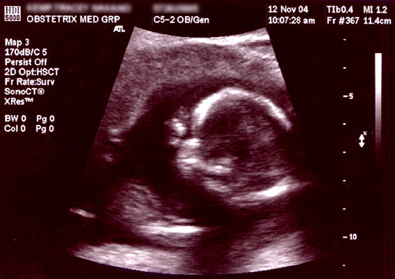
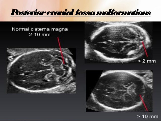
Fetal anatomy scan. Also known as an anomaly scan or anatomic survey an anatomy scan is the most extensive ultrasound exam carried out on the fetus during pregnancy. In the axial scan the characteristic lemon sign and banana sign are seen. The second trimester extends from 13 weeks and 0 days to 27 weeks and 6 days of gestation although the majority of these studies are performed between 18 and 23 weeks.
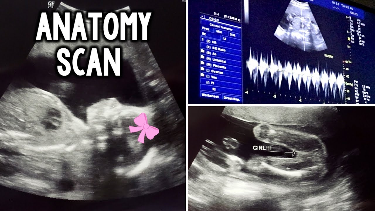
Anatomy scan with power bi directional colour doppler of both fetal kidneys at 18 weeks of pregnancy to detect renal agenesis. The second trimester scan is a routine ultrasound examination in many countries that is primarily used to assess fetal anatomy and detect the presence of any fetal anomalies. Those who want to can find out the sex of the baby if desired.
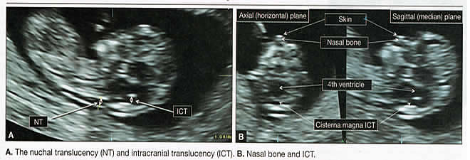
This scan is especially recommended for women with a family history of heart abnormalities or where increased nuchal translucency had been found at the 12 week scan. Lastly we look at the spine to make sure its closed on every level. Heart scan in special cases a detailed examination of the fetal heart and connecting vessels may be done at 20 weeks by a fetal cardiologist.
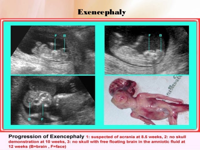
It is performed between weeks 18 and 22 and is the one most people are referring to when they talk about their routine pregnancy scan or their 20 week scan. Anatomy scan of the fetal head at 20 weeks of pregnancy in a fetus affected by spina bifida. This sonogram is used to determine fetal anomalies the babys size and weight and also to measure growth to ensure that the fetus is developing properly.
Then we scan the extremities hands feet fingers toes. A fetal ultrasound or sonogram is an imaging technique that uses high frequency sound waves to produce images of a baby in the uterus. The anatomy scan is a level 2 ultrasound which is typically performed on pregnant women between 18 and 22 weeks.
Fetal ultrasound images can help your health care provider evaluate your babys growth and development and determine how your pregnancy is progressing. By the 20th week of pregnancy the baby can weigh up to 11 ounces and measure 10 inches outstretched.
 Second Trimester Ultrasound Scan Radiology Reference
 What Is An Anatomy Ultrasound During Pregnancy Babymed Com
 Normal 2nd Trimester Ultrasound How To

 Anomaly Scan Wikipedia

 Week 32 Ultrasound What It Would Look Like Parents
 18w2d Anatomy Scan The Pursuit Of Pregnancy
 Routine Fetal Anatomy Scan At 18 23 Weeks

 Anatomy Scan Over 40 First Time Mom
 The Detection Of Spina Bifida At 11 13 6 Weeks Gestation
 The 20 Week Ultrasound Anatomy Scan Awesome First Sights
 Anatomy Scan Ultrasounds Pregnancy Brianameyer Mrowl
 21 Weeks Anatomy Scan Life As I Know It
Anatomy Scan 21 Weeks Ultrasound
 20 Week Ultrasound Scan Centre In Chennai Fetal Anomaly Scans
 Obstetric Ultrasonography Wikipedia
 What Is An Anatomy Scan Anatomy Scan Results Procedure Risks
 Routine Fetal Anatomy Scan At 18 23 Weeks
 18 Weeks Fetal Anatomy Scan Babycenter
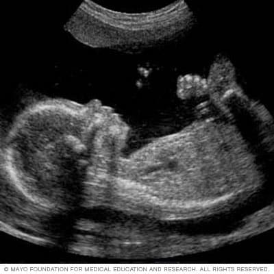 Fetal Ultrasound Mayo Clinic
 Routine Fetal Anatomy Scan At 18 23 Weeks

 Fetal Hydrops Cancer Therapy Advisor
Belum ada Komentar untuk "Fetal Anatomy Scan"
Posting Komentar