Fovea Anatomy
It refers to a pit or depression in a structure. Plural foveae ˈ f oʊ v i i is a term in anatomy.
 Fovea Centralis Wikipedia
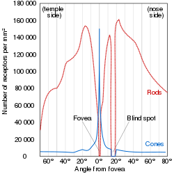
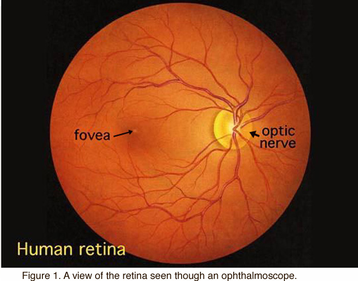
The fovea centralis a pit at the rear of the retina which contains no rods and has the densest concentration.
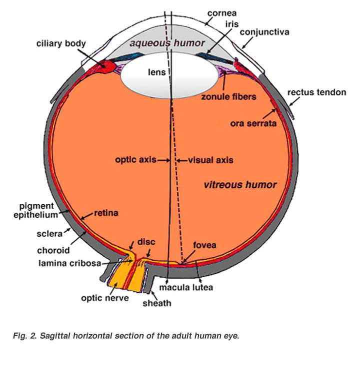
Fovea anatomy. Anatomy anatomy any small pit or depression in the surface of a bodily organ or part. Macula area of depression in the centre of the macula lutea. Central fovea of retina fovea centralis retinae a small pit in the center of the macula lutea composed of slim elongated cones.
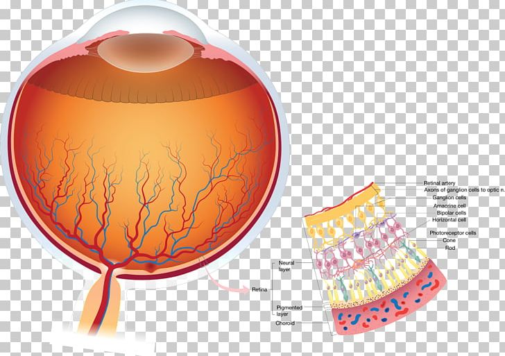
Only in the fovea are the layers of the retina spread aside to let light fall directly on the cones the cells that give the sharpest image. The fovea is of course free of a nerve fiber layer as the inner retina and ganglion cells are pushed away to the foveal slope. The macula contains mostly cones and few rods and the fovea centralis contains only cones and no rods.
Fovea of the femoral head. In the eye a tiny pit located in the macula of the retina that provides the clearest vision of all. Pterygoid fovea of the mandible neck.
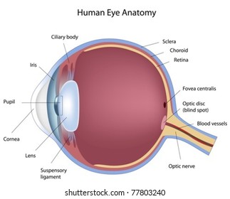
The retina by the development of the fovea centralis a localized region of the retina close to the. In the eye disease known as age related macular degeneration or amd the cones are damaged by a buildup of toxic products of eye metabolism called drusin. Diameter 15mm 1 disc diameter about 5 deg of vf.
Branch retinal vein occlusion. The fovea is situated between the ulnar styloid process and the flexor carpi ulnaris tendon. Ligamentous structures in the fovea region when the forearm is in a neutral position form the foveal attachments of the conjoined palmar and dorsal radioulnar ligaments and the ulnocarpal ligaments.
Eye anatomy and function in retina concentrate at two sites. The central ganglion cell fibers run around the foveal slope and sweep in the direction of the optic nerve. Trochlear fovea of the frontal bone.
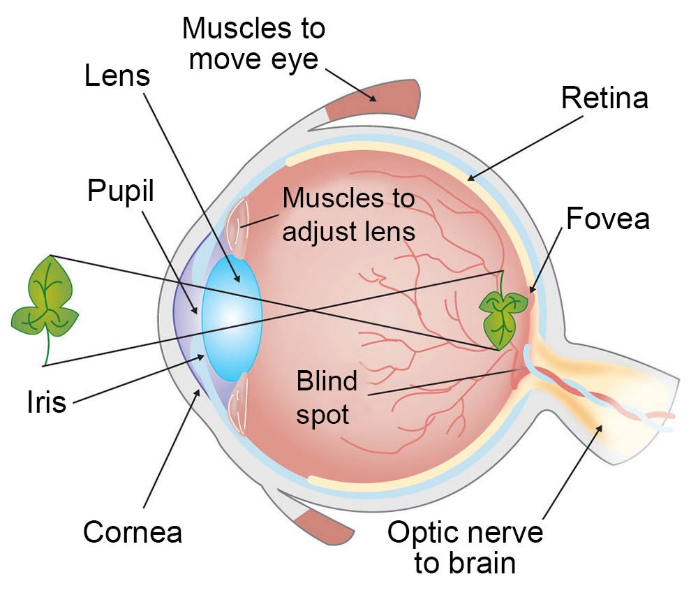
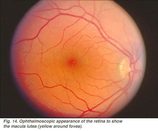
Central retinal vein occlusion. A number of eye problems can affect the fovea and can lead to vision loss if they are not treated. It is the area of clearest vision because here the layers of the retina are spread aside permitting light to fall directly on the cones.
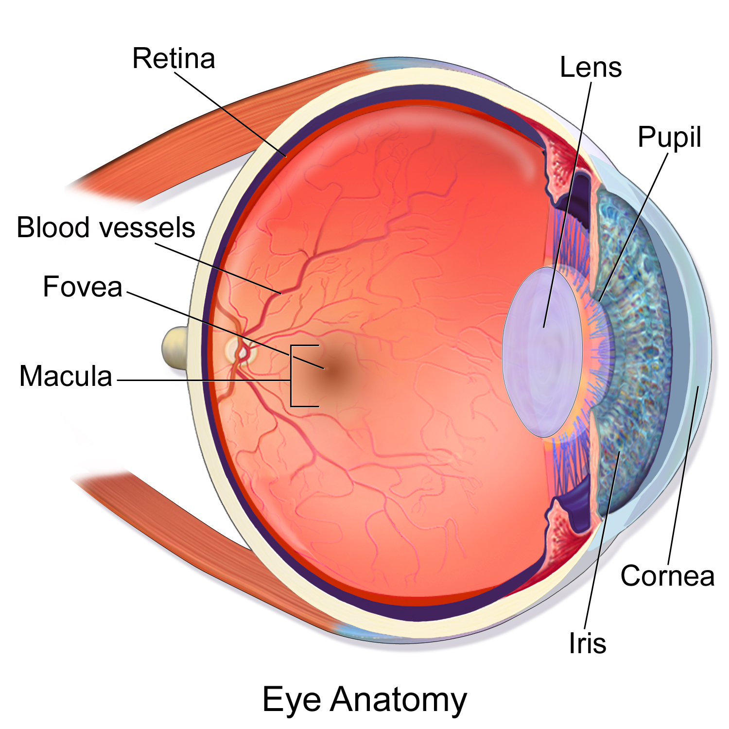
In the center of the macula is the fovea centralis. Anatomy see fovea centralis. Anatomical fovea fovea centralis clinical.
The depression in the very center of the macula where eyesight is sharpest. It is also called the fovea centralis. Fovea ˈ f oʊ v i ə latin for pit.
Fovea centralis of the retina.

 Macula Of Retina Wikipedia
 Fovea Images Stock Photos Vectors Shutterstock
Fovea American Academy Of Ophthalmology
 Anatomy Of The Eye 101 Eyecheck

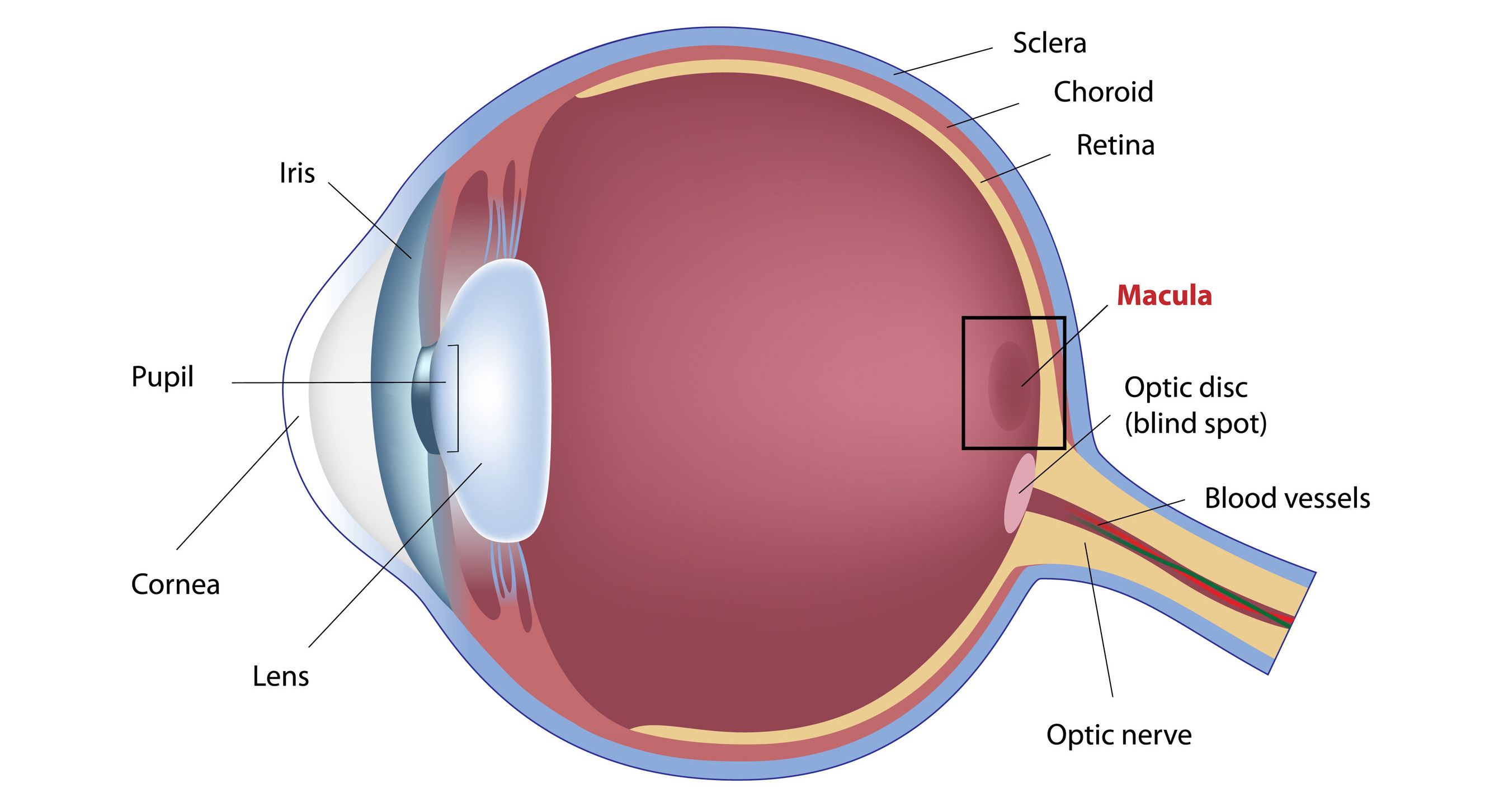 What Is The Macula

 Anatomy Of V1 In Marmosets A Schematic Diagram Of The
 Fovea Psychology Wiki Fandom
 What Is The Fovea Centralis Definition Function Study Com
 Fovea Images Stock Photos Vectors Shutterstock
 Anatomy And Structure Of The Eye Brightfocus Foundation
 Fovea Stock Photos Fovea Stock Images Alamy

 Retina Human Eye Anatomy Visual Perception Png Clipart
 Bi242 Anatomy Lab 6 Diagram Quizlet
 Simple Anatomy Of The Retina By Helga Kolb Webvision
 Set Of 3 Eye Anatomy Prints Of Fovea Retina And Rods And Cones Eye Anatomy Watercolor Optometry And Ophthalmology Art
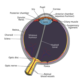
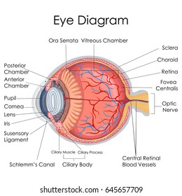
 Eye Anatomy
 Detection Of Macula And Fovea For Disease Analysis In Color

 Bee Anatomy Hutchings Bee Service
 The Primate Fovea Structure Function And Development
 Fovea Wine For Soul
 View Large Accessmedicine Mcgraw Hill Medical
 Fovea Centralis

How Do We See Light Ask A Biologist
Belum ada Komentar untuk "Fovea Anatomy"
Posting Komentar