Veins Of The Leg Anatomy
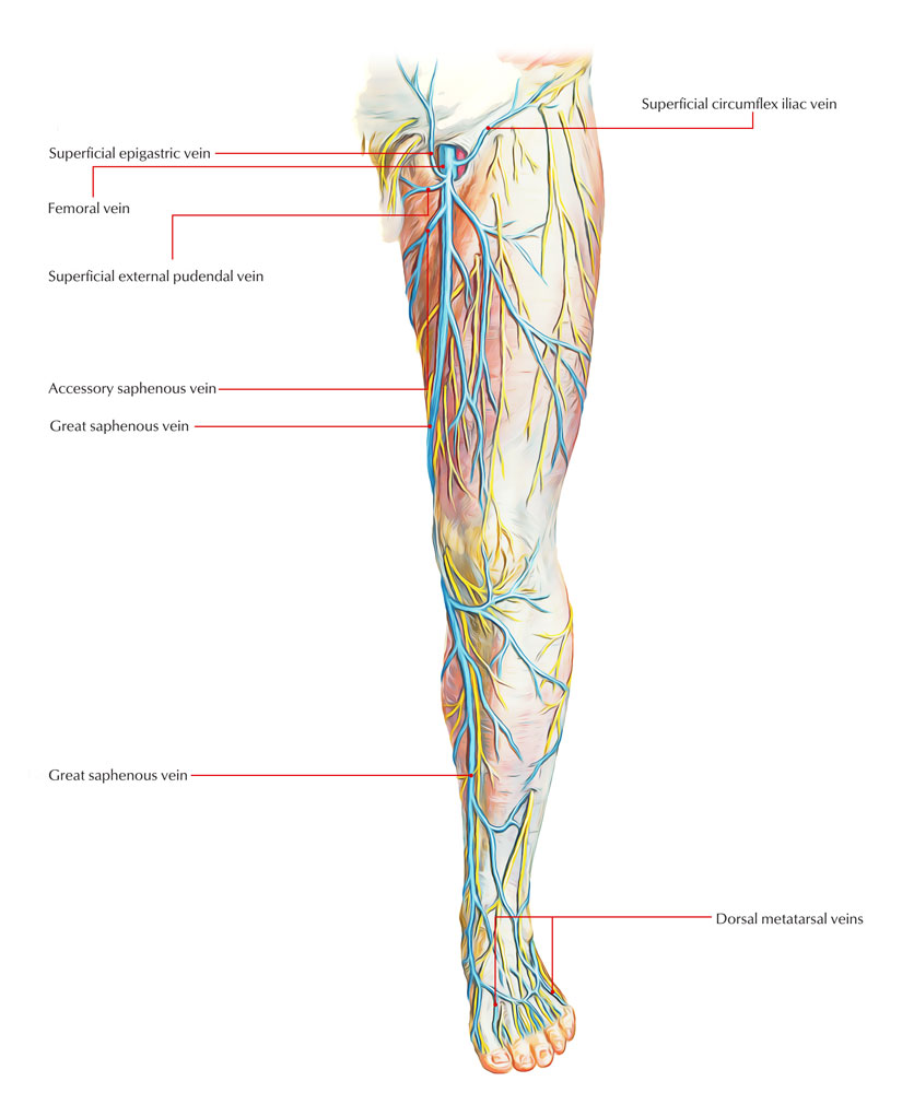
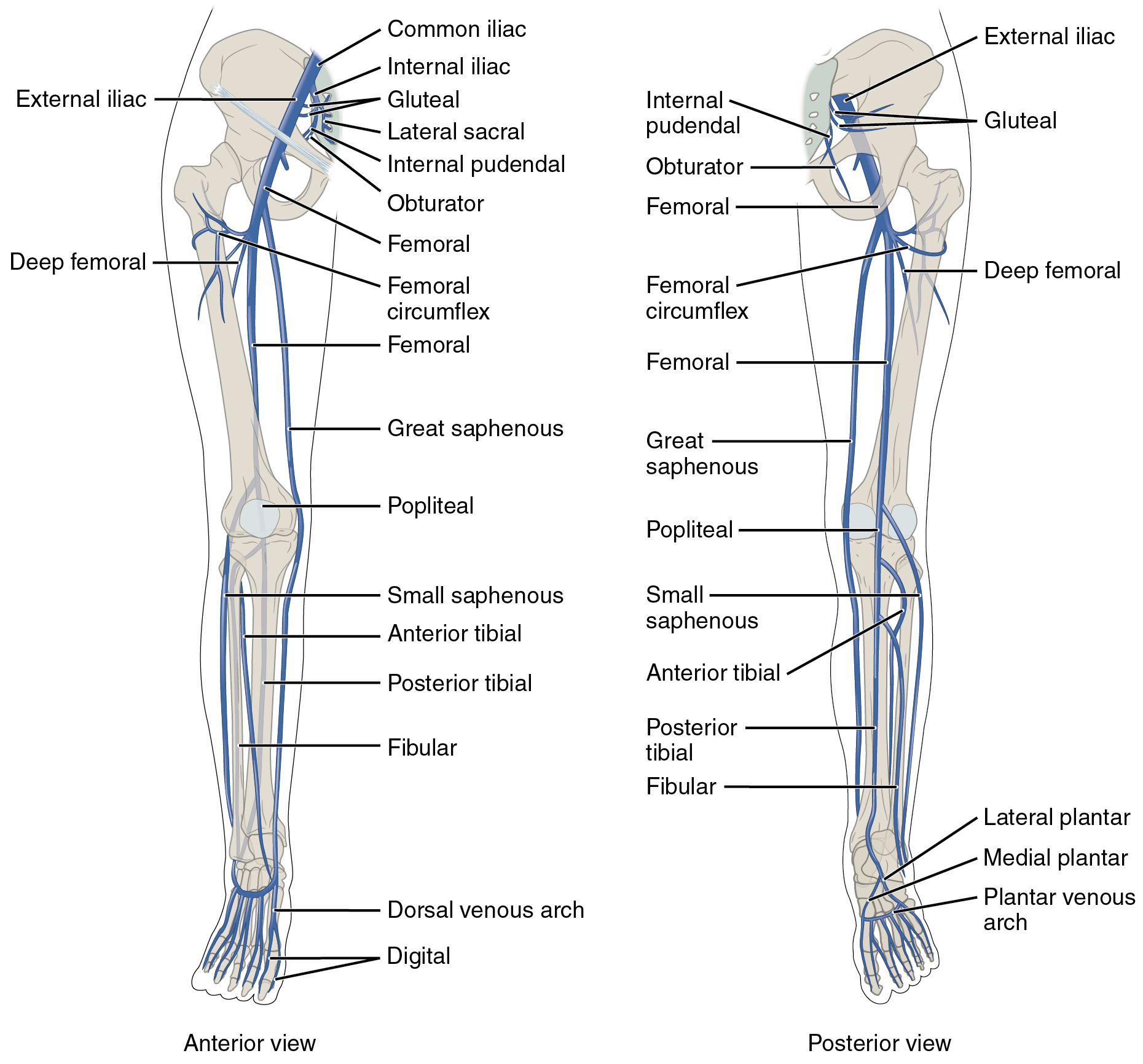
The deep veins of the leg accompany the arteries of the same name and are generally paired in the calf. Veins of the lower limb.
 Veins Of The Lower Limb An Overview Sciencedirect Topics
Posterior tibial vein and fibular vein also known as the peroneal vein which form from the medial and lateral plantar veins.

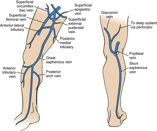
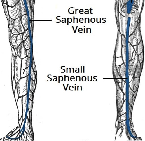
Veins of the leg anatomy. Also included are the soleal sinusoids the dorsal venous foot arch and the saphenous veins to name a few. This article will discuss the anatomy and tributaries of the veins of the lower limb in detail followed by any related clinical notes. The deep veins of the leg lie in the tight fascial compartment along the arteries.
Anterior tibial vein which receives blood from the dorsal venous arch. There are three main deep veins in the lower leg. However their junctions are not paired and their locations are variable.
They also contain tributaries other veins which drain into them. Deep veins of the sole eg medial and lateral plantar veins. The foot and leg.
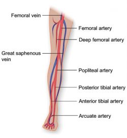
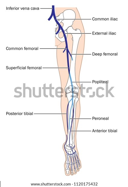
The main veins of the leg include the femoral vein the iliac veins the popliteal vein the tibial veins and the posterior arch vein. Some veins from the arch penetrate deep into the leg forming the anterior tibial vein. Venae comitantes convoying the dorsalis pedis anterior tibial and posterior tibial arteries.
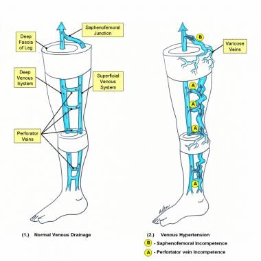
Often the artery and vein are located within the same vascular sheath so that the arterial pulsations aid the venous return. Both types of veins contain venous valves to prevent reflux of blood distally but they are more numerous in the deep veins. The main venous structure of the foot is the dorsal venous arch which mostly drains into the superficial veins.
In the context of diagnosing a deep vein thrombosis the posterior tibial and peroneal veins are the most frequently affected. The major deep veins of the lower limb are as follows.
 About Vein Disease Alaska Vein Clinic
 Veins Of Lower Limb Earth S Lab
 Lower Extremity Veins Radiology Key
 Human Body Anatomy Hip Legs And Hands Skeleton With Veins
 Lower Extremity Veins Human Anatomy Organs
 Treatment Of Varicose Veins And Telangiectatic Lower
 Skeletal Muscle Pump For Leg Veins Illustration Stock
 Glands Veins Of The Leg Pelvis Sternum Intestine Testicle Blood Supply 1880s Antique Anatomy Print Colour Anatomical Print
 Urgo Medical Anatomy Of The Normal Venous System In The
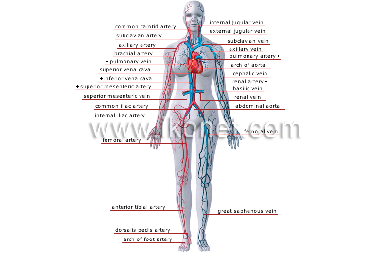 Human Being Anatomy Blood Circulation Principal Veins
 Varicose Vein Surgery Practice Essentials Anatomy
 Anatomy Of The Lower Extremity Veins Varicose Veins
 Circulatory Routes Boundless Anatomy And Physiology
Cardiovascular System Of The Leg And Foot
 Vasculature Of The Leg Texas Heart Institute
 Leg Picture Image On Medicinenet Com
 Main Veins Leg Foot Inferior Vena Stock Vector Royalty Free
 Femoral Vein Wikipedia
 Varicose Veins Clinical Features Management
 Varicose Veins Clumsy Lost Medical Student That Can T Find
 Figure 1 From Lower Extremity Venous Anatomy Semantic Scholar
 Veins Of The Lower Extremity Preview Human Anatomy Kenhub
 20 5 Circulatory Pathways Anatomy And Physiology
 Vein Health Treatment Options Nanticoke Cardiology
 Veins Anatomy Exhibits


Belum ada Komentar untuk "Veins Of The Leg Anatomy"
Posting Komentar