Anatomy Of The Eye Drawing
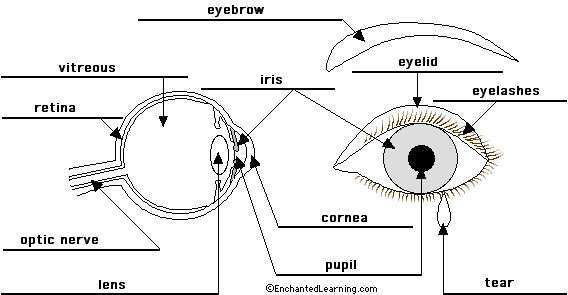
Anatomy of the eye. The eyeball is covered by a top and bottom lid.
Anatomy of the eye the anatomy and physiology of the human eye is an important part of many courses eg.
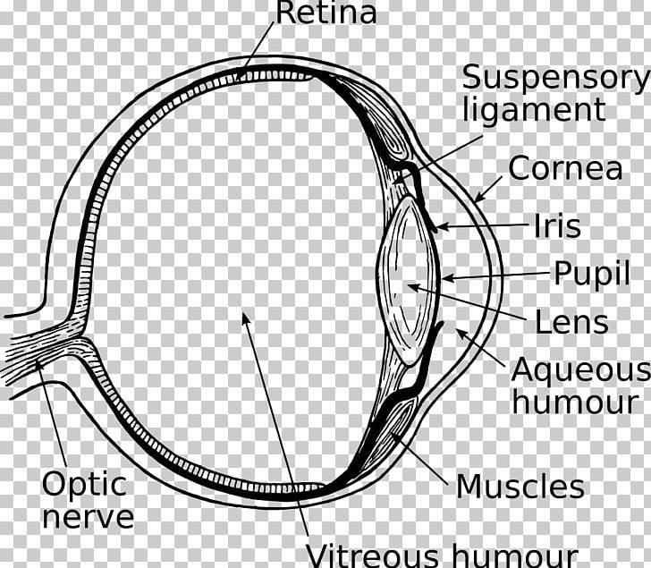
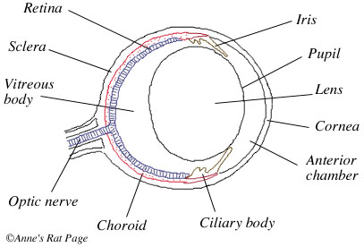
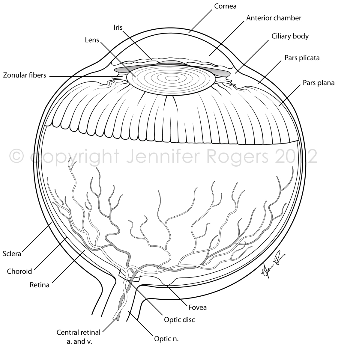
Anatomy of the eye drawing. The eye is surrounded by the orbital bones and is cushioned by pads of fat within the orbital socket. Extraocular muscles help move the eye in different directions. The iris the colored part of the eye controls how much light the pupil lets in.
Light projects through your pupil and lens to the back of the eye. Most of the eye is filled with a clear gel called the vitreous. The cornea is shaped like a dome and bends light to help the eye focus.
How to draw eyes structure. Nerve signals that contain visual information are transmitted through the optic nerve to the brain. Light is focused primarily by the cornea the clear front surface of the eye which acts like a camera lens.
If using a line you can use broken or implied line. In biology human biology physics and practical courses in medicine nursing and therapies. The inside lining of the eye is covered by special light sensing.
Some of this light enters the eye through an opening called the pupil pyoo pul. The top edge of this hole has a ridge thats called the brow ridge. At the inner side of the lids there is a tear duct.
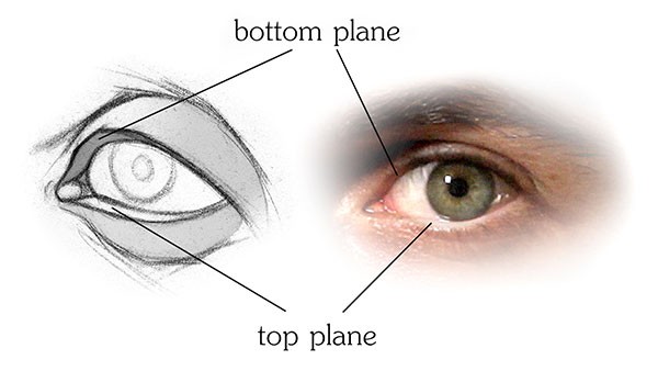
The iris of the eye functions like the diaphragm of a camera controlling the amount of light reaching the back of the eye by automatically adjusting the size of the pupil aperture. Draw the subtle changes in tone which indicate the eye socket and the planes of the nose and brows which help to set the eyes into the face. This simple introduction the subjects of the eye and visual optics includes a simple diagram of the eye together with definitions of the parts of the eye labelled in the illustration.
The lens works together with the cornea to focus light correctly on the retina. Abdominal musculature 12 photos of the abdominal musculature abdominal manual muscle testing abdominal muscle belt abdominal muscle hernia after tummy tuck abdominal muscle nearest the midline abdominal muscle youtube human anatomy abdominal manual muscle testing abdominal muscle belt abdominal muscle hernia after tummy tuck abdominal muscle nearest the midline abdominal muscle youtube. Next light passes through the lens a clear inner part of the eye.
Lets start by going over some common terminology. All the best eye anatomy drawing 35 collected on this page. The eye socket refers to the hole in the skull where the eyeball sits.
 Unlabeled Eye Anatomy Diagram
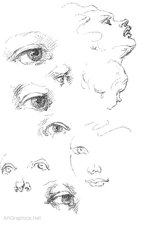
 How To Draw Eyes Structure Proko
 Single Eye Sketching Exercise Found This In Deviantart By

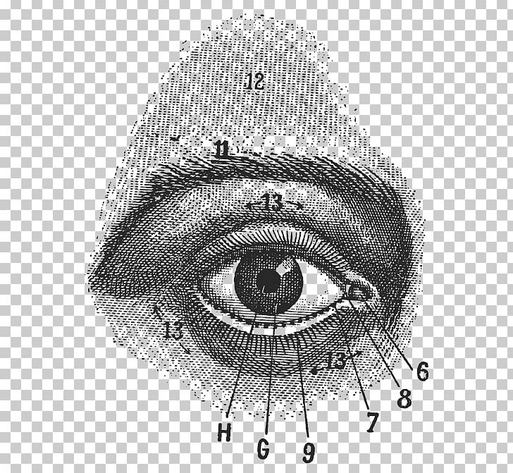 Human Eye Drawing Anatomy Stock Photography Png Clipart
 Pin By Connie Jackson Williams On Eyes Eyeball Drawing
 Sculpting Eyes Drawings Art Drawings Eye Anatomy

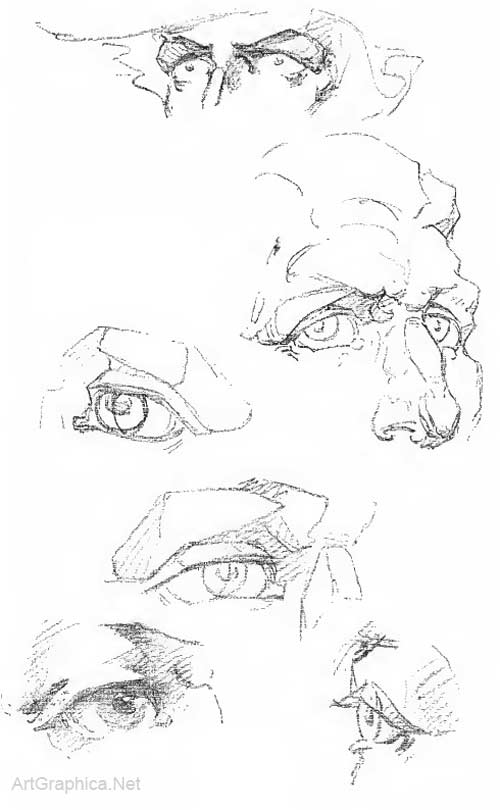
 How To Draw Animals Horses Their Anatomy And Poses
 Eyes Print Anatomy Print Black Eye Tattoo Print Eye Drawing Illustration Eye Anatomy Primt Eye Tattoo Drawing Eye Illustration
 Images Eye Sketches Realistic Eye Anatomy Infographics
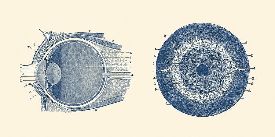 Human Eye Anatomy Diagram Dual View
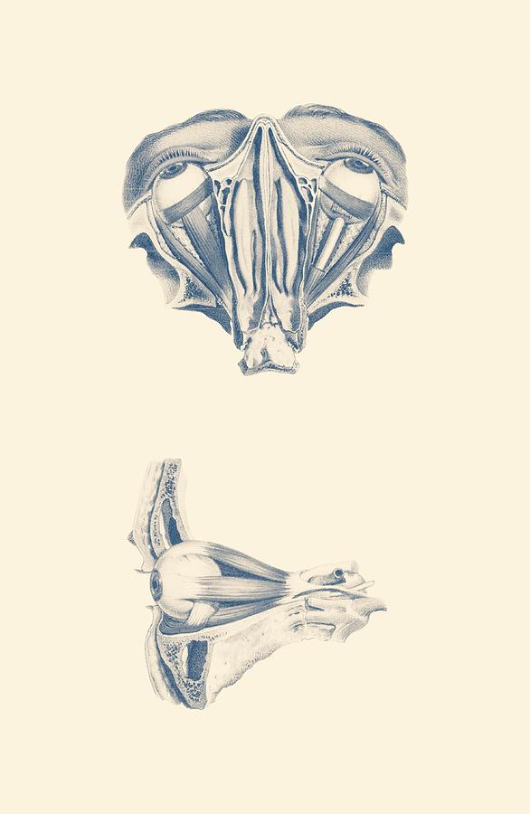
 The Eye As Art Anatomy And Vision In The 18th Century
Human Eye Drawing Png Clipart Anatomy Angle Area Auto

 Character Design Collection Eyes Anatomy
 Human Eye Anatomy Dual View
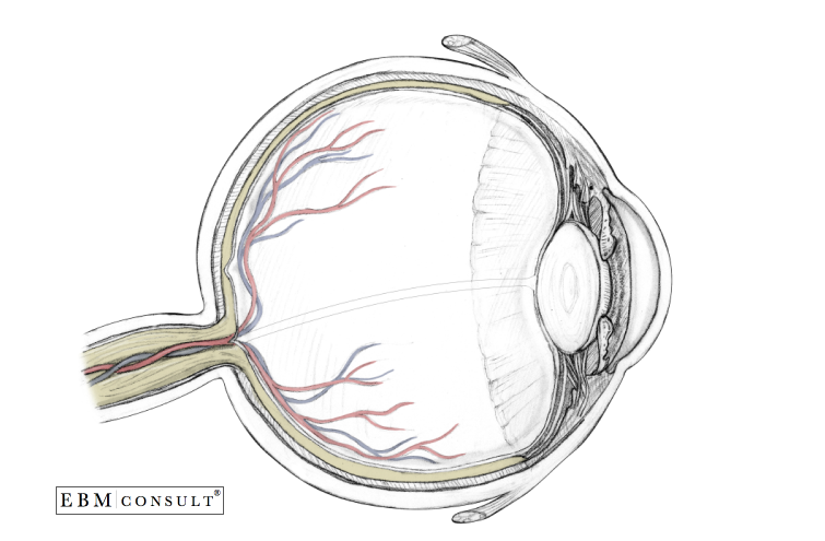 Anatomy Eyeball Sagittal View
 7 Best Anatomy Structure Of Eyes Images Anatomy Drawing
 Royalty Free Human Eye Anatomy Stock Images Photos
 Anatomy For Artists Eye Anatomy
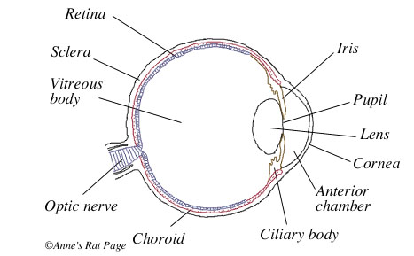
 Eye Anatomy Diagram Enchantedlearning Com
Belum ada Komentar untuk "Anatomy Of The Eye Drawing"
Posting Komentar