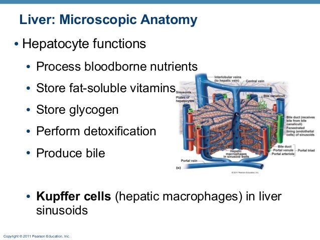
Microscopic Anatomy Of The Liver
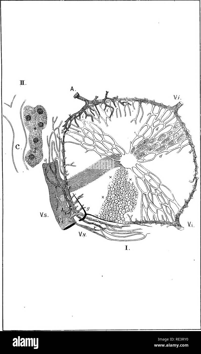
Such structural and functional organization allows assessment of diffuse disease processes in small representative biopsy specimens. The liver is a complex three dimensional structure that consists of epithelial and mesenchymal elements arranged in repetitive microscopic units.
 Visceral Organs Advanced Anatomy 2nd Ed
The liver is a complex threedimensional structure that consists of epithelial and mesenchymal elements arranged in repetitive microscopic units.

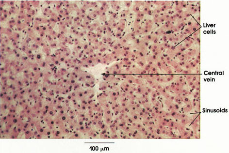
Microscopic anatomy of the liver. Start studying correctly label the following microscopic anatomy of the liver. The liver is a roughly triangular organ that extends across the entire abdominal cavity just inferior to the diaphragm. Such structural and functional organization allows assessment of diffuse disease processes in small representative biopsy specimens.
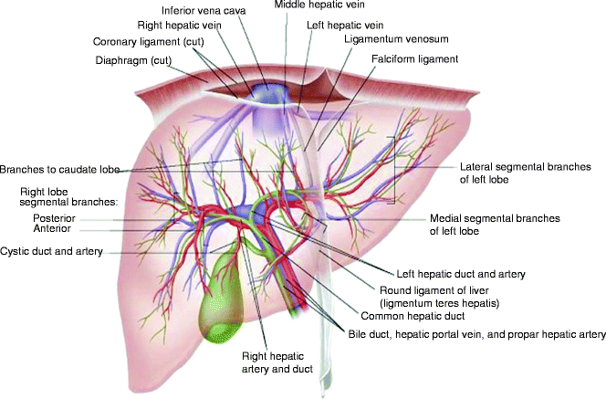
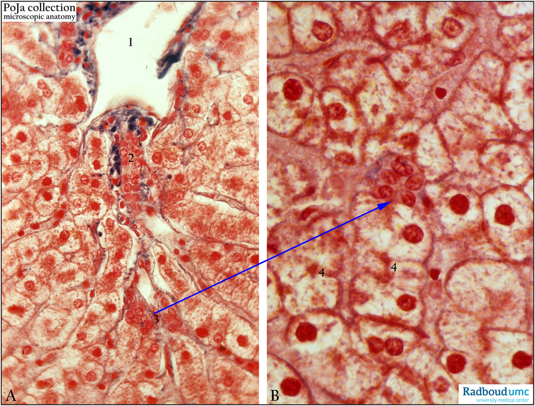
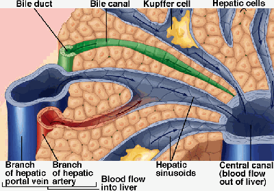
The other major cell of the liver the kupffer cell adheres to the wall of the sinusoid and projects into its lumen. Anatomy of the liver. At the porta hepatis the glisson capsule travels along the portal tracts.
Normal histology of the liver defines the clear shape of the terminal hepatic venule thvcentral vein cv and sinusoids whereas that of the kidney defines clear shape of the bowman capsule and. Microscopic anatomy of the liver murli krishna md. Histologically speaking it has a complex microscopic structure that can be viewed from several different angles.
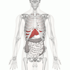
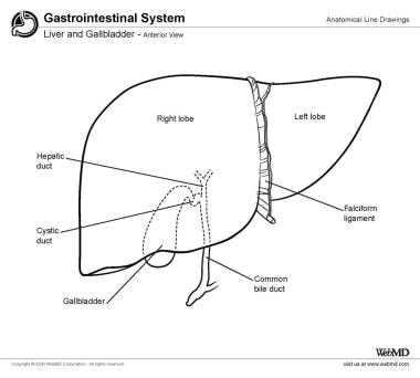
Anatomy of the liver gross anatomy. Anatomically the liver consists of four lobes. The liver is made of very soft pinkish brown tissues encapsulated by a connective tissue capsule.
Learn vocabulary terms and more with flashcards games and other study tools. Microscopic anatomy the surface of the liver is covered by visceral peritoneum serosa with a glisson capsule underneath. The macroscopic and microscopic anatomy of the liver is difficult to understand partly because of its inherently complicated three dimensional structure and partly because of the recent trend to replace simple but misleading morphological descriptions with more accurate but less obvious functional descriptions.
It functions as a phagocyte a cell that engulfs and destroys foreign material or other cells. Small spaces disse spaces are present in places between the hepatocyte and the sinusoidal endothelium. Most of the livers mass is located on the right side of the body where it descends inferiorly toward the right kidney.
Two larger ones right and left and two smaller ones quadrate and caudate.
 Physiology And Anatomy Of The Liver Springerlink
![]() Liver And Gallbladder Anatomy Location And Functions Kenhub
Liver And Gallbladder Anatomy Location And Functions Kenhub
 Microscopic Anatomy Of Liver Tissue Viewed Under 109
 B Str 301 Study Guide Midterm Guide Rectus Femoris Muscle Subscapularis Muscle Cervical Vertebrae
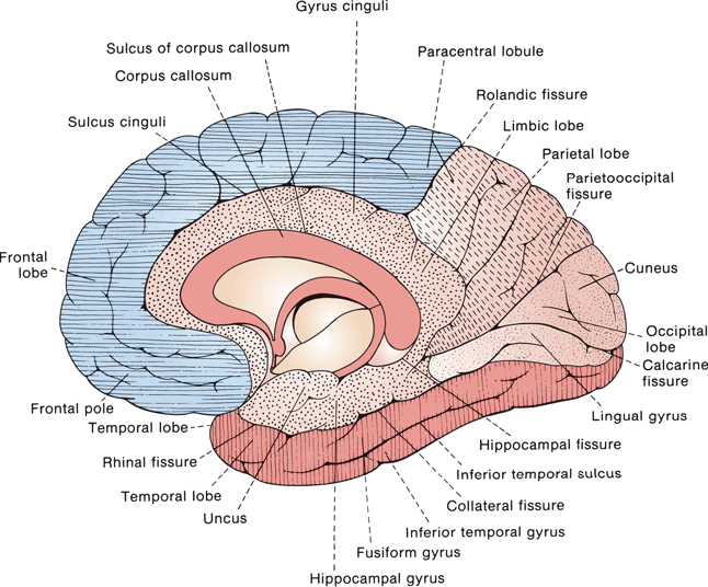 Gross And Microscopic Anatomy Of The Cerebral Hemispheres
 Ou Human Physiology Accessory Organs In Digestion The
The Liver Gallbladder And Pancreas Unity Companies
 Exemplary Microscopic Anatomy Of The Liver And Hepatic
 Anatomy Atlases Atlas Of Microscopic Anatomy Section 1 Cells

 Macroscopic Anatomy An Overview Sciencedirect Topics
 Microscopic Anatomy An Overview Sciencedirect Topics
 Liver Wikipedia
 Duodenal Anatomy Overview Gross Anatomy Microscopic Anatomy
 Anatomy And Function Of The Liver
 Anatomy And Physiology Hepatitisliver
 Microscopic Anatomy Liver Labeling Diagram Quizlet
 Liver Anatomy
 Digest Ii Online
 Microscopic Anatomy Of The Liver Diagram Quizlet
 Liver Anatomy Overview Gross Anatomy Microscopic Anatomy
![]() Liver And Gallbladder Anatomy Location And Functions Kenhub
Liver And Gallbladder Anatomy Location And Functions Kenhub
 Special Report On Diseases Of Cattle Cattle Diseases Of
 Liver Anatomy Overview Gross Anatomy Microscopic Anatomy
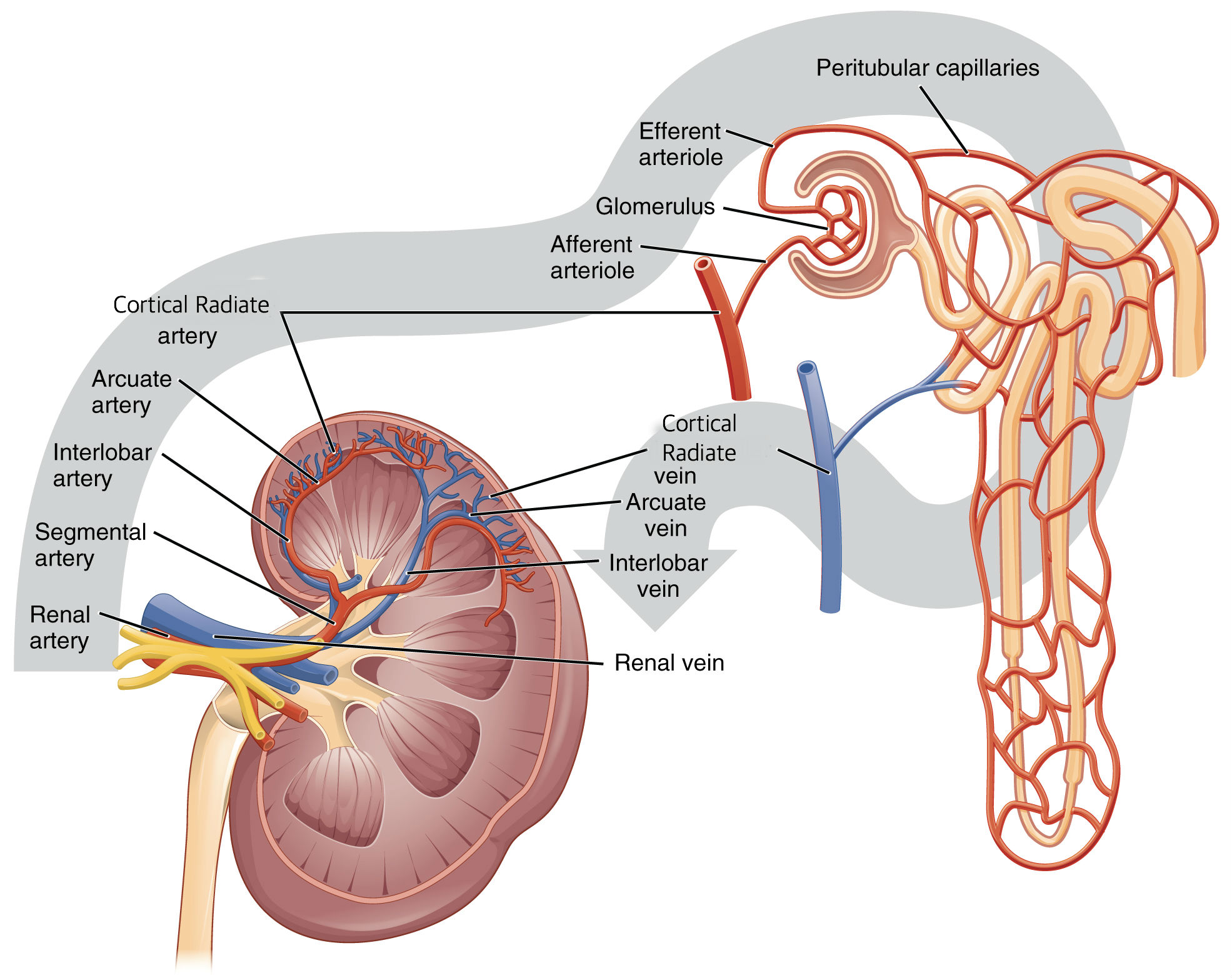
 25 1 Internal And External Anatomy Of The Kidney Anatomy
Belum ada Komentar untuk "Microscopic Anatomy Of The Liver"
Posting Komentar